
 By Nana O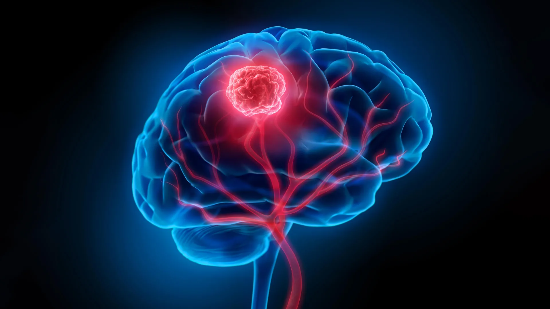
Glioblastoma, a formidable and aggressive form of brain cancer, presents one of the most significant challenges in modern oncology. Its insidious nature lies in its ability to infiltrate surrounding healthy brain tissue, rendering complete surgical removal a complex and often impossible task. This infiltration, coupled with the formidable barrier imposed by the blood-brain barrier, severely limits the efficacy of conventional treatments like chemotherapy and radiotherapy. Consequently, the grim reality for patients diagnosed with glioblastoma is a five-year survival rate that hovers around a mere 7 percent, underscoring the urgent need for innovative therapeutic strategies.
In a significant stride towards addressing these critical limitations, researchers from the University of Technology Sydney (UTS), in collaboration with Harvard University and Henan University, have unveiled a groundbreaking ‘double-punch’ nanozyme platform. This sophisticated system, detailed in the prestigious journal Science Translational Medicine, ingeniously employs a single, intelligent nanoparticle design to tackle both the surgical and therapeutic hurdles posed by glioblastoma.
A Paradigm Shift in Glioblastoma Treatment: The Double-Punch Nanoparticle
The core innovation of this new platform lies in its ability to perform two distinct, sequential functions, guided by the same external trigger. "We’ve engineered a single material that does two jobs in sequence," explained Dr. Bingyang Shi, Chair Professor of nanomedicine from the School of Electrical, Mechanical and Biomedical Engineering at UTS and a lead author on the study. "It’s a precise guide for the surgeon during the operation, and then a targeted clean-up treatment afterward." This dual-action capability represents a significant departure from conventional single-purpose therapeutic agents.
At the heart of this revolutionary system is an exceptionally thin, two-dimensional sheet. This material is meticulously constructed by placing individual atoms, one by one, utilizing a method adapted from advanced semiconductor manufacturing techniques. This atomic-level precision imbues the material with the remarkable ability to switch between its dual roles. Initially, it acts as a highly sensitive imaging agent, enabling surgeons to visualize cancerous tissues with unprecedented clarity. Following surgical resection, the same nanoparticle platform can be reactivated to deliver a targeted therapeutic intervention, all initiated and controlled by a single wavelength of near-infrared light. This unified activation mechanism simplifies the treatment protocol and enhances its precision.
Enhancing Surgical Precision: Visualizing Microscopic Tumors
The initial phase of the nanozyme platform’s application focuses on augmenting surgical precision. During an operation, the nanoparticles function as a potent imaging agent. "During surgery, it functions as a highly sensitive imaging agent," stated Professor Shi. A specially engineered fluorescent dye integrated into the nanoparticle sheet emits a distinct glow when exposed to near-infrared light, a wavelength invisible to the human eye. This allows surgeons to discern individual tumor cell clusters as small as 44 micrometers, a level of detail far exceeding the capabilities of current clinical imaging technologies.
Furthermore, the nanoparticles are equipped with a specific targeting molecule. This crucial component enables the nanoparticles to actively navigate the challenging terrain of the blood-brain barrier, a natural defense mechanism that prevents many therapeutic agents from reaching their intended targets in the brain. Once past this barrier, the targeting molecule ensures that the nanoparticles selectively accumulate within glioma cells, concentrating their imaging capabilities precisely where they are needed most. This targeted accumulation minimizes off-target effects and maximizes the potential for accurate tumor delineation.
The ability to visualize such minuscule tumor clusters is paramount in glioblastoma surgery. Even the most skilled surgeons can struggle to differentiate between cancerous and healthy brain tissue, particularly when the tumor has infiltrated extensively. The advanced imaging provided by this nanoparticle platform offers a visual roadmap, guiding the surgeon to remove as much of the tumor as possible while preserving vital brain functions. This enhanced visualization has the potential to significantly reduce the risk of incomplete resection, a major contributor to tumor recurrence.
Post-Surgical Intervention: Eliminating Residual Cancer Cells
Once the visible tumor has been surgically excised, the nanozyme platform seamlessly transitions to its therapeutic role. The same material, now residing within the surgical cavity, is reactivated using the identical near-infrared light. This activation triggers a two-pronged attack on any remaining microscopic cancer cells that may have eluded surgical removal.
"After the visible tumor is removed, the same material is administered into the surgical cavity and reactivated with the same wavelength of light for postoperative phototherapy," Professor Shi elaborated. The platinum atoms embedded within the nanoparticle structure play a critical role. They catalyze a reaction with the tumor’s inherent hydrogen peroxide, converting it into oxygen. This process is vital because glioblastoma tumors often create a hypoxic, or low-oxygen, microenvironment. This low-oxygen state typically shields cancer cells from conventional treatments, rendering them more resistant. By increasing oxygen levels, the nanoparticles disrupt this protective shield, making the residual cancer cells more susceptible to therapy.
Simultaneously, the near-infrared light activation generates heat and reactive oxygen species (ROS). These highly potent molecules are cytotoxic, meaning they are toxic to cells. They work to destroy any microscopic cancer cells that may have remained in the surgical bed, preventing them from proliferating and initiating a recurrence of the tumor. This targeted post-operative therapy aims to eradicate the microscopic disease that is often the root cause of treatment failure and patient mortality in glioblastoma.
Addressing the Challenge of Tumor Recurrence
Tumor recurrence is the Achilles’ heel of glioblastoma treatment. Even after seemingly successful surgery and adjuvant therapies, microscopic cancer cells left behind can lie dormant before initiating a devastating resurgence. The dual-action nanoparticle platform is specifically designed to confront this persistent problem. By offering both enhanced visualization for maximal tumor removal and a targeted therapeutic intervention to eliminate residual disease, it presents a comprehensive strategy to combat recurrence.
Pre-Clinical Efficacy: Promising Results in Animal Models
The potential of this innovative platform has been rigorously tested in pre-clinical studies using mouse models of glioblastoma. The results have been highly encouraging, demonstrating a significant reduction in tumor recurrence following surgical intervention when the nanoparticle treatment was administered. In a critical comparison, mice treated with the nanoparticle approach following surgery showed a median survival of 60 days. In stark contrast, mice that underwent surgery alone had a median survival of only 42 days. This represents a substantial improvement in survival rates in the animal models.
Furthermore, extensive follow-up testing in these models revealed no detectable neurological or motor impairments associated with the treatment. This is a crucial finding, as any new therapeutic intervention for brain cancer must prioritize the preservation of neurological function and quality of life. The absence of adverse side effects in the pre-clinical setting bodes well for the potential translation of this technology to human patients.
The Road Ahead: From Bench to Bedside
While the findings are undeniably promising, the researchers are careful to temper expectations with a clear understanding of the current stage of development. "The results are very encouraging, but this is still early-stage research carried out in mouse models, not in people – and that distinction is important," Professor Shi emphasized. He further noted that the imaging and therapeutic performance of the nanoparticles will need to be rigorously validated at the scale of a human brain, which is considerably larger and more complex than a mouse brain.
The journey from laboratory discovery to clinical application is a long and arduous one, involving multiple phases of rigorous testing, regulatory approval, and potential manufacturing scale-up. However, the potential impact of this nanozyme platform is substantial. If it continues to demonstrate efficacy and safety through the subsequent stages of research and clinical trials, it could fundamentally alter the landscape of glioblastoma treatment.
The hope is that, in the future, surgeons will be equipped with tools that allow them to "see more of the tumor during an operation and treat more of what’s left behind afterward," as Professor Shi articulated. This represents a meaningful step towards overcoming the persistent challenge of glioblastoma recurrence, offering a glimmer of hope for patients facing this devastating diagnosis and their families. The development of such sophisticated, multi-functional nanomedicines underscores the accelerating pace of innovation in cancer research and the potential for nanotechnology to revolutionize the way we diagnose and treat complex diseases. The successful translation of this platform could mark a significant turning point in the fight against one of the deadliest forms of cancer.
