
 By Suherman Candra Maholtra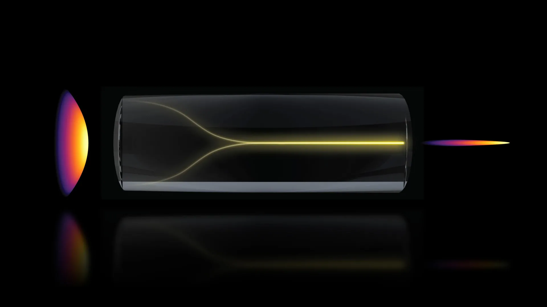
In a significant departure from long-held principles in optical physics, researchers at the Massachusetts Institute of Technology (MIT) have uncovered a self-organizing behavior in laser light that enables the creation of a highly focused "pencil beam." This discovery, documented in a recent study published in the journal Nature Methods, provides a pathway toward imaging living tissue with unprecedented speed and detail. By harnessing this unexpected effect, the research team has demonstrated the ability to produce 3D images of the human blood-brain barrier (BBB) at speeds approximately 25 times faster than current gold-standard techniques, all while maintaining the high resolution necessary for cellular-level analysis.
The discovery challenges the prevailing scientific consensus regarding high-power laser behavior in optical fibers. Traditionally, physicists have operated under the assumption that increasing the power of a laser traveling through a multimode fiber would inevitably lead to increased chaos, scattering, and signal disorder. However, the MIT team, led by Sixian You, an assistant professor in the Department of Electrical Engineering and Computer Science (EECS), found that under precise conditions, the light does not succumb to disorder but instead self-organizes into a stable, narrow beam. This phenomenon allows for deep-tissue imaging without the need for the complex and often expensive beam-shaping hardware typically required to correct optical aberrations.
The Physics of Self-Organizing Light
The foundation of this breakthrough lies in the interaction between high-intensity light and the material properties of optical fibers. Most modern bioimaging relies on single-mode fibers for precision or multimode fibers for power, but both have limitations. Multimode fibers are capable of carrying significant energy, but the various paths (or modes) that light takes through the fiber usually result in a scrambled, unusable output.
The research began when Honghao Cao, an EECS graduate student and the paper’s lead author, was testing the limits of a custom-built fiber shaper. As Cao gradually increased the laser power, the team expected to see the signal degrade into a chaotic "speckle" pattern—a common problem in fiber optics where interference creates a grainy, disordered output. Instead, as the power levels approached the threshold that would normally damage the fiber, the light underwent a phase transition. The nonlinearity of the glass material began to counteract the intrinsic disorder of the fiber, causing the light to collapse into a singular, ultra-sharp "pencil beam."
"Disorder is intrinsic to these fibers," Professor You explained. "The light engineering you typically need to do to overcome that disorder, especially at high power, is a longstanding hassle. But with this self-organization, you can get a stable, ultrafast pencil beam without the need for custom beam-shaping components."
To achieve this state, the researchers identified two non-negotiable parameters: a strict zero-degree alignment of the laser as it enters the fiber and a power level high enough to trigger nonlinear interactions within the glass. This specific combination allows the system to bypass the standard trade-off between power and clarity, resulting in a beam that lacks the "sidelobes" or blurred halos that typically plague high-intensity optical signals.
A New Era for Blood-Brain Barrier Research
The practical applications of this discovery were immediately tested on one of the most challenging structures in human biology: the blood-brain barrier. The BBB is a semi-permeable border of endothelial cells that prevents solutes in the circulating blood from non-selectively crossing into the central nervous system. While this barrier is essential for protecting the brain from toxins and pathogens, it represents a massive hurdle for modern medicine, as it also blocks more than 98% of small-molecule drugs and nearly 100% of large-molecule drugs.
Current methods for studying the BBB involve capturing multiple 2D "slices" of tissue and digitally reconstructing them into a 3D model. This process is time-consuming and often fails to capture the dynamic, real-time movement of molecules across the barrier. The MIT team’s new approach eliminates this bottleneck. Because the self-organized pencil beam possesses both high resolution and a large depth of focus, it can capture volumetric data much faster than traditional point-scanning systems.
In laboratory trials, the team used the pencil beam to monitor how individual cells within a human BBB model absorbed proteins and drugs. The system operated 25 times faster than existing gold-standard methods, such as two-photon microscopy, providing a real-time view of molecular transport.
Implications for Drug Development and Neurodegenerative Disease
The ability to watch cells absorb treatments in real time has profound implications for the pharmaceutical industry, particularly in the development of therapies for Alzheimer’s disease, Amyotrophic Lateral Sclerosis (ALS), and brain tumors. One of the primary reasons many promising neurological drugs fail in clinical trials is the inability to confirm if the treatment actually reached the intended neurons in sufficient concentrations.
Roger Kamm, the Cecil and Ida Green Distinguished Professor of Biological and Mechanical Engineering at MIT and a co-author of the study, emphasized the importance of using human-based models in this research. "The pharmaceutical industry is especially interested in using human-based models to screen for drugs that effectively cross the barrier, as animal models often fail to predict what happens in humans," Kamm noted.
A critical advantage of the MIT method is that it is "label-free." Traditional imaging often requires cells or drugs to be tagged with fluorescent dyes to make them visible under a microscope. However, these tags can alter the chemical properties of the drug or the biological behavior of the cell, leading to inaccurate data. The pencil-beam technique allows for high-contrast imaging without these artificial markers, providing a more authentic look at biological processes.
"For the first time, we can now visualize the time-dependent entry of drugs into the brain and even identify the rate at which specific cell types internalize the drug," Kamm added.
Chronology of the Discovery and Research Team
The development of this technique was an iterative process that combined expertise from electrical engineering, biological engineering, and clinical medicine. The research was conducted at MIT’s Research Laboratory for Electronics (RLE) and involved a multi-disciplinary team.
- System Design: The team initially developed a precise fiber shaper to control light in multimode fibers, aiming to solve the "speckle" problem through manual engineering.
- Observation of Anomaly: During high-power stress tests, lead author Honghao Cao observed the sudden transition from a scattered signal to a concentrated beam.
- Physical Validation: The team spent months identifying the exact physical requirements—alignment and power thresholds—needed to make the effect reproducible.
- Biological Application: In collaboration with Subhash Kulkarni of Harvard University and Beth Israel Deaconess Medical Center, the team applied the beam to engineered human tissue models.
- Data Analysis: Comparison with existing 3D imaging standards confirmed a 25-fold increase in acquisition speed without loss of image integrity.
The research paper, titled "Self-organized nonlinear fiber lithography for high-speed volumetric imaging," lists a diverse group of contributors, including postdocs Sarah Spitz, Francesca Michela Pramotton, and Federico Presutti, as well as several graduate students. The work reflects a growing trend in "convergence" research, where physical sciences and engineering are applied to solve complex biological puzzles.
Technical Analysis: Breaking the Resolution-Depth Trade-off
In traditional optics, researchers are generally forced to choose between high resolution and a large depth of field. A lens that provides a very sharp image usually has a very narrow plane of focus, meaning only a thin "slice" of a sample is clear at any given time. To image a 3D object, the microscope must move the focus up and down, taking hundreds of pictures.
The MIT "pencil beam" effectively breaks this trade-off. Because the beam is self-contained and non-diverging over a significant distance, it maintains its focus throughout a much deeper volume of tissue. This allows the system to gather 3D information in a single "sweep" rather than through the laborious layer-by-layer approach.
Furthermore, the stability of the beam is a result of "spatial beam cleaning," a nonlinear process where the most intense part of the light beam essentially "pulls" the surrounding energy into its path, cleaning up the peripheral noise that usually causes blurring. This results in images that are not only faster to acquire but are also "cleaner" and free of the optical artifacts that often complicate the interpretation of biological data.
Future Outlook and Scalability
While the current study focused on the blood-brain barrier, the researchers are confident that the technology can be adapted for a wide range of medical and industrial uses. Sarah Spitz, a postdoc on the team, noted that the approach enables the "time-resolved tracking of diverse compounds and molecular targets across engineered tissue models," making it a versatile tool for general biological engineering.
The next phase of research will focus on two fronts: deepening the understanding of the underlying physics and miniaturizing the hardware. The researchers hope to eventually develop portable or endoscopic versions of this imaging system, which could allow surgeons to use high-speed 3D imaging during operations to distinguish between healthy tissue and tumors in real time.
The study was supported by a diverse array of organizations, including the National Science Foundation (NSF), the Silicon Valley Community Foundation, the Diacomp Foundation, the Harvard Digestive Disease Core, and MIT startup funds. Additional support came from the MathWorks Fellowship and the Claude E. Shannon Award.
As neurodegenerative diseases continue to affect millions of people worldwide, the development of tools that can accurately track the efficacy of new treatments is becoming a global health priority. By turning a previously avoided "nuisance" of optical physics—nonlinear chaos—into a precision tool, the MIT team has provided the scientific community with a powerful new weapon in the fight against brain-related illnesses. The discovery underscores the value of "embracing uncertainty" in scientific inquiry, leading to a solution that is as elegant as it is impactful.
