
 By George Ongkojoyo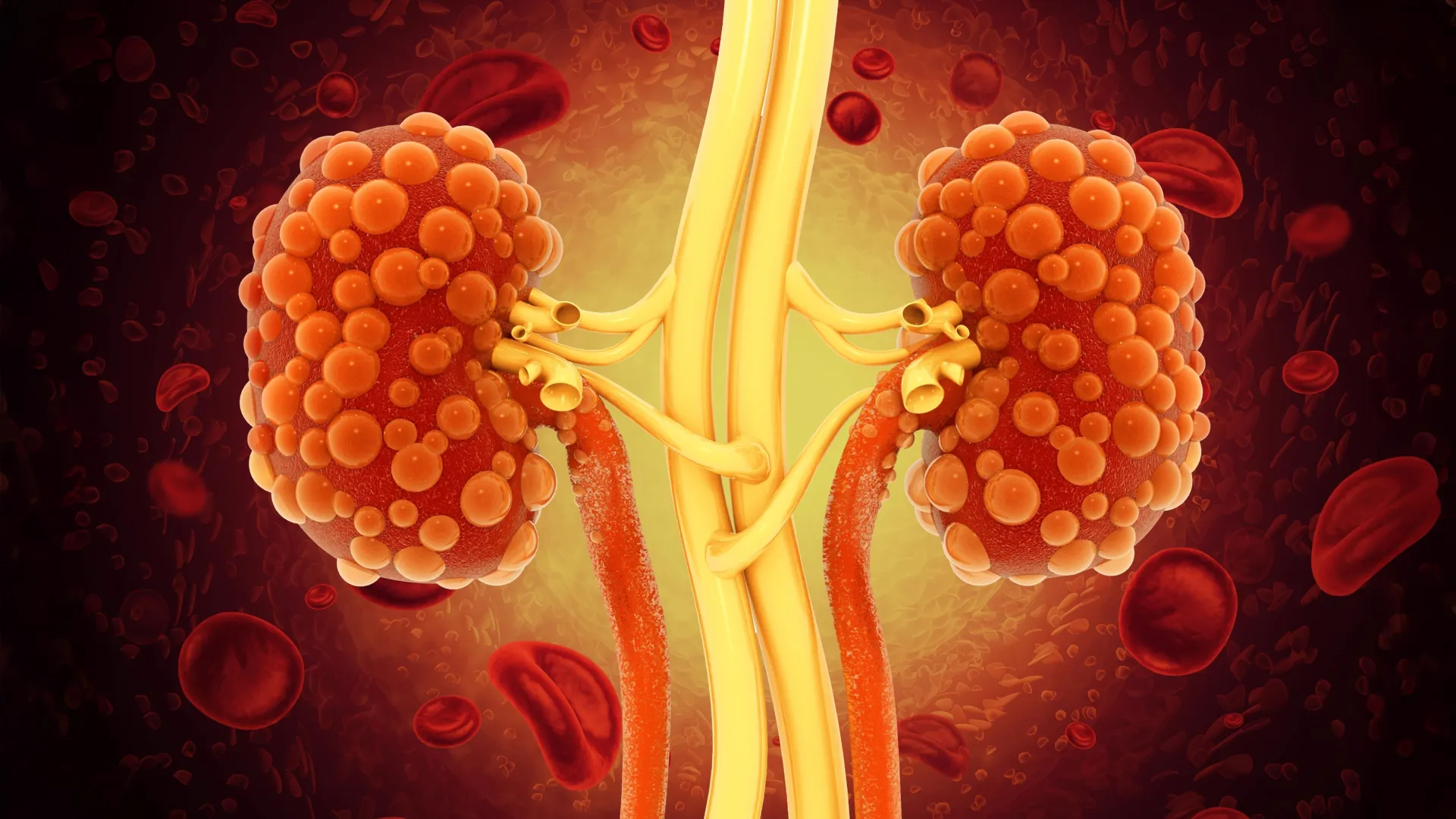
In a significant advancement for renal medicine, researchers at the University of California, Santa Barbara (UCSB) have unveiled a novel therapeutic approach to treating Polycystic Kidney Disease (PKD), a debilitating and currently incurable genetic disorder. The study, published in the prestigious journal Cell Reports Medicine, details how the team successfully engineered a specialized class of monoclonal antibodies capable of penetrating the interior of kidney cysts to halt their progression. This breakthrough addresses a long-standing hurdle in nephrology: the inability of standard large-molecule drugs to reach the specific biological sites where PKD-driven damage occurs.
Polycystic Kidney Disease is characterized by the relentless growth of fluid-filled cysts within the kidneys. As these clusters expand, they displace healthy renal tissue, eventually leading to organ failure. For many of the millions affected worldwide, the disease is a slow-motion crisis that culminates in the need for lifelong dialysis or a kidney transplant. The UCSB research, led by senior author and biologist Thomas Weimbs, offers a potential shift from managing symptoms to actively disrupting the cellular mechanisms that drive cyst proliferation.
Understanding Polycystic Kidney Disease: A Global Health Challenge
PKD is one of the most common inherited disorders, affecting approximately 1 in 400 to 1 in 1,000 people globally. It is primarily categorized into two forms: Autosomal Dominant PKD (ADPKD), which usually manifests in adulthood and accounts for the vast majority of cases, and the rarer Autosomal Recessive PKD (ARPKD), which can be fatal in infancy or early childhood.
The pathology of the disease is rooted in genetic mutations—most commonly in the PKD1 or PKD2 genes—that disrupt the normal signaling of epithelial cells lining the kidney tubules. This disruption causes the cells to proliferate abnormally and secrete fluid, forming "balloons" or cysts. Over decades, a kidney that should be the size of a human fist can swell to the size of a football, weighing up to 30 pounds. This massive enlargement causes chronic pain, hypertension, and a steady decline in glomerular filtration rate (GFR).
Despite the prevalence of the condition, the therapeutic landscape has remained starkly limited. For decades, treatment focused almost exclusively on blood pressure management and dietary restrictions. The economic burden is equally staggering; in the United States alone, the cost of treating end-stage renal disease (ESRD) resulting from PKD and other conditions exceeds billions of dollars annually, much of it covered by Medicare.
The Limitations of Current Pharmacological Interventions
Until recently, the only FDA-approved drug specifically for slowing ADPKD progression was Tolvaptan, a vasopressin V2-receptor antagonist. While Tolvaptan has been shown to reduce the rate of cyst growth and preserve kidney function in some patients, it is far from a "silver bullet."
According to Professor Weimbs, current small-molecule treatments like Tolvaptan often come with a heavy price in terms of patient quality of life. The drug works by blocking the action of vasopressin, which leads to massive water loss. Patients on Tolvaptan must consume several liters of water a day and experience frequent urination, which can be socially and professionally debilitating. More concerning is the risk of hepatotoxicity (liver damage), which requires patients to undergo regular blood monitoring.
The challenge for researchers has been to find a way to stop the "runaway train" of cyst expansion without harming the rest of the body. While small-molecule drugs can enter cells easily, they often lack the specificity needed to target only the diseased tissue. Conversely, traditional monoclonal antibodies—the gold standard in modern cancer immunotherapy—are highly specific but have historically been unable to access the interior of kidney cysts.
The Biological Barrier: Why Standard Immunotherapy Fails PKD Patients
Monoclonal antibodies, specifically the Immunoglobulin G (IgG) class, have revolutionized the treatment of autoimmune diseases and various cancers. However, the unique anatomy of a kidney cyst presents a formidable barrier to these proteins.
In PKD, cysts are essentially sealed chambers. They are lined by a single layer of epithelial cells that create a tight barrier, preventing large molecules from passing from the bloodstream or the surrounding interstitial tissue into the cyst fluid. "IgG antibodies never cross the cell layers and they can never make it inside the cysts," Weimbs explained. This is a critical failure because the "engine room" of the disease is located inside these chambers.
The cells lining the cysts function in an autocrine and paracrine loop; they secrete growth factors into the cyst fluid, which then bind back to receptors on the same cells, triggering further growth and fluid secretion. This self-sustaining cycle of activation is what causes the cysts to grow "endlessly." To stop this, a drug must be able to cross the epithelial barrier, enter the cyst fluid, and neutralize either the growth factors or the receptors.
Engineering a Solution: The Transition from IgG to dIgA
The UCSB team’s innovation lies in changing the "delivery vehicle" of the antibody. Rather than using the common IgG structure, they turned to Dimeric Immunoglobulin A (dIgA).
In the human body, dIgA is a natural component of the mucosal immune system. It is found in tears, saliva, and the lining of the gut. Crucially, dIgA has a unique biological property: it is designed to be transported across epithelial cell layers. This process, known as transcytosis, is mediated by the polymeric immunoglobulin receptor (pIgR). When dIgA binds to pIgR on one side of a cell, the receptor "shuttles" the antibody through the cell and releases it on the other side.
The UCSB researchers hypothesized as early as 2015 that they could hijack this natural transport system. By re-engineering a therapeutic antibody to have a dIgA "backbone," they believed they could trick the kidney cyst cells into pulling the drug inside the cyst.
The Mechanism of Action: Targeting the cMET Receptor
To test their theory, the researchers focused on a specific target: the mesenchymal-epithelial transition (cMET) receptor. cMET is a well-known driver of cell proliferation and is found in high concentrations on the surface of cyst-lining cells in PKD patients.
The research process involved several complex steps:
- Genetic Engineering: The team took the DNA sequence of an existing IgG antibody and modified its "backbone" to match the structure of a dIgA antibody.
- Verification: They confirmed that this newly designed dIgA could still recognize and bind to the cMET receptor with high affinity.
- Transport Testing: Using mouse models of PKD, they demonstrated that the dIgA antibody successfully utilized the pIgR pathway to move from the blood into the interior of the kidney cysts.
Once inside the cyst, the antibody performed exactly as hoped. It bound to the cMET receptors, effectively "plugging" them and preventing growth factors in the cyst fluid from activating the cells.
Experimental Findings and Preclinical Success
The results of the study, as reported in Cell Reports Medicine, were highly encouraging. By blocking the cMET receptor, the dIgA treatment significantly reduced the intracellular signaling pathways that lead to cyst expansion.
Most notably, the treatment appeared to trigger "selective apoptosis." Apoptosis is a form of programmed cell death. In the treated mouse models, the cyst-lining cells began to die off, but the surrounding healthy kidney tissue remained completely unaffected. This selectivity is the "holy grail" of PKD therapy, as it suggests the disease could be reversed or halted without the systemic toxicity associated with chemotherapy or current small-molecule drugs.
"We saw a dramatic onset of apoptosis in cyst epithelial cells," the researchers noted, emphasizing that no noticeable harmful side effects were observed in the healthy parts of the kidney or other organs. This suggests that the pIgR-mediated delivery system is highly localized to the areas where the receptor is active—namely, the diseased cysts.
Chronology of Development and Future Research Directions
The path to this discovery has been nearly a decade in the making.
- 2015: Thomas Weimbs and his colleagues publish a theoretical paper proposing that dIgA could be used to penetrate kidney cysts via the pIgR pathway.
- 2016-2022: The team secures funding from the National Institutes of Health (NIH) and the U.S. Department of Defense (DoD). They begin the arduous task of protein engineering and developing mouse models that mimic human ADPKD.
- 2023-2024: The team successfully demonstrates the efficacy of the dIgA-cMET antibody, leading to the publication of their findings.
Despite the excitement surrounding these results, the researchers cautioned that the work is still in the preclinical stage. Moving from mouse models to human clinical trials is a process that typically takes several years. The next steps involve optimizing the manufacturing of dIgA antibodies—which is more complex and expensive than producing standard IgG—and identifying other growth factors that could be targeted.
"In the literature, there are dozens of growth factors that have been shown to be active in these cyst fluids," Weimbs said. He suggested that the future of PKD treatment might involve a "cocktail" of different dIgA antibodies, each targeting a different receptor (such as EGFR or HER2) to ensure the growth cycle is completely broken from multiple angles.
Economic and Clinical Implications for Nephrology
The potential impact of a successful dIgA therapy cannot be overstated. For patients, it could mean a future where PKD is a manageable condition rather than a precursor to organ failure. From a clinical perspective, a targeted antibody therapy would likely require fewer doses and result in fewer side effects than daily oral medications, potentially improving patient compliance.
For the healthcare system, the long-term savings could be immense. The cost of a single kidney transplant can exceed $400,000, and the annual cost of dialysis is approximately $90,000 per patient. If a targeted therapy can delay the onset of ESRD by even a decade, it would save the global healthcare economy billions of dollars while preserving the productivity and well-being of patients.
Furthermore, this "Trojan Horse" delivery method using dIgA could have applications beyond the kidney. Other conditions involving epithelial-lined cysts or barriers, such as polycystic liver disease or certain types of glandular cancers, might eventually be treated using similar antibody-shuttling techniques.
Conclusion: A New Era for Targeted Renal Therapy
The work conducted by Margaret F. Schimmel, Bryan C. Bourgeois, and the rest of the UCSB team represents a pivotal moment in the fight against Polycystic Kidney Disease. By looking toward the body’s own mucosal defense mechanisms, they have found a way to bypass a biological wall that has frustrated drug developers for decades.
While the journey toward a commercially available treatment remains long, the proof of concept established in this study provides a clear roadmap. The transition from "stop-gap" treatments to targeted, molecular interventions offers a new sense of hope for the millions of families affected by PKD. As the researchers seek partners for clinical development, the medical community will be watching closely to see if this lab-made protein can finally deliver a knockout blow to one of the world’s most persistent genetic challenges.
