
 By George Ongkojoyo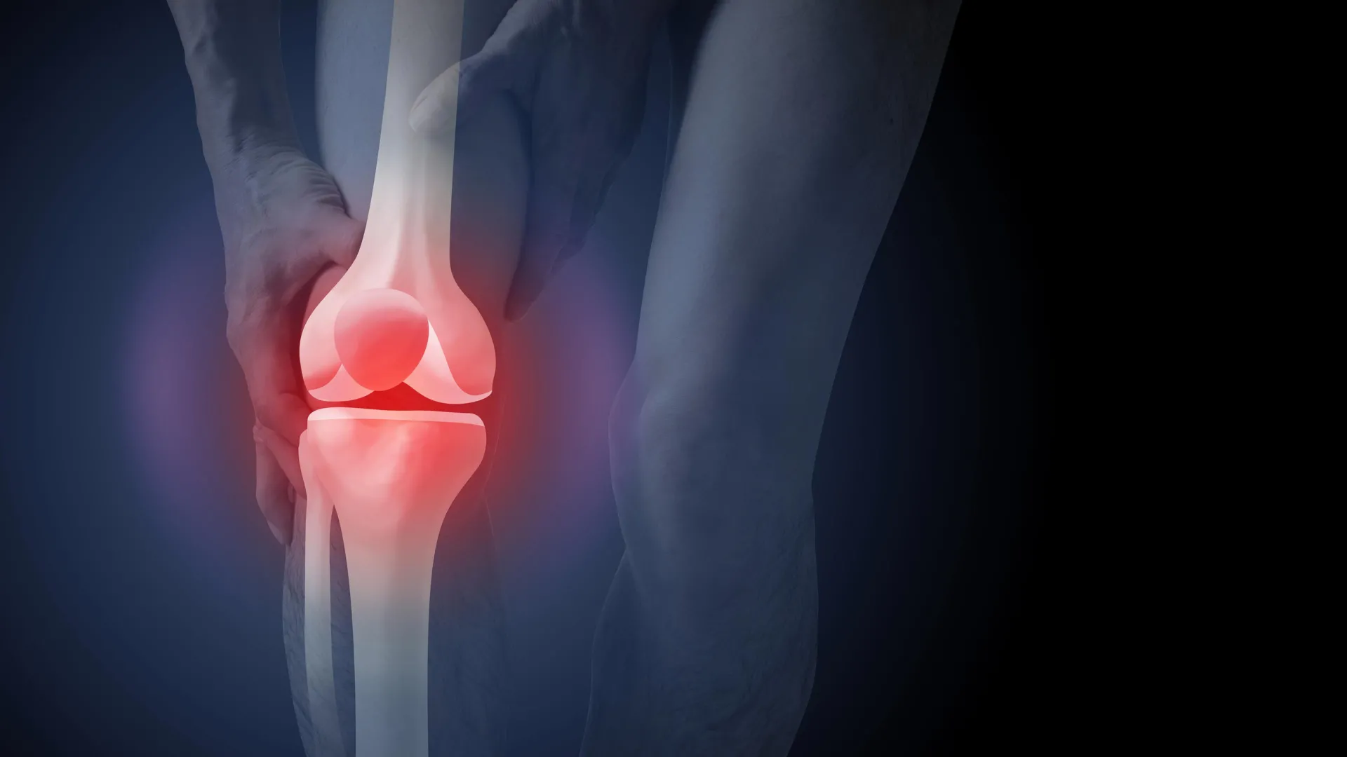
The Australian healthcare system is currently facing a significant disconnect between clinical guidelines and actual medical practice regarding the diagnosis of knee osteoarthritis. While peak health bodies and clinical standards explicitly recommend against the use of routine X-rays for diagnosing this common condition, nearly half of all new patients presenting to general practitioners with knee pain are still referred for imaging. This trend not only places a substantial financial burden on the public purse—costing an estimated A$104.7 million annually for imaging alone—but also fundamentally alters how patients perceive their health. A landmark study now suggests that the visual evidence provided by an X-ray can inadvertently drive patients toward invasive and potentially unnecessary surgeries, such as total knee replacements, while fostering a debilitating fear of physical activity.
The Disconnect Between Diagnostic Guidelines and Clinical Practice
Knee osteoarthritis is a chronic condition characterized by joint changes where the body works to repair itself following various stressors. It is a complex disease involving the entire joint environment, including bone, cartilage, ligaments, and muscle tissue. Despite the sophisticated nature of the disease, the criteria for diagnosis are remarkably straightforward. According to the Australian Commission on Safety and Quality in Health Care, a "clinical diagnosis" is the gold standard. This involves assessing patients aged 45 and over who experience activity-related joint pain and either no morning stiffness or stiffness that resolves within 30 minutes.
However, data indicates a persistent reliance on diagnostic imaging. Of the thousands of Australians who visit their GP for knee pain each year, approximately 50% are sent for X-rays. This practice persists despite overwhelming evidence that the structural changes visible on an X-ray—such as joint space narrowing or bone spurs—do not correlate accurately with a patient’s level of pain or physical disability. Many individuals with significant "wear and tear" on an image experience no symptoms, while others with "clean" X-rays suffer from chronic, debilitating pain.
A Deep Dive into the Psychological Impact of Imaging
A new study published in PLOS Medicine has shed light on why the over-utilization of X-rays is more than just a financial concern. Researchers recruited 617 participants from across Australia to investigate how different diagnostic methods influence a patient’s belief system regarding their recovery. The study utilized a controlled experimental design where participants were randomly assigned to watch one of three hypothetical consultation videos.
In the first scenario, a GP provided a clinical diagnosis based solely on age and symptoms, without ordering an X-ray. In the second and third scenarios, an X-ray was used to confirm the diagnosis, with one group being shown the actual images and the other merely being told the results. The findings were stark: those who received an X-ray-based diagnosis and were shown their images reported a 36% higher perceived need for knee replacement surgery compared to the clinical diagnosis group.
Furthermore, the study revealed that seeing the "damage" on an X-ray led to increased kinesiophobia—the fear of movement. These participants were significantly more likely to believe that exercise and physical activity would cause further harm to their joints. They also expressed higher levels of anxiety regarding the progression of their condition, viewing their knees as "worn out" rather than as living tissue capable of adaptation and repair.
The Economic Reality of Osteoarthritis in Australia
The financial implications of these diagnostic choices are immense. Beyond the A$104.7 million spent on initial imaging, the downstream costs of osteoarthritis management are a major driver of healthcare expenditure. In the 2020–21 financial year, hospital services related to osteoarthritis cost the Australian system approximately $3.7 billion. This expenditure is primarily driven by joint replacement surgeries.
In 2021–22, more than 53,000 Australians underwent knee replacement surgery. While these procedures can be life-changing for patients with end-stage disease who have exhausted all other options, the rising numbers suggest that surgery is increasingly being viewed as a first-line or inevitable intervention rather than a last resort. The new research suggests that the "visual confirmation" provided by an X-ray plays a pivotal role in this surgical pipeline, convincing patients that their condition is a mechanical failure that can only be fixed through hardware replacement.
Debunking the "Wear and Tear" Narrative
One of the most significant barriers to modern osteoarthritis management is the pervasive myth that the condition is caused by simple "wear and tear." This terminology implies that the joint is like a car tire that has reached the end of its mileage. However, biological joints are dynamic; they respond to loading and movement by strengthening the surrounding structures.
The research highlights that focusing on structural damage visible on an X-ray reinforces the "wear and tear" misconception. When a patient sees a narrowed joint space on a screen, they often internalize a "bone-on-bone" narrative. This leads to a cycle of inactivity, which in turn leads to muscle weakness, weight gain, and increased pain—the very factors that accelerate the need for surgery.
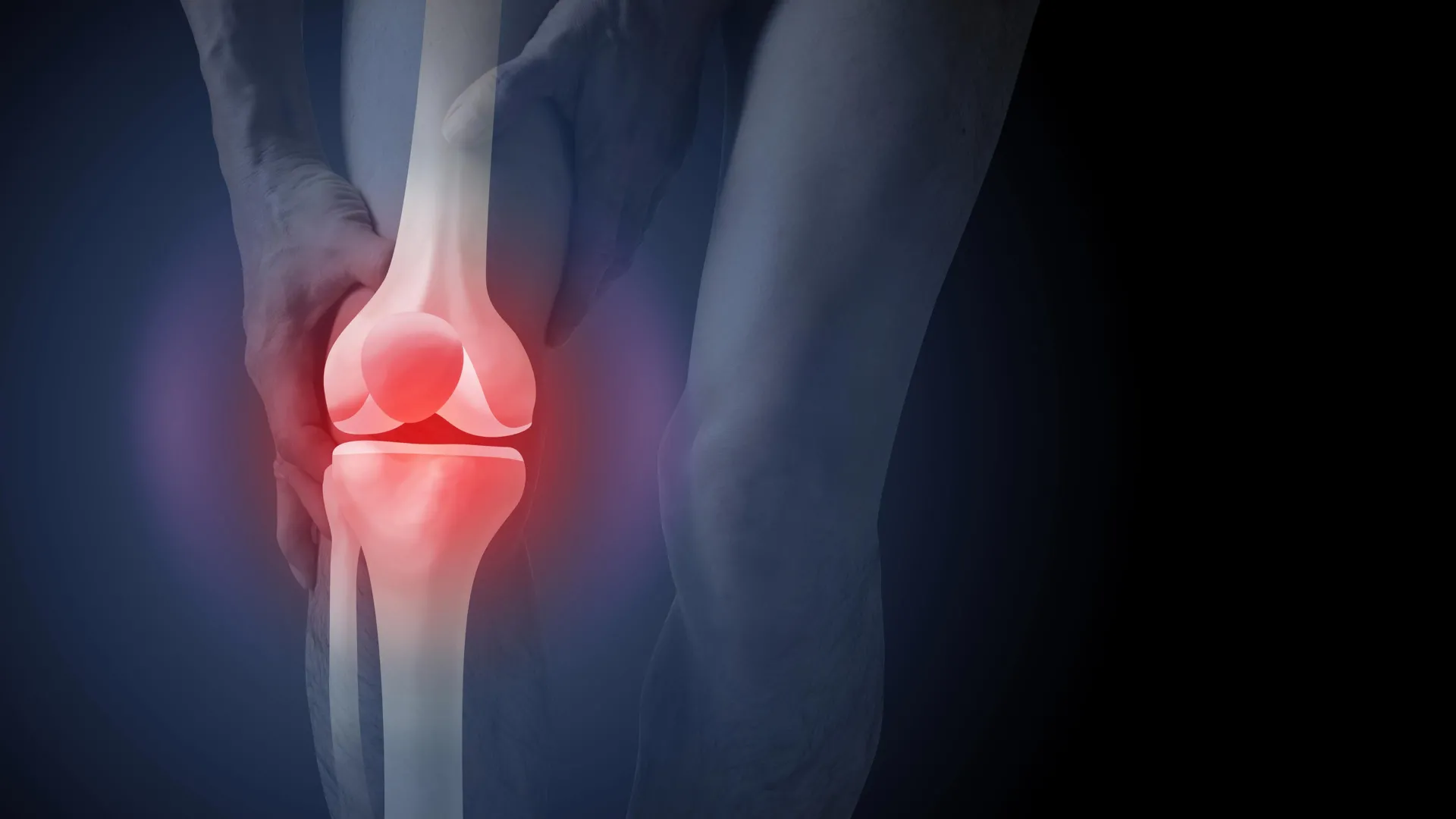
Clinical guidelines emphasize that the extent of joint changes seen on an image does not predict how a person’s symptoms will change over time. By moving away from imaging, clinicians can refocus the conversation on functional improvement and self-management, rather than structural "defects."
The Risks of Premature Surgical Intervention
The push toward knee replacement surgery is not without risk. While often successful, total knee arthroplasty is a major operation that carries the potential for serious adverse events, including deep vein thrombosis (blood clots), pulmonary embolism, and surgical site infections. Furthermore, clinical data suggests that approximately 20% of patients remain dissatisfied with the results of their knee replacement, often continuing to experience chronic pain or restricted mobility.
For the majority of patients, non-surgical management remains the most effective and safest route. This includes:
- Exercise and Physical Activity: Tailored programs to strengthen the muscles supporting the knee.
- Weight Management: Reducing the mechanical load on the joint through dietary interventions.
- Education: Empowering patients to understand that pain does not always equal damage.
By opting for an X-ray early in the diagnostic process, patients may bypass these evidence-based treatments in favor of a surgical solution that they might not have needed if their initial diagnosis had been handled clinically.
Broader Implications for Musculoskeletal Health
The findings of this study resonate beyond the knee. Similar patterns have been observed in the management of chronic back and shoulder pain. In these areas, routine imaging (such as MRIs for lower back pain) has been shown to lead to worse outcomes, higher rates of surgery, and increased psychological distress.
The phenomenon of "over-diagnosis" via imaging is a global concern. When health professionals focus on age-related changes that are often normal—such as disc bulges in the spine or minor rotator cuff tears in the shoulder—it can medicalize the aging process and make patients feel more fragile than they actually are. This latest study adds to a growing body of evidence suggesting that "less is more" when it comes to diagnostic imaging for musculoskeletal conditions.
The Path Forward: Changing the Clinical Conversation
Reducing unnecessary X-rays requires a two-pronged approach involving both healthcare providers and patients. For GPs, there is often pressure to order tests to satisfy patient expectations or to provide a sense of "thoroughness." The study noted that patients were slightly more satisfied with an X-ray-based diagnosis, likely because it aligned with their preconceived notions of how medicine should work.
However, true clinical excellence involves educating the patient on why an X-ray might be harmful to their long-term recovery. Health systems may need to consider policy changes, such as revising Medicare reimbursement criteria for knee X-rays to align more closely with clinical care standards.
For the patient, the message is clear: an X-ray is not a prerequisite for effective treatment. In fact, avoiding the image might be the best way to keep moving. By focusing on symptoms and functional goals rather than a grayscale image of a joint, patients can engage in treatments that reduce pain and improve quality of life without the risks associated with surgery and radiation exposure.
As Australia’s population ages and the prevalence of osteoarthritis rises, the sustainability of the healthcare system will depend on shifting toward high-value, evidence-based care. Eliminating routine X-rays for knee pain is a critical step in ensuring that patients receive the right treatment at the right time, rather than being led down a path of unnecessary anxiety and intervention.
