
 By Nana O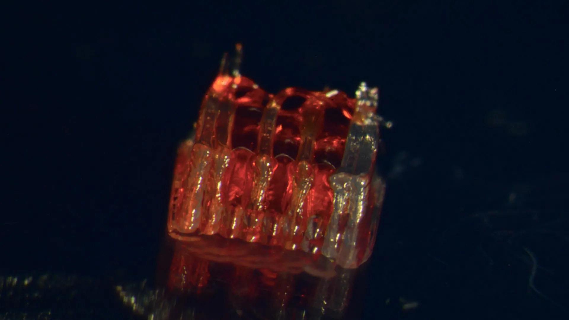
A groundbreaking fusion of 3D printing technology, advanced stem cell biology, and the creation of lab-grown tissues has been successfully demonstrated for the first time by a research team at the University of Minnesota Twin Cities, offering a beacon of hope for individuals suffering from spinal cord injuries. This pioneering work, detailed in the latest issue of the peer-reviewed scientific journal Advanced Healthcare Materials, represents a significant leap forward in the quest to reverse the devastating effects of spinal cord damage and paralysis.
The Unmet Challenge of Spinal Cord Injury
Spinal cord injuries (SCIs) represent a profound public health crisis, impacting hundreds of thousands of lives annually. In the United States alone, the National Spinal Cord Injury Statistical Center reports that over 300,000 people live with the consequences of these injuries. The fundamental challenge in treating SCIs lies in the inherent limitations of the central nervous system: the death of critical nerve cells and the remarkable inability of nerve fibers, or axons, to regenerate across the site of injury. This biological barrier has long thwarted efforts to restore function and has left a vast population with permanent paralysis. The University of Minnesota’s innovative approach directly confronts this long-standing obstacle by creating a novel pathway for neural regeneration.
A Synergistic Approach: 3D Printing, Stem Cells, and Organoid Scaffolds
The core of this research lies in the ingenious design and application of a specialized 3D-printed framework, termed an "organoid scaffold." This scaffold is meticulously engineered with microscopic channels, providing a precise architectural blueprint for the development of lab-grown neural tissues. The critical step involves populating these intricate channels with regionally specific spinal neural progenitor cells (sNPCs). These sNPCs are not just any cells; they are derived from human adult stem cells, endowing them with the remarkable plasticity to divide and differentiate into various specialized mature cell types, including the vital neurons essential for spinal cord function.
Dr. Guebum Han, a former postdoctoral researcher in mechanical engineering at the University of Minnesota and the lead author of the study, currently with Intel Corporation, elaborated on the significance of this directed growth. "We use the 3D printed channels of the scaffold to direct the growth of the stem cells, which ensures the new nerve fibers grow in the desired way," Dr. Han explained. "This method creates a relay system that when placed in the spinal cord bypasses the damaged area." This elegantly simple yet profound concept highlights the precision offered by 3D printing in guiding cellular behavior, a feat difficult to achieve with conventional methods. The engineered scaffold acts as a biological bridge, facilitating the reconnection of severed neural pathways.
Pre-Clinical Success in Animal Models
To rigorously test their innovative approach, the research team transplanted these sophisticated organoid scaffolds into rats that had sustained completely severed spinal cords. The results of these pre-clinical trials were highly encouraging. The sNPCs within the scaffolds not only survived but successfully differentiated into functional neurons. Crucially, these newly formed nerve fibers exhibited robust growth, extending in both the rostral (towards the head) and caudal (towards the tail) directions. This bi-directional growth is essential for re-establishing communication across the injury site and integrating with the host’s existing neural network.
Over time, the newly generated nerve cells demonstrated remarkable integration with the host spinal cord tissue. This seamless integration was accompanied by significant functional recovery in the treated rats, indicating a tangible restoration of motor and sensory capabilities. While the precise metrics of functional recovery would be detailed in the full publication, the observation of restored function in a model of complete spinal transection is a powerful testament to the potential of this therapeutic strategy.
A New Era in Regenerative Medicine for SCI
The implications of this research extend far beyond the laboratory. Ann Parr, a professor of neurosurgery at the University of Minnesota and a senior figure in the study, expressed enthusiasm for the future trajectory of this work. "Regenerative medicine has brought about a new era in spinal cord injury research," Professor Parr stated. "Our laboratory is excited to explore the future potential of our ‘mini spinal cords’ for clinical translation." The term "mini spinal cords" aptly describes the engineered constructs, highlighting their ability to mimic key aspects of the native spinal cord’s structure and function.
The path from laboratory discovery to clinical application is often a lengthy and complex one, but this research represents a pivotal early step. The team acknowledges that the current study is in its nascent stages, but it undeniably opens a new and promising avenue of hope for the millions affected by spinal cord injuries worldwide. The immediate goals for the researchers include scaling up the production of these organoid scaffolds and continuing the rigorous development of this multi-faceted technology, with an ultimate aim of translating these findings into effective clinical treatments for human patients.
A Collaborative Endeavor with Broad Support
This significant scientific advancement was the product of a highly interdisciplinary collaboration, bringing together expertise from various departments at the University of Minnesota and beyond. Key contributors to the study included:
- From the University of Minnesota Department of Mechanical Engineering: Hyunjun Kim and Michael McAlpine, alongside Dr. Guebum Han.
- From the University of Minnesota Department of Neurosurgery: Nicolas S. Lavoie, Nandadevi Patil, and Olivia G. Korenfeld.
- From the University of Minnesota Department of Neuroscience: Manuel Esguerra.
- From Virginia Commonwealth University, Department of Physics: Daeha Joung.
The research received crucial financial backing from several esteemed organizations, underscoring the recognized importance of this line of inquiry:
- The National Institutes of Health (NIH)
- The State of Minnesota Spinal Cord Injury and Traumatic Brain Injury Research Grant Program
- The Spinal Cord Society
Future Directions and Potential Impact
The success of this research hinges on its ability to overcome the biological barriers that have historically impeded spinal cord repair. The precise control afforded by 3D printing allows for the creation of intricate microarchitectures that can guide cell growth and axonal extension in a manner that mimics the natural organization of the spinal cord. By using sNPCs derived from adult stem cells, the researchers leverage the body’s own regenerative potential, aiming to create a biocompatible and effective therapeutic solution.
The implications of this work are far-reaching. A successful clinical translation could offer individuals living with paralysis the possibility of regaining lost function, improving their quality of life, and reducing the immense personal and societal costs associated with spinal cord injuries. This includes not only motor function but also potentially sensory perception and autonomic functions, which are often compromised following SCI.
The "mini spinal cords" developed by the University of Minnesota team represent a significant step towards personalized regenerative medicine. The ability to create patient-specific or regionally matched scaffolds and cell populations could further enhance the efficacy and safety of such treatments.
Looking Ahead: From Bench to Bedside
The journey from a laboratory breakthrough to a widely available clinical treatment is rigorous and demanding. The next phases of research will likely involve:
- Long-term studies: Assessing the durability and sustained functional recovery in animal models.
- Immunological compatibility: Further investigation into how the host immune system interacts with the transplanted scaffolds and cells.
- Optimization of cell sourcing and differentiation: Refining protocols for generating large quantities of high-quality sNPCs.
- Exploration of different injury models: Testing the efficacy of the technology in more complex and clinically relevant SCI models.
- Regulatory pathways: Engaging with regulatory bodies to chart a course for human clinical trials.
The publication of the full paper, "3D-Printed Scaffolds Promote Enhanced Spinal Organoid Formation for Use in Spinal Cord Injury," on the Advanced Healthcare Materials website provides a comprehensive account of the methodology, findings, and future outlook of this transformative research. As scientists continue to unravel the complexities of neural regeneration and harness the power of emerging technologies, the prospect of effective treatments for spinal cord injuries moves closer to reality, offering renewed hope to millions.
