
 By Nana O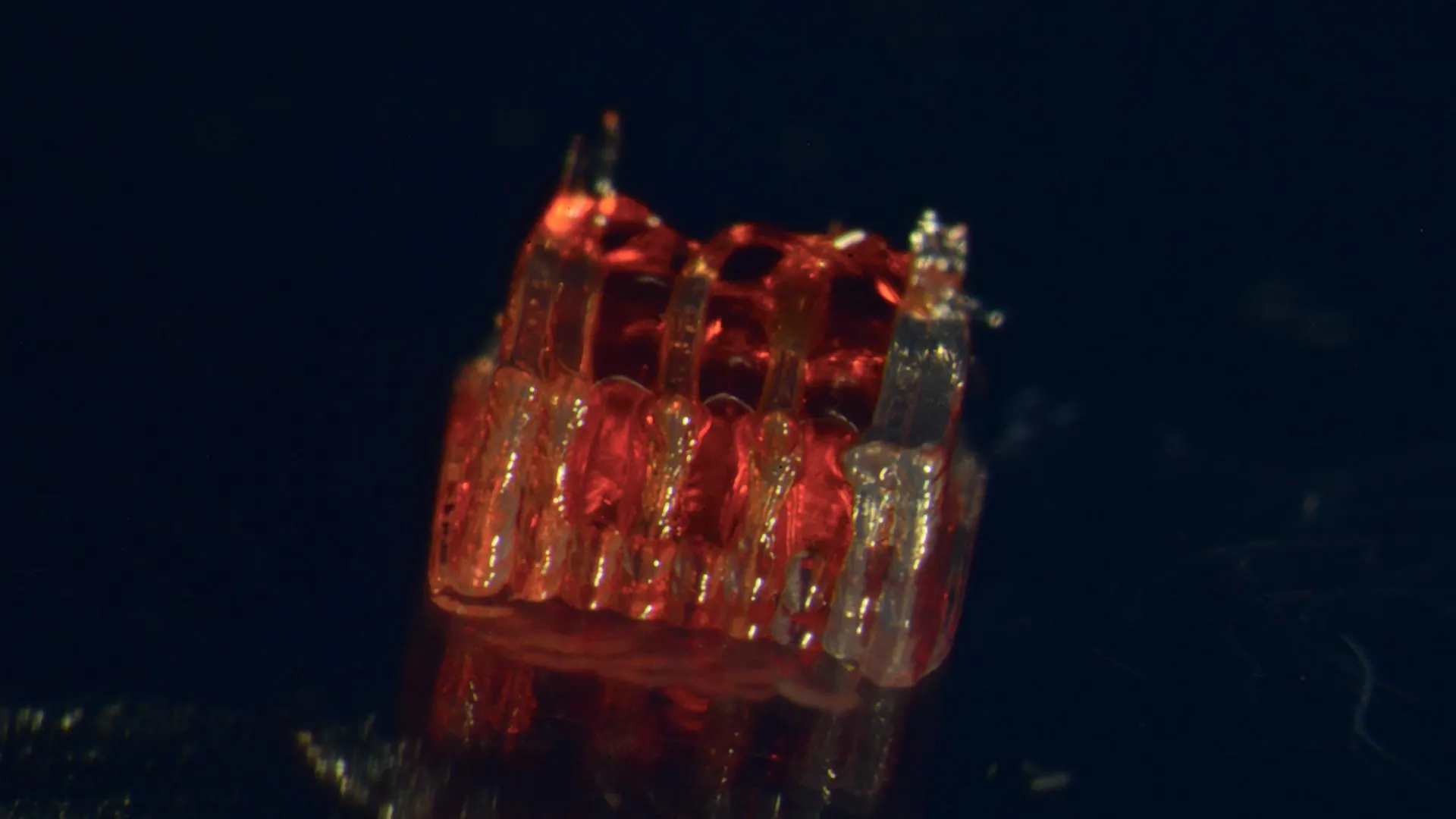
A groundbreaking advancement in regenerative medicine has emerged from the University of Minnesota Twin Cities, where a research team has successfully pioneered a novel process that synergistically integrates 3D printing, stem cell biology, and the creation of lab-grown tissues to address the profound challenges of spinal cord injury recovery. This pioneering work, detailed in a recent publication in the esteemed peer-reviewed journal Advanced Healthcare Materials, offers a beacon of hope for millions worldwide affected by this devastating condition.
The Unmet Need in Spinal Cord Injury Treatment
Spinal cord injuries (SCIs) represent a significant public health crisis, with the National Spinal Cord Injury Statistical Center reporting that over 300,000 individuals in the United States alone live with these injuries. The devastating consequences often include paralysis, loss of sensation, and a myriad of secondary health complications, profoundly impacting the quality of life for patients and their families. Despite decades of intensive research, a definitive cure or complete reversal of SCI-induced damage and paralysis remains elusive. A fundamental obstacle in achieving functional recovery lies in the inherent inability of damaged nerve cells to regenerate effectively and the failure of nerve fibers to bridge the gap created by the injury site. This new research directly confronts these critical limitations, proposing a transformative approach to foster neural regrowth and restore lost function.
A Novel Bio-Integrated Approach: The Organoid Scaffold
The core of this innovative methodology lies in the creation of a sophisticated, bio-integrated system. Researchers engineered a unique 3D-printed framework, termed an "organoid scaffold," designed with intricate microscopic channels. This scaffold serves as a meticulously crafted environment to guide the development and organization of lab-grown tissues.
Crucially, these microscopic channels are then meticulously populated with regionally specific spinal neural progenitor cells (sNPCs). These sNPCs are derived from human adult stem cells, possessing the remarkable capacity to self-renew and differentiate into various specialized cell types, including the vital neurons that form the intricate network of the spinal cord.
Dr. Guebum Han, a former postdoctoral researcher in mechanical engineering at the University of Minnesota and the study’s lead author, who now contributes his expertise to Intel Corporation, elaborated on the ingenious design. "We leverage the precisely engineered 3D printed channels within the scaffold to actively direct the growth patterns of the stem cells," Dr. Han explained. "This controlled guidance is paramount, ensuring that the nascent nerve fibers extend and orient themselves in the most effective and desired manner. The ultimate aim is to construct a functional relay system that, when implanted into the spinal cord, can effectively bypass the compromised or severed area, re-establishing communication pathways."
Pre-Clinical Success: Demonstrating Functional Recovery in Animal Models
To rigorously test the efficacy of their approach, the research team conducted a series of critical pre-clinical trials. In their study, these innovative scaffolds, teeming with differentiated neural cells, were surgically transplanted into rats that had sustained complete spinal cord transections – a severe form of injury that typically results in irreversible paralysis.
The results were profoundly encouraging. The transplanted cells not only survived but also successfully differentiated into mature neurons within the spinal cord environment. Furthermore, these newly formed neurons exhibited remarkable regenerative capabilities, extending their axons (nerve fibers) in both directions – rostrally (towards the head) and caudally (towards the tail). This bidirectional growth is essential for bridging the lesion site and reconnecting with the host’s existing neural circuitry.
Over time, a striking observation was the seamless integration of these new nerve cells into the host spinal cord tissue. This integration was not merely structural; it translated into significant functional recovery in the experimental animals. The rats demonstrated a marked improvement in motor function and coordination, indicating that the bio-engineered "mini spinal cords" were effectively restoring communication across the injury site.
A Paradigm Shift in Regenerative Medicine
This breakthrough represents a significant leap forward in the field of regenerative medicine, an area dedicated to developing therapies that repair, replace, or regenerate damaged cells, tissues, or organs. The ability to precisely control cell growth and organization using 3D printing technology, combined with the inherent regenerative potential of stem cells, opens up unprecedented possibilities for treating conditions previously considered intractable.
Professor Ann Parr, a distinguished neurosurgery professor at the University of Minnesota and a key contributor to the research, expressed palpable excitement about the findings and their future implications. "Regenerative medicine has truly ushered in a new and transformative era in spinal cord injury research," Professor Parr stated. "Our laboratory is immensely enthusiastic about exploring the future potential of these ‘mini spinal cords,’ particularly their translation into clinical applications that can directly benefit human patients."
The Path Forward: From Lab to Clinic
While this research is still in its nascent stages, it provides a powerful new avenue of hope for individuals living with the debilitating effects of spinal cord injuries. The research team is actively focused on scaling up production of these complex bio-engineered constructs and further refining the combination of technologies. Their ultimate goal is to develop this innovative approach into a viable and effective therapeutic strategy for future clinical applications.
The interdisciplinary nature of this research is underscored by the diverse expertise of the team involved. In addition to Dr. Han and Professor Parr, the study benefited from the contributions of Hyunjun Kim and Michael McAlpine from the University of Minnesota’s Department of Mechanical Engineering; Nicolas S. Lavoie, Nandadevi Patil, and Olivia G. Korenfeld from the University of Minnesota’s Department of Neurosurgery; Manuel Esguerra from the University of Minnesota’s Department of Neuroscience; and Daeha Joung from the Department of Physics at Virginia Commonwealth University.
The significant undertaking of this research was made possible through substantial funding from prestigious organizations, including the National Institutes of Health, the State of Minnesota Spinal Cord Injury and Traumatic Brain Injury Research Grant Program, and the Spinal Cord Society. Their commitment to advancing SCI research has been instrumental in bringing this promising technology closer to reality.
The complete findings of this groundbreaking study are available for review in the Advanced Healthcare Materials journal, under the title, "3D-Printed Scaffolds Promote Enhanced Spinal Organoid Formation for Use in Spinal Cord Injury." This publication marks a pivotal moment in the ongoing quest for effective treatments for spinal cord injuries, offering a tangible glimpse into a future where paralysis may no longer be a permanent sentence.
Broader Implications and Future Directions
The implications of this research extend beyond the immediate application to spinal cord injuries. The fundamental principles of using 3D-printed scaffolds to guide stem cell differentiation and tissue organization hold immense potential for a wide range of regenerative medicine applications. This includes the repair of damaged heart tissue after a heart attack, the regeneration of nerves in cases of peripheral nerve injury, and potentially even the development of functional tissues for organ transplantation.
The precision offered by 3D printing allows for the creation of highly customized scaffolds, tailored to the specific anatomical and cellular requirements of different tissues and organs. This level of control is a significant advancement over previous methods, which often lacked the ability to replicate the complex three-dimensional architecture of biological tissues.
Furthermore, the use of regionally specific sNPCs addresses another critical challenge in regenerative medicine: ensuring that transplanted cells differentiate into the correct cell types and integrate appropriately into the existing tissue. By selecting progenitor cells that are already predisposed to becoming spinal cord neurons, the researchers increase the likelihood of successful regeneration and functional recovery.
The successful translation of this technology from animal models to human clinical trials will undoubtedly involve rigorous safety testing, further optimization of the scaffold design and cell culture protocols, and careful consideration of ethical implications. However, the initial success achieved by the University of Minnesota team represents a profound step forward, offering a renewed sense of optimism and a clear path toward a future where the devastating consequences of spinal cord injury can be effectively treated and potentially reversed. The journey from laboratory innovation to patient bedside is often long and complex, but this remarkable achievement marks a significant milestone on that critical path.
