
 By Nana O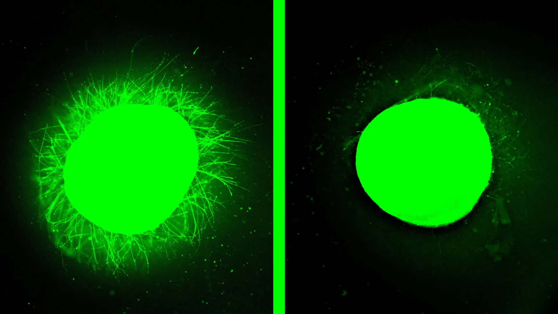
Northwestern University scientists have achieved a significant breakthrough in the quest to understand and treat human spinal cord injuries (SCIs), unveiling the most sophisticated lab-grown model of the human spinal cord to date. This pioneering research, published on February 11 in the prestigious journal Nature Biomedical Engineering, utilizes advanced human spinal cord organoids to meticulously replicate the complex biological aftermath of SCI and critically evaluate a promising regenerative treatment known as "dancing molecules." The implications of this development are profound, potentially accelerating the discovery and validation of therapies that could restore function and improve the lives of millions affected by paralysis.
The research team, led by Samuel I. Stupp, a Board of Trustees Professor at Northwestern with extensive expertise in regenerative materials science, successfully engineered human spinal cord organoids capable of faithfully mimicking the hallmark cellular and molecular responses to traumatic injury. For the first time, these miniature, lab-grown spinal cords exhibited key pathological features observed in human SCIs, including widespread cell death, robust inflammatory responses, and the formation of glial scars. Glial scarring, a dense accumulation of scar tissue, poses a formidable barrier to nerve regeneration, physically and chemically impeding the regrowth of severed nerve fibers.
"One of the most exciting aspects of organoids is that we can use them to test new therapies in human tissue," stated Professor Stupp, the study’s senior author and the visionary behind the "dancing molecules" therapy. "Short of a clinical trial, it’s the only way you can achieve this objective. We decided to develop two different injury models in a human spinal cord organoid and test our therapy to see if the results resembled what we previously saw in the animal model. After applying our therapy, the glial scar faded significantly to become barely detectable, and we saw neurites growing, resembling the axon regeneration we saw in animals. This is validation that our therapy has a good chance of working in humans."
The study’s findings demonstrated that when these injured organoids were treated with Stupp’s innovative "dancing molecules" therapy, the results were remarkably encouraging. This novel therapeutic approach, which had previously shown efficacy in restoring movement and repairing tissue in animal models, led to substantial neurite outgrowth—the regeneration of the long extensions that neurons use to communicate with each other. Concurrently, the therapy significantly reduced the formation of scar-like tissue. These outcomes provide compelling evidence that the "dancing molecules" therapy, which recently garnered Orphan Drug Designation from the U.S. Food and Drug Administration (FDA), holds significant promise for improving recovery in human SCI patients.
The Power of Human Organoids in Injury Research
Organoids, miniature organs grown from induced pluripotent stem cells in a laboratory setting, represent a revolutionary leap in biomedical research. While simplified versions of their full-sized counterparts, they closely recapitulate the intricate structure, diverse cellular makeup, and functional properties of native organs. This remarkable fidelity makes organoids invaluable tools for dissecting disease mechanisms, screening potential treatments, and unraveling developmental processes. Crucially, they offer a faster and more cost-effective alternative to traditional animal experimentation and human clinical trials, allowing for more rapid progress in scientific discovery.
While other research groups have successfully developed spinal cord organoids for fundamental biological studies, the Northwestern team’s model marks a significant advancement specifically for SCI research. These organoids, measuring several millimeters in diameter, were sufficiently mature to accurately model traumatic damage. Over several months, the researchers meticulously guided stem cells to develop into complex spinal cord tissue, incorporating not only neurons but also astrocytes, a crucial type of glial cell. A key innovation in this study was the incorporation of microglia, the resident immune cells of the central nervous system, into the organoid structure. This inclusion was vital for accurately replicating the inflammatory cascade that invariably follows SCI in living organisms.
"It’s kind of a pseudo-organ," Professor Stupp explained. "We were the first to introduce microglia into a human spinal cord organoid, so that was a huge accomplishment. It means that our organoid has all the chemicals that the resident immune system produces in response to an injury. That makes it a more realistic, accurate model of spinal cord injury." The presence of these immune cells allows the organoid model to more closely reflect the dynamic and complex biological environment present after a real-world spinal cord injury, enhancing its predictive power for therapeutic efficacy.
Understanding "Dancing Molecules": A Novel Therapeutic Strategy
The "dancing molecules" therapy, first introduced by Stupp’s laboratory in 2021, is a groundbreaking approach that leverages controlled molecular motion to promote tissue repair and potentially reverse paralysis following traumatic SCI. This therapy belongs to a broader category of supramolecular therapeutic peptides (STPs). STPs function by forming large molecular assemblies, often comprising over 100,000 molecules, which interact with cell receptors to activate the body’s inherent repair signaling pathways. Notably, the underlying concept of supramolecular therapies shares similarities with current GLP-1 drugs used for weight loss and diabetes management, an area Professor Stupp’s lab explored nearly fifteen years ago, highlighting the enduring relevance of his foundational research.
Delivered as a simple liquid injection, the "dancing molecules" therapy rapidly self-assembles into a three-dimensional nanofiber scaffold. This structure closely mimics the extracellular matrix of the spinal cord, the supportive network that surrounds cells. The innovation lies in the dynamic movement of the molecules within this scaffold. By precisely tuning the molecular motion, researchers can optimize how effectively these therapeutic molecules interact with cell receptors, which are themselves in constant motion.
"Given that cells themselves and their receptors are in constant motion, you can imagine that molecules moving more rapidly would encounter these receptors more often," Professor Stupp elaborated in his earlier work. "If the molecules are sluggish and not as ‘social,’ they may never come into contact with the cells." This principle of enhanced molecular mobility translates directly into improved bioactivity and more effective cellular signaling.
Previous animal studies have provided compelling evidence for the therapy’s effectiveness. A single injection administered just 24 hours after a severe SCI in mice enabled them to regain mobility within four weeks. Furthermore, formulations exhibiting faster molecular motion consistently outperformed slower versions, underscoring the critical role of dynamic movement in driving therapeutic outcomes.
Simulating Spinal Cord Trauma in a Lab Setting
To rigorously test the "dancing molecules" therapy, the Northwestern team developed two distinct injury models within their human spinal cord organoids. These models were designed to simulate common types of SCI encountered in clinical practice. The first model involved a precise scalpel incision, replicating the type of laceration that might occur during surgery or a penetrating injury. The second model utilized a compressive contusion injury, mimicking the blunt force trauma often sustained in severe accidents, such as car crashes or falls.
Both simulated injury types reliably induced the critical pathological hallmarks of SCI in the organoids. Researchers observed significant cell death and the characteristic formation of glial scars, mirroring the biological consequences seen in human patients. Professor Stupp noted the ability to differentiate between the astrocytes that constitute healthy tissue and those that become hypertrophied and densely packed within the glial scar. "We also detected the production of chondroitin sulfate proteoglycans, which are molecules in the nervous system that respond to injury and disease," he added. These molecular markers are key indicators of the body’s response to damage.
Following treatment with the "dancing molecules" therapy, the treated organoids displayed a marked reduction in inflammation and a significant shrinkage of glial scarring. Crucially, the therapy stimulated substantial neurite extension, with neurons exhibiting organized growth patterns. Neurites include axons, the long projections of nerve cells that are frequently severed in SCIs, disrupting neural communication and leading to paralysis and sensory loss. The promotion of neurite regrowth is therefore a critical step toward reconnecting damaged neural pathways and potentially restoring lost function.
The Critical Role of Molecular Motion in Healing
Professor Stupp attributes the remarkable efficacy of the "dancing molecules" therapy to the principle of supramolecular motion. This refers to the ability of the therapeutic molecules to move dynamically within the nanofiber network, and even to momentarily detach and reattach. This intrinsic mobility allows them to actively seek out and engage with cell receptors.
Further reinforcing this hypothesis, experiments conducted on healthy, undamaged organoids yielded striking results. "Before we even developed the injury model, we tested the therapy on a healthy organoid," Professor Stupp recounted. "The dancing molecules spun out all these long neurites on the surface of the organoid but, when we used molecules that had less or no motion, we saw nothing. This difference was very vivid." This experiment clearly demonstrated that the therapeutic effect, including neurite outgrowth, is directly linked to the dynamic motion of the molecules, not just their presence.
Looking ahead, the Northwestern team is committed to further refining their organoid models. Future research will focus on engineering even more advanced organoids capable of replicating chronic, long-standing SCIs, which are characterized by thicker and more persistent scar tissue. The potential for personalized medicine is also a significant long-term goal. Professor Stupp envisions the possibility of generating implantable tissue from a patient’s own stem cells, thereby minimizing the risk of immune rejection and paving the way for highly individualized treatment strategies. This groundbreaking work, supported by the Center for Regenerative Nanomedicine at Northwestern University and a generous gift from the John Potocsnak Family for spinal cord injury research, marks a pivotal moment in the fight against spinal cord injuries, offering tangible hope for a future where recovery and functional restoration are attainable realities.
