
 By Suherman Candra Maholtra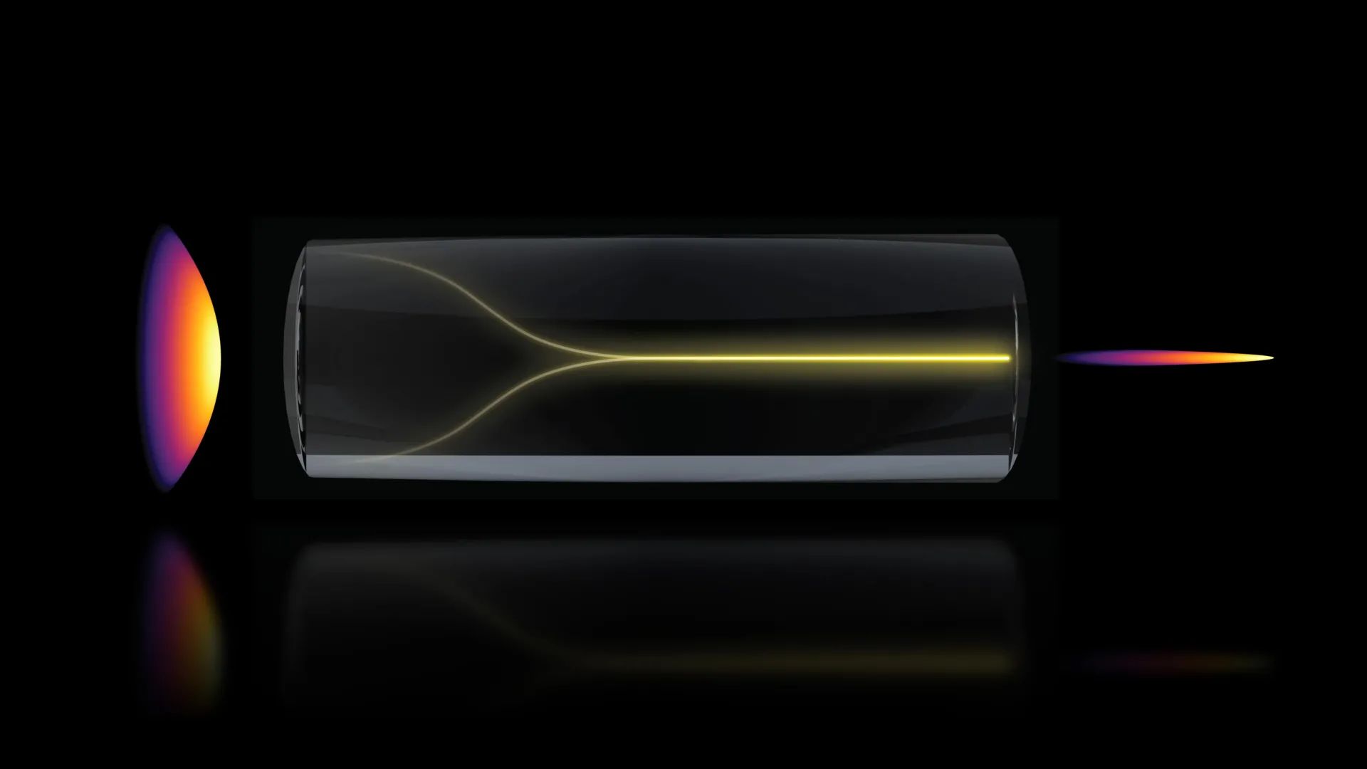
In a significant departure from long-held principles in optical physics, a research team at the Massachusetts Institute of Technology (MIT) has discovered a self-organizing laser phenomenon that promises to transform the field of biological imaging. By harnessing an unexpected "pencil beam" effect within multimode optical fibers, the researchers have developed a method to image living tissue, specifically the human blood-brain barrier, at speeds approximately 25 times faster than current industry-standard techniques. This breakthrough, published today in the journal Nature Methods, offers a new window into the microscopic interactions between drugs and the brain’s protective structures, potentially accelerating the development of treatments for neurodegenerative diseases such as Alzheimer’s and Amyotrophic Lateral Sclerosis (ALS).
The discovery challenges the fundamental assumption that increasing the power of a laser traveling through a disordered medium, such as a standard optical fiber, inevitably leads to chaotic scattering. Instead, the MIT team demonstrated that under precise conditions of alignment and intensity, laser light can undergo a process of self-organization, resulting in a stable, ultra-narrow beam that maintains high resolution over a significant depth of focus. This "pencil beam" allows for rapid 3D reconstruction of complex biological systems without the need for traditional fluorescent labeling, a development that members of the pharmaceutical and bioengineering communities are calling a significant leap forward.
The Physics of Self-Organizing Light
To understand the magnitude of this discovery, one must consider the historical limitations of optical fibers in high-power applications. Multimode optical fibers are designed to carry large amounts of light energy, but they are intrinsically disordered. As photons travel through these fibers, they bounce off internal imperfections, leading to a phenomenon known as modal dispersion. For decades, the consensus in optical engineering was that "cranking up" the power would only exacerbate this disorder, resulting in a blurred, unusable signal.
The MIT research team, led by Sixian You, an assistant professor in the Department of Electrical Engineering and Computer Science (EECS), began their investigation by testing the limits of a "fiber shaper"—a device they had previously engineered to control light propagation. During the experiments, lead author and graduate student Honghao Cao observed a counterintuitive result: as the laser power approached the threshold where the fiber was expected to sustain damage, the scattered light did not become more chaotic. Instead, it suddenly collapsed into a singular, highly focused beam.
"Disorder is intrinsic to these fibers," explained Professor You, who is also a member of the Research Laboratory for Electronics. "The light engineering you typically need to do to overcome that disorder, especially at high power, is a longstanding hassle. But with this self-organization, you can get a stable, ultrafast pencil beam without the need for custom beam-shaping components."
The mechanism behind this transformation is rooted in nonlinear optics. When the laser intensity reaches a critical point, the light begins to interact directly with the glass material of the fiber. This nonlinearity creates a self-focusing effect that effectively "cancels out" the intrinsic disorder of the fiber. The result is a beam that remains tightly focused over a long distance, resembling a thin pencil rather than the traditional cone-shaped focus used in standard microscopy.
Chronology of the Discovery and Experimental Validation
The path to this discovery was marked by a series of rigorous tests designed to reproduce the "pencil beam" effect reliably. The team identified two non-negotiable requirements for the phenomenon to occur. First, the laser must enter the multimode fiber at a perfectly aligned, zero-degree angle—a degree of precision that exceeds standard laboratory practices. Second, the power must be tuned to a specific "Goldilocks" zone where the nonlinear effects are strong enough to organize the light but not so intense as to melt the silica fiber.
Once the conditions were perfected, the team compared their self-organized beam against conventional Gaussian beams and other high-resolution imaging modalities. They found that their beam was remarkably "clean," lacking the "sidelobes" or blurred halos that often plague high-resolution optical systems. These sidelobes typically introduce artifacts into images, reducing clarity and making it difficult to distinguish between closely packed cellular structures.
Following the physical validation of the beam, the researchers transitioned to biological applications. They chose the human blood-brain barrier (BBB) as their primary subject—a choice driven by both the complexity of the tissue and its immense clinical relevance. The BBB is a semi-permeable border of endothelial cells that prevents solutes in the circulating blood from non-selectively crossing into the central nervous system. While it protects the brain from toxins, it also serves as a formidable obstacle for therapeutic drugs.
Supporting Data: Speed, Resolution, and Label-Free Imaging
The data produced by the MIT system highlights a significant shift in the efficiency-resolution trade-off that has long governed microscopy. In traditional 3D imaging, a microscope must scan a sample plane by plane (2D slices) and then stack those slices to create a 3D volume. This process is time-consuming and can lead to phototoxicity or photobleaching if the sample is exposed to light for too long.
According to the study, the new pencil-beam approach achieved the following metrics:
- Imaging Speed: The system generated 3D images of engineered human BBB models 25 times faster than the current gold-standard multiphoton microscopy.
- Depth of Focus: Unlike traditional lenses that have a very shallow "sweet spot" for focus, the pencil beam maintains high resolution across a much larger depth, allowing for deeper penetration into tissue without loss of detail.
- Real-Time Tracking: The speed of the system allowed researchers to observe the time-dependent entry of proteins and drug mimics into cells.
- Label-Free Detection: The method does not require cells to be tagged with fluorescent dyes. This is a critical advantage, as dyes can sometimes alter the natural behavior of cells or interfere with the chemical properties of the drugs being tested.
The ability to perform label-free, high-speed imaging is particularly valuable for the pharmaceutical industry. Currently, many drug candidates for neurological disorders fail in clinical trials because animal models—which are often used for initial screening—do not accurately reflect the permeability of the human blood-brain barrier. By using the MIT team’s method on human-derived engineered tissue models, researchers can gain a more accurate prediction of how a drug will behave in a human patient.
Professional Reactions and Industry Implications
The implications of this research extend far beyond the laboratory. Roger Kamm, the Cecil and Ida Green Distinguished Professor of Biological and Mechanical Engineering at MIT and a co-author of the paper, emphasized the importance of this tool for drug screening.
"The pharmaceutical industry is especially interested in using human-based models to screen for drugs that effectively cross the barrier," Kamm stated. "That this new method doesn’t require the cells to have a fluorescent tag is a game-changer. For the first time, we can now visualize the time-dependent entry of drugs into the brain and even identify the rate at which specific cell types internalize the drug."
Postdoctoral researcher Sarah Spitz noted that the versatility of the "pencil beam" makes it applicable to a wide range of biological engineering tasks. "This approach is not limited to the blood-brain barrier but enables time-resolved tracking of diverse compounds and molecular targets across engineered tissue models," Spitz added. This suggests that the technology could eventually be used to study everything from gut-microbiome interactions to the way cancer cells metastasize through vascular walls.
From a technical standpoint, the simplicity of the setup is perhaps its most disruptive feature. Because the beam self-organizes based on the physics of the fiber rather than complex external hardware, it could be integrated into existing optical systems with relative ease. Professor You noted that the method can be executed with "a normal, optical setup and without much domain expertise," which lowers the barrier to entry for biological laboratories that may not have specialized physics departments.
Analysis of Broader Impact and Future Directions
The discovery of self-organizing light in multimode fibers represents a pivotal moment in the intersection of nonlinear physics and biology. Traditionally, nonlinear effects were seen as obstacles to be managed or suppressed. The MIT team has instead leaned into these effects, turning a "nuisance" of high-power optics into a powerful tool for discovery.
In the short term, this technology is likely to be adopted by research institutions focusing on "Organ-on-a-Chip" technologies. These platforms rely on high-fidelity imaging to validate that their lab-grown tissues are behaving like their real-world counterparts. The ability to monitor these tissues 25 times faster than before means that experiments that previously took weeks could potentially be completed in days, significantly shortening the R&D cycle for new therapeutics.
Looking further ahead, the team plans to investigate whether this self-organizing effect can be achieved in different types of fibers or through different mediums, such as those found deeper within the body. There is also potential for this technology to be used in neuroimaging to watch the firing of neurons in three dimensions in real-time, providing new insights into the functional connectivity of the brain.
The research was supported by a diverse coalition of funders, reflecting its cross-disciplinary appeal. Contributors included the National Science Foundation (NSF), the Silicon Valley Community Foundation, the Diacomp Foundation, and the Harvard Digestive Disease Core, among others. As the team continues to refine the physics of the "pencil beam," the focus will shift toward commercialization and the development of user-friendly interfaces that will allow the broader scientific community to harness the power of self-organizing light.
By bridging the gap between theoretical optical physics and practical biomedical engineering, the MIT researchers have not only solved a technical challenge but have also provided a new lens through which we can observe the most guarded secrets of the human body. The "pencil beam" may soon become a standard tool in the fight against some of the most complex diseases of our time.
