
 By Suherman Candra Maholtra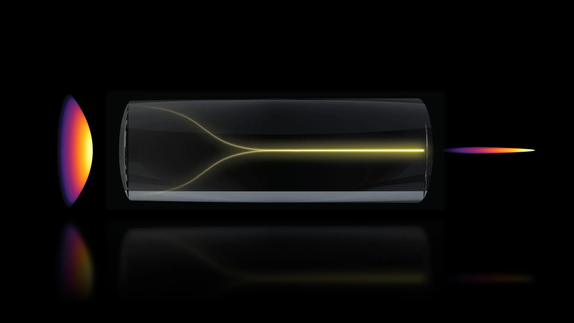
Researchers at the Massachusetts Institute of Technology (MIT) have documented a previously unknown phenomenon in optical physics that allows laser light to spontaneously reorganize itself into a highly focused, stable "pencil beam." This discovery, detailed in a recent publication in the journal Nature Methods, challenges long-standing assumptions regarding the behavior of high-power light in disordered media and offers a transformative approach to biological imaging. By harnessing this self-organizing effect, the research team has demonstrated the ability to produce three-dimensional images of the human blood-brain barrier at speeds approximately 25 times faster than current gold-standard techniques, all while maintaining the high resolution required to observe individual cellular interactions in real time.
The implications of this breakthrough extend far beyond the laboratory, offering a potential paradigm shift in how pharmaceutical companies evaluate the efficacy of treatments for neurodegenerative diseases such as Alzheimer’s and Amyotrophic Lateral Sclerosis (ALS). For the first time, scientists can visualize the movement of drugs across the blood-brain barrier and their subsequent absorption by specific brain cells without the need for invasive fluorescent tagging. This label-free, high-speed imaging capability addresses a critical bottleneck in drug development, where the inability to confirm whether a compound has reached its intended target often leads to the failure of clinical trials.
The Challenge of Optical Disorder and High-Power Lasers
In traditional optical physics, the behavior of light within multimode optical fibers—fibers designed to carry large amounts of power—is characterized by inherent disorder. As laser light travels through these fibers, it encounters microscopic imperfections and variations in the glass material. Under normal circumstances, increasing the power of the laser intensifies this disorder, causing the light to scatter into a chaotic, speckled pattern that is unsuitable for high-precision imaging.
For decades, the prevailing scientific consensus has been that high-power laser signals are fundamentally unstable in such environments. To overcome this, engineers have historically relied on complex "beam-shaping" components—expensive and delicate hardware designed to manually correct the light’s path. However, the MIT team, led by Sixian You, an assistant professor in the Department of Electrical Engineering and Computer Science (EECS), discovered that under a specific set of conditions, the light does not succumb to chaos. Instead, it undergoes a process of self-organization, effectively "solving" the problem of disorder on its own.
"The common belief in the field is that if you crank up the power in this type of laser, the light will inevitably become chaotic," Professor You noted. "But we proved that this is not the case. We followed the evidence, embraced the uncertainty, and found a way to let the light organize itself into a novel solution for bioimaging."
Chronology of the Discovery: From Fiber Shaping to Self-Organization
The discovery originated from an experimental setup involving a "fiber shaper," a device developed by the MIT team to provide granular control over laser light as it passes through multimode fibers. Lead author Honghao Cao, a graduate student in the EECS department, was conducting stress tests on the fiber to determine its operational limits.
The experimental process followed a rigorous timeline of observation and verification:
- Initial Calibration: The team utilized a standard multimode fiber, which is generally preferred for its high power-handling capabilities but criticized for its tendency to produce distorted signals.
- Incremental Power Escalation: Cao began increasing the laser power, expecting to see the light scatter further as the energy levels approached the fiber’s damage threshold.
- The Emergence of the "Pencil Beam": At a critical power level—just below the point where the glass fiber would physically degrade—the scattered light suddenly and unexpectedly condensed into a singular, razor-sharp beam.
- Replication and Parameter Identification: To ensure the phenomenon was not a localized anomaly, the researchers spent months isolating the variables required to trigger the self-organization.
The team identified two non-negotiable requirements for the effect to occur. First, the laser must be injected into the fiber at a "zero-degree" angle of alignment, a precision requirement far stricter than what is typically practiced in optical engineering. Second, the power must reach a specific "nonlinear" threshold where the light begins to interact directly with the molecular structure of the fiber’s glass.
"At this critical power, the nonlinearity can counter the intrinsic disorder, creating a balance that transforms the input beam into a self-organized pencil beam," Cao explained. This balance allows for a stable, ultrafast beam that requires no external correction, simplifying the optical setup significantly.
Technical Superiority: Overcoming the Resolution-Depth Tradeoff
The "pencil beam" produced by this self-organizing effect offers several technical advantages over conventional imaging beams, such as Bessel beams or Gaussian beams. One of the primary obstacles in high-resolution microscopy is the trade-off between lateral resolution (the clarity of the image) and the depth of focus (how much of the sample stays in focus at once).
Standard imaging techniques often produce "sidelobes"—secondary rings of light that surround the main beam. These sidelobes create blurred halos, or artifacts, in the final image, which can obscure minute cellular structures. The self-organized pencil beam discovered at MIT is remarkably "clean," lacking these sidelobes. This purity allows for a large depth of focus without sacrificing resolution, enabling the system to probe deep into thick tissue samples while maintaining cellular-level detail.
In comparative testing, the MIT system demonstrated the ability to capture 3D volumes of tissue at speeds 25 times faster than the current gold-standard multiphoton microscopy. This speed is crucial for "time-resolved" imaging, where researchers need to observe biological processes as they happen, rather than capturing a static snapshot of a dead or frozen sample.
Real-World Application: Mapping the Blood-Brain Barrier
To demonstrate the practical utility of the new imaging method, the researchers applied it to one of the most challenging structures in human biology: the blood-brain barrier (BBB). The BBB is a highly selective semipermeable border of endothelial cells that prevents solutes in the circulating blood from non-selectively crossing into the central nervous system. While this barrier protects the brain from toxins, it also serves as a formidable obstacle for drug delivery.
Currently, the pharmaceutical industry relies heavily on animal models to test whether drugs can penetrate the BBB. However, animal models frequently fail to replicate human physiology accurately, leading to high failure rates in human trials. The MIT team used their pencil beam to image human-based engineered tissue models of the BBB.
Roger Kamm, the Cecil and Ida Green Distinguished Professor of Biological and Mechanical Engineering at MIT and a co-author of the study, highlighted the significance of the "label-free" nature of the technique. "That this new method doesn’t require the cells to have a fluorescent tag is a game-changer," Kamm said. "For the first time, we can now visualize the time-dependent entry of drugs into the brain and even identify the rate at which specific cell types internalize the drug."
By removing the need for fluorescent dyes, which can sometimes alter the behavior of the cells or the drugs being studied, the researchers can observe a more natural and accurate representation of drug absorption. This provides a powerful tool for screening compounds intended to treat neurological disorders.
Broader Impact and Future Directions
The implications of this research extend into the broader field of biological engineering. Sarah Spitz, a postdoc and co-author on the paper, noted that the approach is not limited to the brain. It can be utilized for time-resolved tracking of various compounds and molecular targets across a wide array of engineered tissue models, including liver, kidney, and gut-on-a-chip systems.
The research team, which included a diverse group of specialists from MIT, Harvard University, and Beth Israel Deaconess Medical Center, is now focusing on the underlying physics of the self-organization. Understanding the exact mechanisms that allow light to find this "stable solution" within a disordered medium could lead to the development of new types of lasers and communication technologies.
Furthermore, the team plans to adapt the technology for imaging neurons in vivo, which could provide new insights into how brain circuits function and how they are disrupted by disease. Because the setup uses standard optical components and does not require specialized expertise in beam shaping, the researchers believe the technology can be rapidly adopted by other laboratories and industrial partners.
The study, "Self-organized nonlinear pencil beams for label-free high-speed 3D microscopy," was supported by a variety of prestigious institutions, including the National Science Foundation (NSF), the Silicon Valley Community Foundation, and the Diacomp Foundation. Additional support was provided by the Harvard Digestive Disease Core, a MathWorks Fellowship, and the Claude E. Shannon Award.
As the pharmaceutical industry continues to seek more efficient ways to bridge the gap between laboratory research and clinical success, the MIT "pencil beam" stands as a testament to the power of fundamental discovery. By embracing the unexpected behavior of light, the researchers have provided a new lens through which we can observe the most complex processes of the human body, potentially accelerating the path to cures for some of the world’s most devastating diseases.
