
 By George Ongkojoyo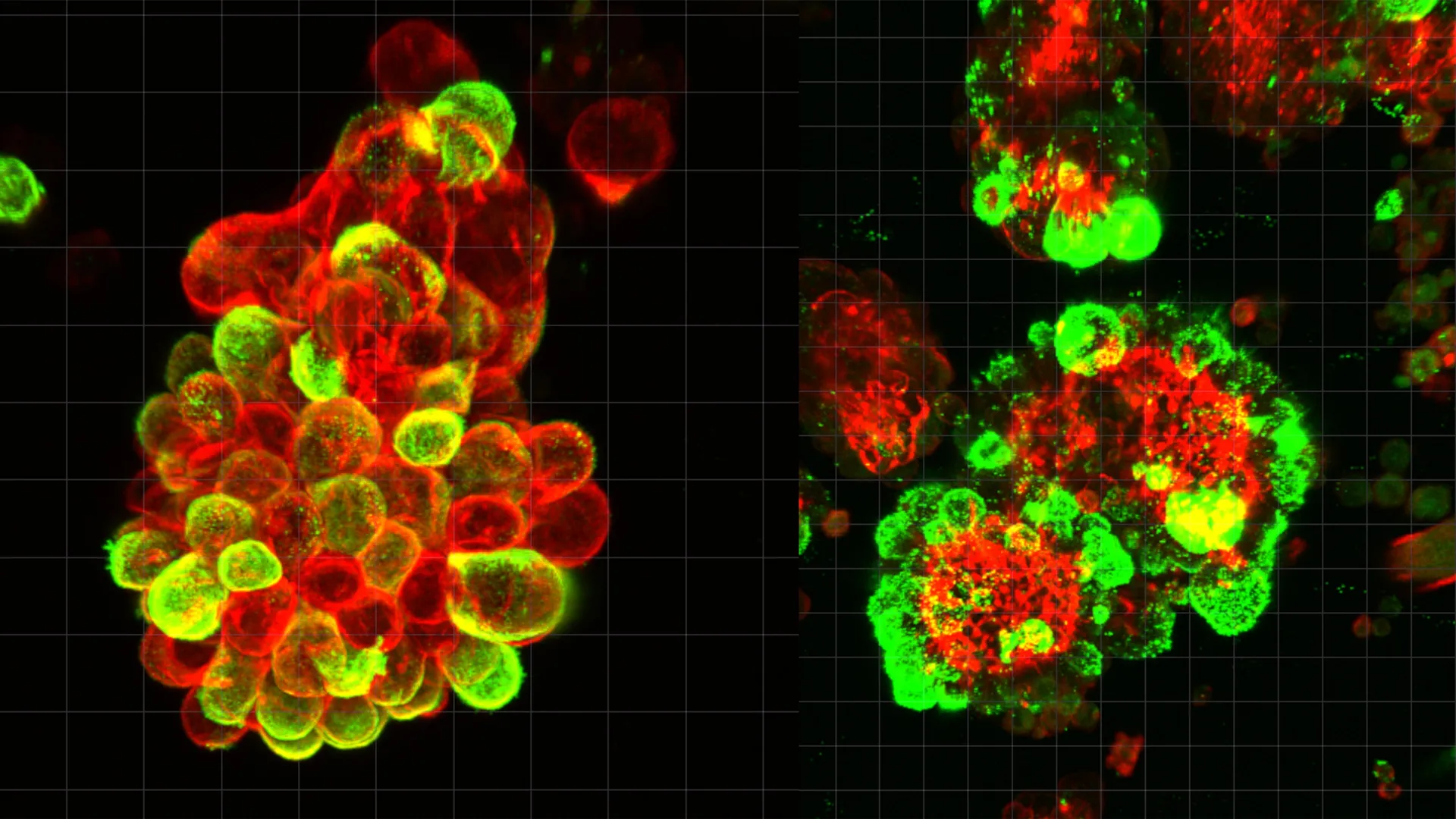
The landscape of gynecological oncology has long been overshadowed by the "silent killer" reputation of ovarian cancer, a disease that remains the most lethal malignancy of the female reproductive system. For decades, clinicians and researchers have grappled with a fundamental paradox: why does ovarian cancer progress with such devastating speed once it enters the abdominal cavity, and why do current treatments so often fail to prevent recurrence? A groundbreaking study led by a team of researchers at Nagoya University in Japan, recently published in the prestigious journal Science Advances, has provided a transformative answer to these questions. The research reveals that ovarian cancer cells do not act as solitary invaders but instead "recruit" healthy mesothelial cells to act as biological pioneers, carving paths through tissue and shielding the cancer from the effects of chemotherapy.
The Mechanism of Co-opted Metastasis
Ovarian cancer is unique in its primary mode of dissemination. Unlike breast or lung cancers, which typically spread through the lymphatic system or the bloodstream (hematogenous spread), ovarian cancer cells frequently detach from the primary tumor and enter the peritoneal fluid—the lubricating liquid that fills the abdominal cavity. Once in this fluid, the cancer cells are subject to the mechanical forces of the body, including respiratory movements and intestinal peristalsis, which distribute the malignant cells throughout the abdomen.
The Nagoya University study, spearheaded by Dr. Kaname Uno, found that during this "floating phase," cancer cells undergo a sophisticated behavioral shift. Rather than drifting as individual units, they actively seek out and bind to mesothelial cells. These mesothelial cells normally form a smooth, protective monolayer known as the mesothelium, which lines the abdominal cavity and organs to reduce friction. When cancer cells encounter these cells—either those still attached to the lining or those that have naturally shed into the fluid—they form hybrid, multicellular spheres.
The researchers analyzed abdominal fluid (ascites) from human patients and discovered that approximately 60% of the observed cancer cell clusters were these hybrid spheres. This finding fundamentally challenges the traditional view of ovarian cancer metastasis as a process driven solely by the intrinsic mutations of the cancer cell itself. Instead, it suggests a cooperative model where the cancer cell "outsources" the mechanical labor of invasion to a non-malignant accomplice.
The Role of TGF-β1 and the Formation of Invadopodia
The biochemical driver of this partnership is a signaling molecule known as Transforming Growth Factor-beta 1 (TGF-β1). The Nagoya University team identified that the cancer cells secrete high levels of TGF-β1, which acts as a molecular "reprogramming" agent for the recruited mesothelial cells. Under the influence of this protein, the mesothelial cells undergo a transition that changes their physical structure and behavior.
The most significant change is the development of "invadopodia." These are specialized, actin-rich protrusions that function like microscopic drills or spikes. In a healthy physiological context, mesothelial cells do not possess these structures. However, once integrated into the hybrid sphere and activated by TGF-β1, the mesothelial cells use these invadopodia to penetrate the extracellular matrix of the abdominal organs.
Dr. Uno’s research demonstrates that the cancer cells themselves remain relatively passive during the initial breach of new tissue. By manipulating the mesothelial cells to do the "heavy lifting" of tissue invasion, the cancer cells can migrate through the openings created by their hosts. This strategy allows the cancer to spread rapidly without requiring the complex genetic mutations usually necessary for a cell to become independently invasive.
A Physician’s Motivation: The Story of Dr. Kaname Uno
The impetus for this research was not merely academic but deeply rooted in clinical tragedy. Dr. Kaname Uno, the study’s lead author, served as a practicing gynecologist for eight years before transitioning into full-time research at Nagoya University’s Graduate School of Medicine. His career shift was motivated by a specific patient whose case highlighted the catastrophic limitations of modern diagnostic technology.
The patient in question had undergone routine gynecological screenings that returned completely normal results. However, only three months later, she presented with advanced-stage ovarian cancer that had already metastasized throughout her abdominal cavity. Despite aggressive intervention, the disease’s rapid progression outpaced the available medical tools. This experience drove Dr. Uno to investigate the "hidden" phase of ovarian cancer spread—the period between a clear scan and the sudden appearance of widespread tumors.
"I wanted to understand why this cancer is so difficult to catch early and why it moves with such incredible speed," Dr. Uno stated regarding his research. His findings suggest that the hybrid spheres allow the cancer to "hide in plain sight" within the peritoneal fluid, utilizing the body’s own cells to facilitate a rapid, stealthy expansion that current imaging and blood tests are not designed to detect.
Supporting Data and Methodological Rigor
To validate their findings, the research team employed a multi-disciplinary approach that combined clinical observation with advanced laboratory techniques. They utilized high-resolution microscopy to observe the behavior of cells taken directly from patient ascites samples, providing real-time evidence of the hybrid sphere formation.
To confirm the role of mesothelial cells in promoting invasion and drug resistance, the team developed mouse models. These experiments showed that tumors formed from hybrid clusters (cancer cells plus mesothelial cells) grew more aggressively and were significantly more resistant to standard chemotherapy agents, such as paclitaxel and carboplatin, compared to tumors formed from cancer cells alone.
Furthermore, the team performed single-cell RNA sequencing to analyze gene activity within the clusters. This genetic profiling confirmed that the mesothelial cells within the spheres were expressing genes associated with tissue remodeling and mesenchymal transitions—traits that are usually associated with highly aggressive cancer cells but were here being expressed by "normal" cells under the cancer’s control.
Implications for Future Treatment and Diagnostics
The discovery of the hybrid cell partnership has profound implications for how ovarian cancer may be treated in the future. Current standard-of-care treatments primarily focus on "cytotoxic" drugs—chemicals designed to kill rapidly dividing cancer cells. However, these drugs often fail to clear the mesothelial "shields" that protect the hybrid clusters, leading to the high rates of recurrence seen in ovarian cancer patients.
The Nagoya University study suggests several new therapeutic avenues:
- Disrupting the Partnership: New drugs could be developed to block the TGF-β1 signaling pathway, preventing cancer cells from "recruiting" mesothelial cells in the first place.
- Targeting Invadopodia: By inhibiting the formation of the spike-like invadopodia on mesothelial cells, doctors might be able to "trap" the cancer cells in the peritoneal fluid, preventing them from embedding into organs.
- Enhanced Monitoring: The presence and concentration of these hybrid spheres in abdominal fluid could serve as a new biomarker. By sampling peritoneal fluid (a procedure known as paracentesis), doctors could better predict a patient’s prognosis or monitor their response to treatment more accurately than through blood tests like CA-125, which can be unreliable.
Broader Context: The Global Burden of Ovarian Cancer
The significance of this research is underscored by the global statistics on ovarian cancer. According to the World Health Organization and the American Cancer Society, ovarian cancer accounts for more deaths than any other cancer of the female reproductive system. Globally, over 300,000 women are diagnosed annually, and nearly 200,000 succumb to the disease.
The high mortality rate is largely attributed to the fact that approximately 70% to 80% of cases are diagnosed at Stage III or IV, when the cancer has already spread beyond the pelvis. The five-year survival rate for late-stage ovarian cancer remains stubbornly low, often hovering around 30%. By identifying the biological mechanism that facilitates this late-stage "explosion" of growth, the Nagoya University team has provided a roadmap for interventions that could finally move the needle on these survival statistics.
Conclusion and Future Directions
The research published in Science Advances marks a pivotal shift in the study of the tumor microenvironment. It moves the focus away from the cancer cell in isolation and toward the complex ecosystem of the abdominal cavity. The realization that ovarian cancer "outsources" its invasion to mesothelial cells explains not only the speed of its spread but also its resilience against current therapies.
As the oncology community digests these findings, the next steps will involve clinical trials aimed at disrupting this cellular cooperation. For patients and families affected by this devastating disease, the work of Dr. Uno and his colleagues offers a new sense of hope—a hope built on the precise understanding of a mystery that has remained unsolved for far too long. The transition from the clinic to the lab has, in this instance, resulted in a discovery that may one day lead patients back from the lab to a healthy life.
