
 By Nana O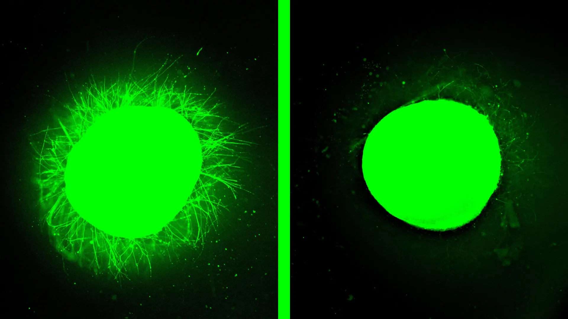
Northwestern University scientists have achieved a significant breakthrough in spinal cord injury research, developing the most advanced lab-grown model to date that accurately replicates the complex biological aftermath of human spinal cord trauma. This innovative model, utilizing human spinal cord organoids, not only reproduces key pathological consequences of injury but also offers a powerful platform for evaluating promising regenerative therapies, such as the "dancing molecules" developed by the research team. The findings, published on February 11th in the prestigious journal Nature Biomedical Engineering, represent a critical step forward in understanding and potentially treating debilitating spinal cord injuries.
Recreating the Devastation of Spinal Cord Injury
The research centers on human spinal cord organoids, miniature, self-organized structures derived from stem cells that mimic the intricate architecture and cellular composition of the human spinal cord. For the first time, these organoids have been engineered to faithfully reproduce the major biological consequences observed following traumatic spinal cord injury (SCI) in humans. This includes critical cellular events such as widespread cell death, an aggressive inflammatory response, and the formation of glial scars.
Glial scarring is a particularly formidable obstacle to nerve regeneration. It involves the dense proliferation of glial cells, primarily astrocytes, which create a physical and chemical barrier that actively inhibits the regrowth of severed nerve fibers, known as axons. The Northwestern team’s organoid model successfully recapitulated this scarring process, providing a realistic in vitro environment for studying its formation and testing strategies to overcome it.
"One of the most exciting aspects of organoids is that we can use them to test new therapies in human tissue," stated Samuel I. Stupp, the study’s senior author and a distinguished Board of Trustees Professor at Northwestern University. Professor Stupp, who is also the inventor of the "dancing molecules" therapy, emphasized the critical advantage of this approach. "Short of a clinical trial, it’s the only way you can achieve this objective. We decided to develop two different injury models in a human spinal cord organoid and test our therapy to see if the results resembled what we previously saw in the animal model."
The "Dancing Molecules" Therapy Shows Remarkable Promise
When the injured spinal cord organoids were treated with Stupp’s "dancing molecules" therapy, the results were nothing short of dramatic. This innovative therapeutic approach, which had previously demonstrated success in restoring movement and repairing tissue in animal studies, induced substantial neurite outgrowth in the damaged organoids. Neurites are the long, branching extensions of neurons that are crucial for transmitting nerve signals. Their regrowth signifies the potential for damaged neural circuits to reconnect.
Furthermore, the treatment significantly reduced the formation of glial scar tissue, rendering it barely detectable in treated organoids. This observed reduction in scarring and promotion of neurite regeneration in human organoids mirrors the positive outcomes seen in animal models, providing strong validation for the therapy’s potential efficacy in human patients. This advancement is particularly timely, as the "dancing molecules" therapy recently received Orphan Drug Designation from the U.S. Food and Drug Administration (FDA), a crucial step in accelerating its development for rare diseases and conditions like spinal cord injury.
Nozomu Takata, a research assistant professor of medicine at Northwestern’s Feinberg School of Medicine and a member of the Center for Regenerative Nanomedicine (CRN), served as the paper’s first author. He contributed significantly to the development and analysis of the organoid models and the assessment of therapeutic outcomes.
The Significance of Human Organoids in Biomedical Research
Organoids, often described as "mini-organs," are complex three-dimensional structures grown in the laboratory from induced pluripotent stem cells (iPSCs). While simplified versions of their in vivo counterparts, they exhibit remarkable fidelity in mimicking the structure, cellular diversity, and functional characteristics of actual human organs. This makes them invaluable tools for a wide range of biomedical research applications, including the study of disease mechanisms, the preclinical testing of novel therapeutic agents, and the investigation of fundamental organ development.
The use of organoids offers several distinct advantages over traditional research methods. They can significantly accelerate the pace of discovery and reduce research costs compared to extensive animal experimentation or the lengthy and expensive process of human clinical trials.
While other research groups have successfully developed spinal cord organoids for basic biological studies, the Northwestern model represents a substantial leap forward specifically for injury research. These organoids are several millimeters in diameter, a size considered mature enough to accurately model the effects of traumatic damage.
Advancements in Organoid Engineering for Injury Modeling
Over a period of several months, the Northwestern team meticulously guided the differentiation of stem cells into complex spinal cord tissue. This intricate process involved the generation of neurons and astrocytes, two key cell types found in the spinal cord. A pivotal innovation in this study was the successful incorporation of microglia, the resident immune cells of the central nervous system, into the organoid model. This inclusion is critical for accurately replicating the robust inflammatory response that invariably follows spinal cord injury in living organisms.
"It’s kind of a pseudo-organ," Professor Stupp explained, highlighting the sophisticated nature of their creation. "We were the first to introduce microglia into a human spinal cord organoid, so that was a huge accomplishment. It means that our organoid has all the chemicals that the resident immune system produces in response to an injury. That makes it a more realistic, accurate model of spinal cord injury."
Unpacking the "Dancing Molecules" Therapy
The "dancing molecules" therapy, first introduced by Stupp’s laboratory in 2021, represents a novel approach to tissue repair. It leverages the controlled dynamic motion of molecules to stimulate cellular regeneration and potentially reverse paralysis following traumatic SCI. This therapy belongs to a broader category of supramolecular therapeutic peptides (STPs). STPs function by forming large molecular assemblies, often comprising over 100,000 individual molecules, which interact with cell receptors to activate the body’s intrinsic repair mechanisms. Interestingly, the underlying concept of supramolecular therapies shares common ground with current GLP-1 drugs used for weight loss and diabetes management, an area that Stupp’s lab explored nearly 15 years prior.
The delivery mechanism for this therapy involves a liquid injection. Upon administration, these molecules rapidly self-assemble into a fine nanofiber network that closely mimics the natural extracellular matrix of the spinal cord. The efficacy of the "dancing molecules" is intricately linked to their dynamic behavior within this scaffold. By fine-tuning the molecular motion – how quickly they move and interact – researchers can optimize their engagement with the constantly shifting cellular receptors on nerve cells.
Professor Stupp elaborated on this principle in 2021: "Given that cells themselves and their receptors are in constant motion, you can imagine that molecules moving more rapidly would encounter these receptors more often. If the molecules are sluggish and not as ‘social,’ they may never come into contact with the cells."
Previous studies involving animal models demonstrated the profound impact of this therapy. A single injection administered just 24 hours after a severe spinal cord injury enabled mice to regain the ability to walk within four weeks. Crucially, formulations exhibiting faster molecular motion consistently outperformed slower-moving versions, underscoring the direct correlation between enhanced molecular dynamics and improved bioactivity and cellular signaling.
Simulating Diverse Spinal Cord Trauma Scenarios
To rigorously test the "dancing molecules" therapy, the Northwestern researchers meticulously recreated two of the most common types of spinal cord injury within the human organoids. The first injury model involved surgically cutting the organoids with a scalpel, simulating a laceration akin to those sustained during surgical procedures or sharp trauma. The second model involved inducing a compressive contusion injury, a more diffuse form of damage comparable to the blunt force trauma experienced in severe car accidents or falls.
Both injury models reliably induced cell death and the formation of glial scars, mirroring the pathological cascade observed in actual human spinal cord injuries. "We could distinguish between the astrocytes that are a part of normal tissue and the astrocytes in the glial scar, which are large and very densely packed," Professor Stupp noted. "We also detected the production of chondroitin sulfate proteoglycans, which are molecules in the nervous system that respond to injury and disease."
Following treatment with the "dancing molecules" therapy, the nanofiber scaffold not only significantly reduced inflammation and shrank the glial scarring but also effectively stimulated neurite extension. Furthermore, the treatment encouraged neurons to grow in more organized and functional patterns, suggesting a restoration of neural circuitry.
Neurites encompass axons, the long projections of nerve cells that are frequently severed in spinal cord injuries. When axons are cut, the vital communication pathways between neurons are disrupted, leading to paralysis, loss of sensation, and other neurological deficits below the injury site. The ability of the "dancing molecules" to promote neurite regrowth therefore holds immense promise for reconnecting these severed pathways and potentially restoring lost function.
The Crucial Role of Molecular Motion in Therapeutic Efficacy
Professor Stupp attributes the remarkable effectiveness of the "dancing molecules" therapy to the principle of supramolecular motion. This refers to the capacity of the therapeutic molecules to move dynamically within the nanofiber network, including transiently detaching and reattaching. This active movement is hypothesized to enhance their ability to interact with cellular targets.
Experiments conducted on healthy, undamaged organoids further reinforced this hypothesis. "Before we even developed the injury model, we tested the therapy on a healthy organoid," Professor Stupp stated. "The dancing molecules spun out all these long neurites on the surface of the organoid but, when we used molecules that had less or no motion, we saw nothing. This difference was very vivid." This observation strongly suggests that the dynamic nature of the molecules is essential for their ability to promote neurite growth even in the absence of injury.
Future Directions and Broader Implications
The Northwestern team is already planning to engineer even more sophisticated organoid models to further refine their understanding of spinal cord injury and treatment. A key future goal is to develop organoids capable of replicating chronic, long-standing injuries. These types of injuries are typically characterized by thicker, more persistent scar tissue, presenting a greater therapeutic challenge.
Looking further ahead, Professor Stupp envisions these miniature spinal cords playing a crucial role in the advancement of personalized medicine. The potential exists to generate implantable spinal cord tissue derived from a patient’s own stem cells. This autologous approach would significantly reduce the risk of immune rejection, a major hurdle in current regenerative medicine strategies.
The groundbreaking study, titled "Injury and therapy in a human spinal cord organoid," received crucial support from the Center for Regenerative Nanomedicine at Northwestern University and a generous gift from the John Potocsnak Family specifically for spinal cord injury research. This collaborative effort underscores the multifaceted support necessary to drive such innovative scientific endeavors from the laboratory bench to potential clinical application. The development of this advanced organoid model and the promising results of the "dancing molecules" therapy mark a significant stride toward a future where spinal cord injuries are no longer considered irreversible.
