
 By Budi Oetomo Narendra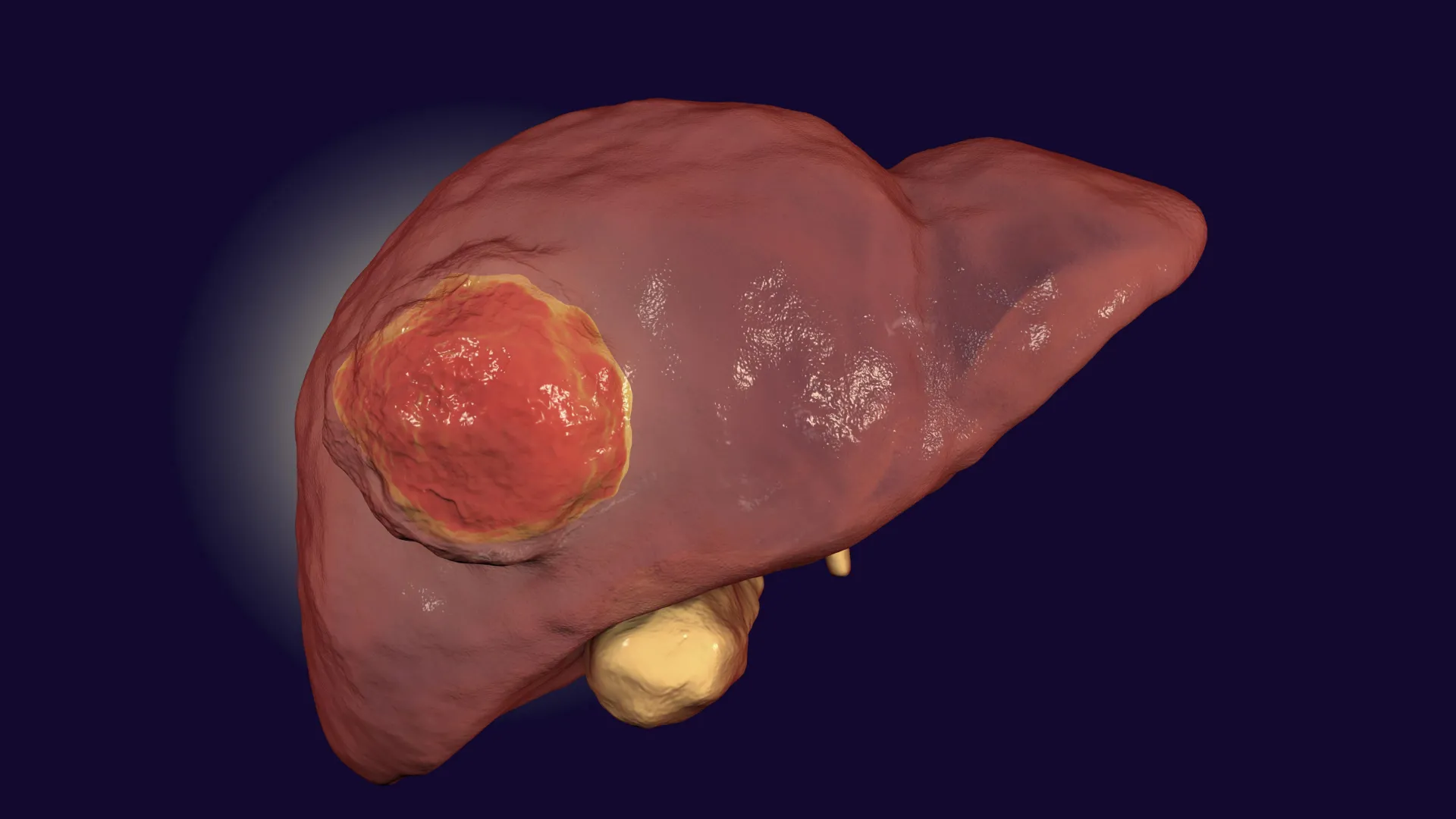
A rare and aggressive form of liver cancer, fibrolamellar carcinoma (FLC), has long presented a formidable challenge to medical science, particularly resisting the transformative power of immunotherapy – a treatment designed to harness the body’s own immune system to combat cancer cells. Now, a collaborative team of researchers has identified a promising pathway to overcome this entrenched resistance, leveraging an existing FDA-approved drug already in use for another medical condition. This discovery offers a significant beacon of hope for patients, predominantly children and young adults, afflicted by this devastating disease.
The pivotal findings, detailed in the prestigious journal Gastroenterology, suggest a novel and immediately actionable treatment strategy for fibrolamellar carcinoma. Unlike the more common forms of liver cancer often associated with chronic liver disease, FLC strikes a younger, healthier demographic, making its aggressive nature and resistance to conventional therapies all the more tragic. It accounts for a small but critical subset of liver cancer cases, estimated at around 1-2% of all liver cancer diagnoses, yet its impact is disproportionately severe due to its high mortality rate and the lack of effective systemic treatments.
The Unmet Need: Understanding Fibrolamellar Carcinoma
Fibrolamellar carcinoma is a distinct entity within the spectrum of liver cancers. While primary liver cancer (hepatocellular carcinoma, HCC) is a global health burden, affecting hundreds of thousands annually, FLC is rarer, with fewer than 1,000 cases diagnosed worldwide each year. This rarity contributes to a significant knowledge gap and fewer dedicated research efforts compared to more prevalent cancers. Patients typically present in their teens or twenties, often with non-specific symptoms such as abdominal pain, weight loss, or a palpable mass, leading to diagnoses frequently made only after the cancer has spread to other parts of the body. At this metastatic stage, the prognosis is particularly grim, with five-year survival rates plummeting to below 20-30%, a stark contrast to the often better outcomes seen with localized disease treated by surgical resection. Currently, surgery remains the only potentially curative option, but recurrence rates are notoriously high, and effective systemic therapies for advanced FLC are virtually non-existent, leaving patients with severely limited options. This critical unmet need underscores the urgency and significance of any potential therapeutic breakthrough.
The Immunotherapy Conundrum: Why FLC Resisted
Immunotherapy, particularly immune checkpoint inhibition (ICI), has revolutionized cancer treatment over the past decade. These therapies work by releasing the "brakes" on the body’s own immune T cells, allowing them to recognize and attack cancer cells more effectively. ICIs have achieved remarkable success in various cancers, including melanoma, lung, kidney, bladder, and even some forms of hepatocellular carcinoma, transforming previously intractable diseases into manageable conditions or even leading to long-term remissions for many patients.
However, a frustrating reality is that many other cancers, often termed "cold" tumors, remain stubbornly resistant to these therapies. Pancreatic, prostate, and certain brain cancers fall into this category, as did, until now, fibrolamellar carcinoma. The scientific community has grappled with understanding why some tumors respond dramatically while others remain impervious. The prevailing hypothesis points to the complex interplay within the tumor microenvironment (TME) – the intricate ecosystem surrounding cancer cells, comprising various immune cells, stromal cells, blood vessels, and extracellular matrix components.
The groundbreaking study published in Gastroenterology meticulously elucidated the precise mechanism by which FLC evades the immune system, shedding critical light on why immunotherapy had previously struggled against this particular cancer. Researchers discovered that FLC tumors actively manipulate their surrounding environment in a way that physically prevents immune T cells from infiltrating the cancerous tissue. Instead of migrating into the tumor core to engage and destroy cancer cells, the T cells become "trapped" or "excluded" elsewhere within the tumor’s periphery or in its surrounding stroma. This phenomenon, known as T-cell exclusion, effectively creates an immunological barrier, rendering the immune system incapable of carrying out its normal cancer-fighting duties, even when activated by immune checkpoint inhibitors.
Advanced Technology Unlocks the Tumor’s Secrets
To unravel this intricate biological puzzle and gain an unprecedented understanding of the FLC tumor microenvironment, the research team employed a cutting-edge technique called single-nucleus transcriptomics. This powerful technology allowed scientists to isolate the nucleus of individual cells within tumor tissue and precisely determine which genes were active in each cell.
"It wasn’t until we were able to use this technology that the picture of the tumor microenvironment began to clear up for us," explained Andreas Stephanou, a co-first author on the study and a Cornell graduate student. This high-resolution approach provided a granular view, enabling the researchers to map the cellular landscape of FLC tumors with exquisite detail, identifying the specific cell types present, their functional states, and the complex molecular conversations taking place between them. Traditional bulk sequencing methods, which average gene expression across millions of cells, would have obscured these crucial cell-specific interactions. Single-nucleus transcriptomics provided the clarity needed to pinpoint the cellular culprits behind FLC’s immune evasion.
The Architects of Exclusion: Fibrous Bands and Altered Stellate Cells
The name "fibrolamellar carcinoma" itself hints at a distinctive histological feature: the presence of thick, lamellar (layered) fibrous bands that crisscross throughout the tumor tissue. For years, the precise role of these prominent fibrous structures in the tumor’s progression and immune evasion remained a mystery. "Despite all of the recent advances in the study of this cancer, we still haven’t pinpointed how these fibrous bands contribute, if at all, to the tumor’s progression," Stephanou noted.
The single-nucleus transcriptomics analysis provided a definitive answer. The researchers discovered that these characteristic fibrous bands are not inert structures but are actively produced by hepatic stellate cells – normal liver cells that undergo a profound transformation when exposed to the cancerous microenvironment. Once altered by the cancer, these stellate cells become activated, transitioning into myofibroblast-like cells. In this activated state, they begin to secrete copious amounts of fibrous proteins, such as collagen, which accumulate to form the dense, characteristic bands within the tumor.
Crucially, the team also uncovered that these activated stellate cells do more than just build physical barriers. Using the same single-cell technology, they identified that these altered stellate cells actively send out specific biochemical signals to nearby T cells. Instead of allowing the immune cells to migrate towards and engage the cancer cells, these signals subtly misdirect the T cells, steering them away from the malignant cells and towards the very fibrous bands produced by the stellate cells. Effectively, the T cells are lured into these dense fibrous traps, becoming sequestered and unable to perform their tumor-killing function. This detailed understanding of the "how" and "who" behind T-cell exclusion was a monumental step forward.
AMD3100: A Repurposed Drug to Restore Immune Access
With a clear understanding of the mechanism of T-cell exclusion, the research team then posed a critical question: "So, then we asked, what if we were to block this signaling in T cells with a compound?" recalled Praveen Sethupathy ’03, professor of physiological genomics and chair of the Department of Biomedical Sciences in the College of Veterinary Medicine, and co-senior author of the study.
The answer came in the form of AMD3100, a drug that is already FDA-approved under the brand name Mozobil (Plerixafor). AMD3100 is currently used in clinical practice for mobilizing hematopoietic stem cells from the bone marrow into the peripheral blood for collection, primarily in patients with non-Hodgkin lymphoma and multiple myeloma undergoing autologous stem cell transplantation. Its mechanism of action involves antagonizing the CXCR4 receptor, which plays a critical role in cell trafficking and homing, including the movement of immune cells.
The researchers hypothesized that by targeting a key receptor involved in the trapping mechanism orchestrated by the stellate cells, AMD3100 could essentially dismantle the tumor’s defensive perimeter and liberate the immune cells. To test this hypothesis, scientists in the laboratory of Dr. Venu Pillarisetty, a surgical oncologist at the University of Washington and co-senior author of the study, conducted sophisticated ex vivo experiments. They utilized slices of patient tumor tissue, a model that preserves the complex architecture and cellular interactions of the tumor microenvironment more accurately than simple cell cultures.
The results were compelling. Treatment with AMD3100 demonstrably guided T cells back into the center of the FLC tumors, effectively reversing the T-cell exclusion. Even more significantly, when AMD3100 was combined with immune checkpoint inhibition – the very therapy FLC had previously resisted – a powerful synergistic effect was observed. This combination led to a substantial increase in T-cell activation and, crucially, a marked rise in tumor cell death. The drug not only allowed T cells to infiltrate but also enhanced their ability to kill cancer cells when combined with conventional immunotherapy.
Expert Perspectives and the Path Forward
Professor Sethupathy emphasized the foundational nature of these findings: "Our results provide among the first indications of why a type of immunotherapy called immune checkpoint inhibition hasn’t worked well in these patients, and even if this particular drug isn’t the end-all-be-all, it teaches us that this T-cell exclusion phenomenon is an important one to tackle in fibrolamellar carcinoma." This sentiment highlights that the discovery of T-cell exclusion as a key resistance mechanism is a critical insight, even beyond the specific drug identified.
The collaborative spirit of the research, involving institutions like Cornell University and the University of Washington, underscored the complexity of the challenge and the multidisciplinary expertise required for such breakthroughs. The study’s co-first authors, Jason Carter and Lindsey Dickerson from the Pillarisetty laboratory, along with Bo Shui from the Sethupathy laboratory, were instrumental in executing the intricate experimental work.
The immediate next step following these compelling preclinical results is to translate these findings into human clinical trials. The researchers are actively seeking collaborations with liver cancer specialists and clinical centers interested in launching Phase 1 and Phase 2 trials to evaluate this novel treatment approach in patients with fibrolamellar carcinoma.
Broader Implications: A Beacon for "Cold" Tumors and Drug Repurposing
The implications of this research extend far beyond fibrolamellar carcinoma. The discovery of T-cell exclusion driven by altered stellate cells and fibrous bands provides a crucial blueprint for understanding resistance in other "cold" tumors that also fail to respond to immunotherapy. Many challenging cancers, including pancreatic and certain colorectal cancers, are characterized by a dense, desmoplastic (fibrous) stroma and poor immune infiltration. This study offers a potential therapeutic strategy for these difficult-to-treat malignancies as well, by targeting the stromal components that act as immune barriers.
Moreover, the fact that AMD3100 is an existing FDA-approved drug is a significant advantage. Drug repurposing, the process of finding new therapeutic uses for already approved drugs, can dramatically reduce the time, cost, and risk associated with drug development. Since AMD3100 has a known safety profile and established manufacturing processes, it can potentially bypass years of preclinical and early-phase clinical testing required for entirely new compounds, significantly accelerating its journey to patients.
"A compelling feature of this work is that AMD3100 is already FDA-approved, which can reduce risks and potentially speed up timelines for clinical trials in fibrolamellar carcinoma," Sethupathy reiterated, emphasizing the practical and immediate impact this could have.
The Fibrolamellar Cancer Foundation, which provided crucial funding for this research, has expressed profound optimism regarding these findings. For a disease with so few therapeutic avenues, the prospect of repurposing an existing drug to overcome a fundamental mechanism of resistance represents a monumental step forward, offering a renewed sense of hope to patients and their families worldwide. This research not only provides a potential new treatment for a rare and devastating cancer but also deepens our understanding of immune evasion, paving the way for future breakthroughs in oncology.
