
 By Nana O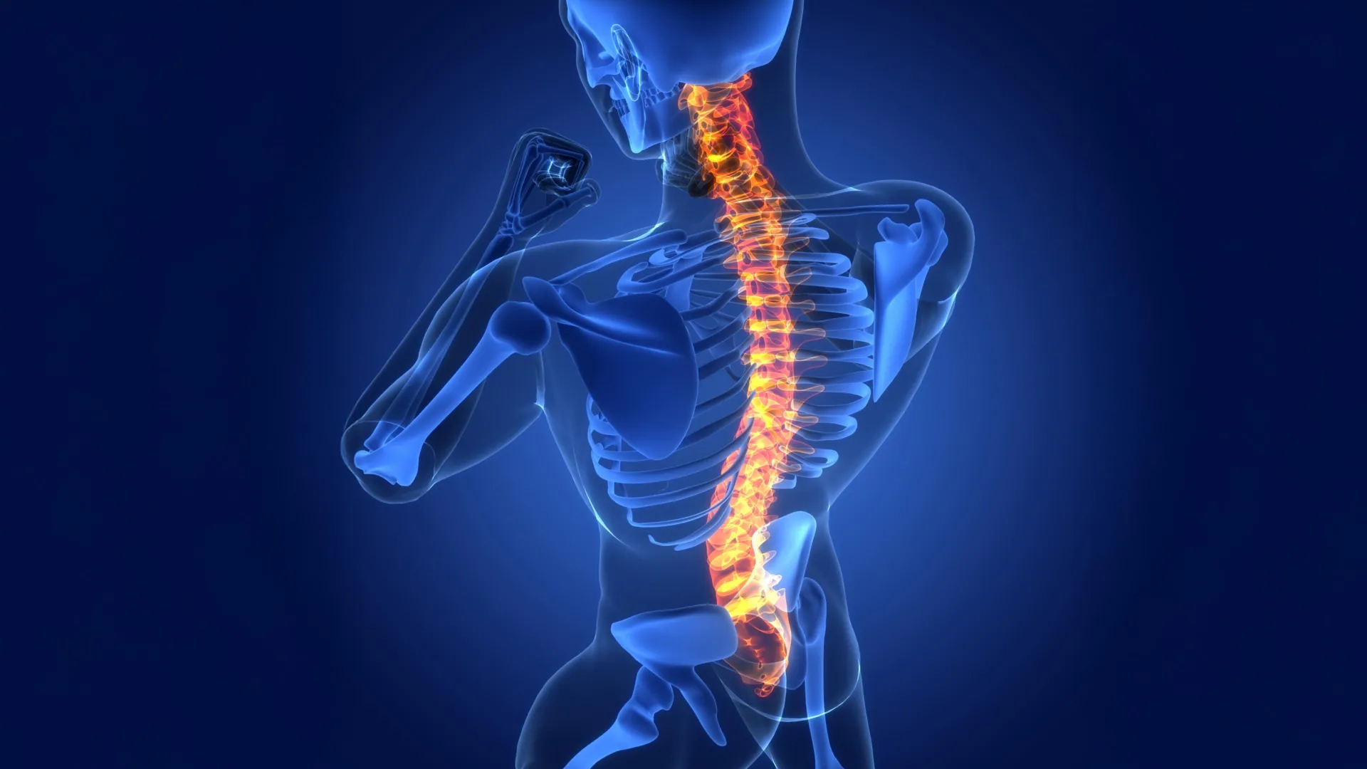
Researchers at Cedars-Sinai have made a groundbreaking discovery, identifying a previously unknown biological repair process within the central nervous system that holds significant promise for developing novel treatments for spinal cord injuries, stroke, and debilitating neurological diseases such as multiple sclerosis. The findings, meticulously detailed in a recent publication in the prestigious scientific journal Nature, reveal an unexpected and critical role for astrocytes, a class of star-shaped glial cells that are fundamental support structures within the brain and spinal cord.
Astrocytes: Beyond Support to Active Repair Agents
"Astrocytes are critical responders to disease and disorders of the central nervous system – the brain and spinal cord," stated neuroscientist Joshua Burda, PhD, assistant professor of Biomedical Sciences and Neurology at Cedars-Sinai and the senior author of the study. "We discovered that astrocytes situated far from the actual site of an injury actually play a crucial role in driving spinal cord repair. Our research has also uncovered a sophisticated mechanism employed by these unique astrocytes to signal the immune system, prompting it to effectively clean up the debris that results from an injury. This debris clearance is an absolutely critical step in the tissue-healing process."
The research team has aptly named these newly identified cells "lesion-remote astrocytes," or LRAs. Furthermore, their investigations have distinguished several distinct subtypes of LRAs, each potentially possessing unique functional capacities. For the first time, this study elucidates how one specific subtype of LRA can effectively detect damage from a considerable distance and subsequently initiate a cascade of responses that actively support tissue recovery. This discovery fundamentally shifts the understanding of astrocytic function from passive support to active participation in repair mechanisms.
Understanding Spinal Cord Injury: A Complex Challenge
The spinal cord, a vital conduit of information, is a long bundle of nerve tissue extending from the brain down the vertebral column. It comprises two primary regions: the inner gray matter, rich in nerve cell bodies and astrocytes, and the outer white matter, composed of astrocytes and long nerve fibers (axons) that transmit signals between the brain and the rest of the body. Astrocytes in this context are essential for maintaining the precise ionic and metabolic environment required for these electrical and chemical signals to travel unimpeded.
When the spinal cord sustains an injury, whether from trauma, disease, or other pathological processes, nerve fibers are inevitably torn apart. This damage can lead to a devastating loss of motor function, resulting in paralysis, and a disruption of sensory perception, including touch, temperature, and pain. The severed nerve fibers undergo degeneration, breaking down into cellular debris. In many other tissues throughout the body, inflammatory responses are typically localized to the immediate vicinity of the injury. However, the intricate, long-distance architecture of the spinal cord presents a unique challenge. Damage and subsequent inflammation can spread far beyond the initial site of injury, potentially affecting a much larger expanse of neural tissue and exacerbating functional deficits. This widespread inflammation can create a hostile environment that impedes natural repair processes and can even lead to secondary damage.
Lesion-Remote Astrocytes: Orchestrating Immune Cleanup
Through a series of rigorous experiments conducted on mouse models with induced spinal cord injuries, the Cedars-Sinai researchers definitively established the pivotal role of LRAs in promoting tissue repair. Critically, their analysis extended beyond animal models, revealing strong evidence that this same astrocytic repair process is also active in spinal cord tissue samples obtained from human patients who have suffered injuries. This cross-species validation significantly enhances the translational potential of their findings.
A key breakthrough in the study was the identification of a specific LRA subtype that produces a crucial protein known as CCN1. This molecule acts as a signaling agent, directly communicating with specialized immune cells within the central nervous system called microglia.
"One of the primary functions of microglia is to serve as the chief garbage collectors within the central nervous system," explained Dr. Burda. "Following tissue damage, these cells are responsible for engulfing and clearing away fragments of nerve fiber debris. This debris is particularly rich in fats, which can pose a metabolic challenge for microglia, essentially causing them a form of ‘indigestion.’ Our experiments demonstrated that the CCN1 produced by astrocytes effectively signals the microglia to alter their metabolic processes, enabling them to more efficiently digest this fatty debris."
According to Dr. Burda, this enhanced debris removal is a critical factor that may help explain why some patients experience partial, spontaneous recovery after spinal cord injury. Conversely, when the researchers experimentally eliminated the production of astrocyte-derived CCN1, the healing process was significantly impaired.
"If we remove astrocyte CCN1, the microglia attempt to consume the debris, but they are unable to properly digest it," Dr. Burda elaborated. "This leads to them calling in more microglia, which also ingest the debris but still cannot digest it effectively. The consequence is the formation of large aggregates of debris-filled microglia, which in turn amplifies inflammation throughout the spinal cord. When this heightened inflammation occurs, the damaged tissue is unable to repair itself as efficiently."
Broader Implications: A New Frontier for Neurological Disease Treatment
The implications of this discovery extend far beyond spinal cord injury. When scientists examined spinal cord samples from individuals diagnosed with multiple sclerosis (MS), a chronic autoimmune disease that affects the central nervous system, they observed the same CCN1-related repair process at play. This suggests that the fundamental mechanisms identified by the Cedars-Sinai team may be broadly applicable to a range of injuries and diseases affecting both the brain and spinal cord.
"The role of astrocytes in central nervous system healing has been remarkably understudied until now," commented David Underhill, PhD, chair of the Department of Biomedical Sciences at Cedars-Sinai. "This work provides compelling evidence that lesion-remote astrocytes represent a highly promising avenue for therapeutic intervention. Harnessing their capabilities could lead to strategies for limiting chronic inflammation, significantly enhancing functionally meaningful regeneration, and ultimately promoting neurological recovery after brain and spinal cord injuries, as well as in various neurological diseases."
The research also opens new avenues for understanding and potentially treating conditions like stroke, where brain tissue damage triggers a complex inflammatory response that can hinder recovery. The ability of LRAs to modulate microglial activity and promote debris clearance could be a key factor in mitigating secondary brain injury following a stroke.
The Road Ahead: Therapeutic Development and Future Research
Dr. Burda is actively engaged in developing therapeutic strategies that aim to harness the CCN1 pathway to improve outcomes for individuals with spinal cord injuries. This could involve the development of drugs or gene therapies designed to stimulate LRA production of CCN1 or to enhance microglial responsiveness to this signal. His team is also investigating how astrocyte-derived CCN1 might influence the inflammatory processes implicated in neurodegenerative diseases, such as Alzheimer’s and Parkinson’s, as well as the broader effects of aging on the nervous system.
The identification of distinct LRA subtypes suggests a level of specialization within these cells that could be further exploited for targeted therapies. Future research may focus on understanding the specific functions of each subtype and how they can be manipulated to promote optimal repair.
The study’s detailed methodology involved advanced imaging techniques, genetic manipulation of animal models, and sophisticated biochemical analyses to track the signaling pathways and cellular interactions. The use of human tissue samples provided crucial validation, bridging the gap between preclinical findings and potential clinical applications.
This significant advancement builds upon decades of research into glial cell biology, which has increasingly highlighted their active roles beyond mere structural support. Early research often viewed astrocytes as passive bystanders or even contributors to damage in the injured nervous system. However, this new work firmly establishes their capacity for active, beneficial intervention.
Funding and Collaboration: A Multidisciplinary Effort
The research was made possible through substantial support from various national and international funding bodies, underscoring the importance and scope of this scientific endeavor. Key funding sources included the US National Institutes of Health (NIH) through multiple grants (5R01NS128094, R00NS105915, K99NS105915 to J.E.B., F31NS129372 to K.S., K99AG084864 to S.M., R35 NS097303 and R01 NS123532 to RD, R01MH128866, U18TR004146, P30 CA023168, and ASPIRE Challenge and Reduction-to-Practice award to G.C.). Additional support came from the Paralyzed Veterans Research Foundation of America (to J.E.B.) and Wings for Life (to J.E.B.). Fellowships and scholarships from Cedars-Sinai Center for Neuroscience and Medicine, the American Academy of Neurology, and the California Institute for Regenerative Medicine also contributed significantly (to S.M.). Further funding was provided by the US Department of Defense through a USAMRAA award (W81XWH2010665) via the Peer Reviewed Alzheimer’s Research Program (to G.C.), and The Arnold O. Beckman Postdoctoral Fellowship (to C.E.R.). The Purdue University Center for Cancer Research, funded by NIH grant P30 CA023168, was also acknowledged for its contribution.
The collaborative nature of this research is evident in the extensive list of contributing authors from Cedars-Sinai and other institutions, including Sarah McCallum, Keshav B. Suresh, Timothy S. Islam, Manish K. Tripathi, Ann W. Saustad, Oksana Shelest, Aditya Patil, David Lee, Brandon Kwon, Katherine Leitholf, Inga Yenokian, Sophia E. Shaka, Jasmine Plummer, Vinicius F. Calsavara, and Simon R.V. Knott from Cedars-Sinai. Additional authors from other institutions include Connor H. Beveridge, Palak Manchandra, Caitlin E. Randolph, Gordon P. Meares, Ranjan Dutta, Riki Kawaguchi, and Gaurav Chopra. This broad collaboration highlights the interdisciplinary approach required to tackle complex neurological research questions.
The implications of this discovery are profound, offering a renewed sense of hope for millions worldwide affected by spinal cord injuries, stroke, and neurodegenerative diseases. By uncovering and understanding the intricate repair mechanisms orchestrated by lesion-remote astrocytes, scientists at Cedars-Sinai have paved the way for the development of targeted therapies that could significantly improve neurological function and quality of life for patients facing these challenging conditions. The ongoing research into CCN1 and LRA function promises to unlock further secrets of neural repair, potentially ushering in a new era of regenerative medicine for the central nervous system.
