
 By George Ongkojoyo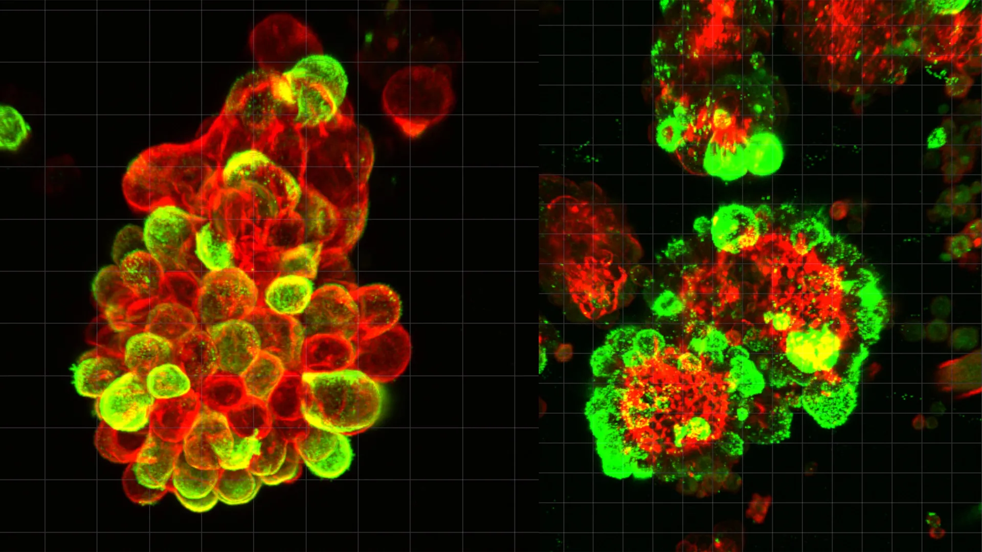
Ovarian cancer has long held a reputation as the "silent killer" of gynecological oncology, characterized by a lack of early-stage symptoms and an aggressive, rapid progression that often leaves medical professionals with limited options by the time of diagnosis. While the medical community has recognized the speed at which this disease spreads, the underlying biological mechanisms that facilitate such efficient metastasis remained a mystery until now. A groundbreaking study led by researchers at Nagoya University in Japan has finally decoded a critical aspect of this process, revealing that ovarian cancer cells do not act in isolation. Instead, they actively hijack the body’s own protective systems, recruiting mesothelial cells to serve as "pathfinders" that drill through healthy tissue, creating a corridor for the cancer to follow.
This discovery, published in the prestigious journal Science Advances, represents a paradigm shift in how scientists understand the transcoelomic spread of cancer—a process where malignant cells travel through the fluid-filled cavities of the body. By identifying the specific signaling pathways and cellular partnerships involved, the research team has opened a new frontier for diagnostic tools and therapeutic interventions that could significantly improve survival rates for a disease that claims the lives of over 13,000 women annually in the United States alone.
The Biological Mechanism of Hybrid Cell Clusters
The cornerstone of the Nagoya University study is the identification of "hybrid cell clusters" found within the abdominal fluid, or ascites, of ovarian cancer patients. For decades, it was assumed that ovarian cancer cells primarily drifted as individual units or small clusters of malignant cells. However, when the research team, led by Dr. Kaname Uno, analyzed fluid samples from human patients, they observed a different reality. The cancer cells were frequently attached to mesothelial cells—the cells that normally form a smooth, protective lining (the peritoneum) over the internal organs and the abdominal wall.
The study estimated that approximately 60% of these floating cancer spheres were not purely cancerous but were instead "hybrid" structures containing recruited mesothelial cells. The interaction begins when the primary tumor releases a signaling molecule known as Transforming Growth Factor-beta 1 (TGF-β1). This protein acts as a molecular "instruction," fundamentally altering the behavior of the mesothelial cells it encounters. Under the influence of TGF-β1, these normally passive protective cells undergo a transformation, developing sharp, spike-like protrusions known as invadopodia.
Invadopodia are actin-rich structures that function like microscopic biological drills. The research demonstrated that these structures are capable of degrading and cutting through the extracellular matrix and the tough membranes of healthy organs. In this partnership, the mesothelial cells effectively do the "heavy lifting" of tissue invasion. The cancer cells, remaining relatively unchanged genetically, simply follow the openings created by their recruited partners. This "outsourcing" of invasive labor allows the cancer to spread with far less energy and biological adaptation than previously thought.
A Departure from Traditional Metastatic Models
The spread of ovarian cancer is fundamentally different from the metastatic pathways utilized by more common cancers, such as breast or lung cancer. In most solid tumors, cancer cells must undergo a complex process to enter the bloodstream or the lymphatic system—a pathway known as hematogenous or lymphatic spread. Because blood flows through a predictable network of vessels, doctors can often use blood-based biopsies (liquid biopsies) to monitor the presence of circulating tumor cells.
Ovarian cancer, however, primarily utilizes "transcoelomic spread." When cells detach from the primary tumor in the ovaries or fallopian tubes, they enter the peritoneal cavity, which is filled with a lubricating fluid. This fluid is in constant motion, driven by the mechanical actions of breathing, digestion, and physical movement. This creates a "conveyor belt" effect, carrying the hybrid clusters to various organs, including the liver, stomach, and diaphragm.
Because these cells bypass the circulatory system during their most aggressive spreading phase, traditional blood tests often fail to detect the progression of the disease. The Nagoya University study clarifies why the disease remains "invisible" to standard screening for so long: the cancer is effectively operating in a "blind spot" of modern diagnostic technology, using the abdominal fluid as a private highway.
The Impact on Chemotherapy Resistance
Beyond facilitating spread, these hybrid cell clusters provide a secondary, more lethal advantage: enhanced resistance to treatment. The study found that when cancer cells are bundled with mesothelial cells, they become significantly more resilient to standard chemotherapy drugs, such as paclitaxel and carboplatin.
This resistance is likely due to the physical structure of the spheres and the protective environment created by the mesothelial cells. The hybrid clusters act as a biological shield, making it difficult for chemotherapeutic agents to penetrate the core of the cluster and reach the malignant cells. This finding explains a common clinical challenge where patients initially respond well to surgery and chemotherapy, only to experience a rapid and more aggressive recurrence of the disease. The "seeds" of the recurrence are often these hybrid spheres that survived the initial chemical onslaught.
From the Clinic to the Lab: Dr. Kaname Uno’s Motivation
The research was driven by the clinical experiences of its lead author, Dr. Kaname Uno. Before transitioning into full-time research as a PhD student and Visiting Researcher at Nagoya University’s Graduate School of Medicine, Dr. Uno spent eight years as a practicing gynecologist. His shift toward molecular biology was sparked by a specific patient whose case highlighted the limitations of current medical technology.
The patient had undergone a routine gynecological screening and received a clean bill of health. However, only three months later, she returned with symptoms that were quickly diagnosed as advanced-stage ovarian cancer. The speed of the progression was staggering, and the existing diagnostic tools had provided a false sense of security.
"She had received normal screening results just three months before doctors diagnosed her with advanced ovarian cancer," Dr. Uno recalled. "Existing diagnostic tools failed to detect the disease early enough to save her life. That experience motivated me to investigate why ovarian cancer spreads so quickly and escapes early detection."
This human element underscores the urgency of the study. By moving from the bedside to the laboratory, Dr. Uno and his colleagues sought to provide the biological answers that clinical observation alone could not offer.
Research Methodology and Real-Time Observation
The research team employed a multi-faceted approach to validate their findings. They utilized advanced live-cell imaging and high-resolution microscopy to watch the formation of hybrid spheres in real-time using fluid samples from human patients. This direct observation was supplemented by:
- Single-Cell RNA Sequencing: This allowed the researchers to analyze gene expression at the individual cell level, confirming the molecular "crosstalk" between TGF-β1 and the mesothelial cells.
- Mouse Models: The team replicated the spread of ovarian cancer in mice, demonstrating that blocking the recruitment of mesothelial cells significantly slowed the rate of tissue invasion and metastasis.
- In Vitro Assays: Using 3D cell culture models, they measured the "drilling" capacity of the invadopodia, proving that the mesothelial cells were the primary drivers of basement membrane degradation.
Future Implications for Treatment and Diagnostics
The discovery of the hybrid cluster mechanism offers several promising avenues for future medical advancement. Current treatment protocols for ovarian cancer are largely "cancer-centric," focusing almost exclusively on killing the malignant cells. The Nagoya University study suggests that a more effective approach might be to target the "accomplices."
New Therapeutic Targets:
Future drugs could be developed to inhibit the TGF-β1 signaling pathway, preventing the cancer cells from being able to "recruit" the mesothelial cells. Alternatively, therapies could target the formation of invadopodia. By stripping the cancer cells of their pathfinders, doctors could potentially "trap" the cancer in the abdominal fluid, making it more susceptible to localized chemotherapy or preventing it from anchoring to vital organs.
Improved Diagnostic Monitoring:
The study also points toward a new method of tracking disease progression. Rather than relying solely on blood markers like CA-125, which can be unreliable, doctors could monitor the concentration and composition of hybrid cell clusters in abdominal fluid. This could provide a "real-time" assessment of how aggressive a patient’s cancer is and how well they are responding to specific treatments.
Refining Surgical Approaches:
Understanding that the mesothelial lining is actively involved in the spread may also influence surgical techniques, emphasizing the need for comprehensive peritoneal management during the removal of primary tumors.
Conclusion
The research conducted by Nagoya University provides a vital missing piece of the puzzle in the fight against ovarian cancer. By revealing that the disease’s rapid spread is a collaborative effort between malignant "seeds" and recruited mesothelial "soil," the study reframes our understanding of metastasis.
While ovarian cancer remains a formidable foe, the identification of the TGF-β1-invadopodia axis offers a concrete target for the next generation of oncology. For the thousands of women diagnosed each year, this research represents a move away from the "silent" progression of the past toward a future where the disease’s own mechanisms are used against it to save lives. As Dr. Uno and his team continue their work, the goal remains clear: to ensure that no more patients fall through the gaps of a diagnostic system that, until now, did not fully understand the enemy it was tracking.
