
 By Suherman Candra Maholtra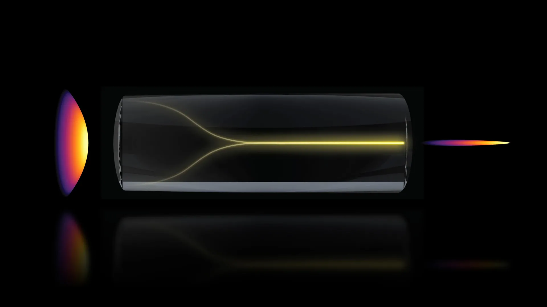
In a significant departure from long-standing principles in optical physics, researchers at the Massachusetts Institute of Technology (MIT) have uncovered a phenomenon where chaotic laser signals spontaneously reorganize into a highly focused, stable "pencil beam." This discovery, detailed in a recent publication in Nature Methods, challenges the conventional wisdom that increasing laser power inevitably leads to increased disorder. By harnessing this self-organizing light, the team has developed a new imaging modality capable of capturing 3D visualizations of the human blood-brain barrier at speeds 25 times faster than current gold-standard techniques, all while maintaining high resolution and eliminating the need for traditional fluorescent markers.
The breakthrough holds profound implications for the pharmaceutical industry and the study of neurodegenerative diseases such as Alzheimer’s and Amyotrophic Lateral Sclerosis (ALS). For the first time, scientists can observe in real-time how individual cells within a complex human tissue model interact with and internalize drug compounds, providing a critical window into the efficacy of treatments that have historically struggled to penetrate the brain’s protective defenses.
The Paradox of Nonlinearity: From Chaos to Order
The foundation of this discovery lies in the behavior of light within multimode optical fibers. Traditionally, these fibers are used to transport high levels of power, but they are notorious for their intrinsic disorder. As light travels through the fiber, imperfections in the glass and the various paths (modes) the light takes cause the signal to scatter, resulting in a distorted and "speckled" output. For decades, the consensus among physicists was that increasing the power input would only exacerbate this chaos, eventually leading to structural damage to the fiber itself.
However, the MIT team, led by Assistant Professor Sixian You and lead author Honghao Cao, observed an anomaly. Using a custom-built "fiber shaper"—a device designed to control light propagation—Cao began incrementally increasing the laser power to test the physical limits of the system. As the power approached the damage threshold, the expected scattering did not occur. Instead, the light underwent a phase transition, collapsing from a disordered state into a single, needle-sharp beam.
"The common belief in the field is that if you crank up the power in this type of laser, the light will inevitably become chaotic," said Sixian You, a senior author and faculty member in the MIT Department of Electrical Engineering and Computer Science (EECS). "But we proved that this is not the case. We followed the evidence, embraced the uncertainty, and found a way to let the light organize itself into a novel solution for bioimaging."
This phenomenon is rooted in nonlinear optics. When the intensity of the light is sufficiently high, it begins to alter the refractive properties of the glass fiber itself. Under specific conditions, this nonlinearity acts as a corrective force, countering the natural disorder of the fiber and forcing the light into a stable, self-reinforcing "pencil beam."
Technical Requirements for Self-Organization
The research team identified two rigorous conditions necessary to trigger this self-organizing effect. The first is a matter of geometric precision: the laser must enter the multimode fiber at a perfectly aligned, zero-degree angle. This requirement is significantly stricter than standard laboratory practices, where slight deviations are usually compensated for by downstream optics.
The second requirement involves the power threshold. The laser must be tuned to a specific intensity where the light-matter interaction becomes "nonlinear." At this stage, the light does not just pass through the glass; it interacts with the material’s molecular structure in a way that balances the scattering effects.
"At this critical power, the nonlinearity can counter the intrinsic disorder, creating a balance that transforms the input beam into a self-organized pencil beam," explained Honghao Cao.
The elegance of the method lies in its simplicity. Because the beam organizes itself through the inherent physics of the fiber, there is no need for the complex and expensive adaptive optics or custom beam-shaping components typically required to produce high-resolution imaging beams. This makes the technology potentially accessible to a wider range of biological laboratories without specialized expertise in advanced laser physics.
Visualizing the Fortress: The Blood-Brain Barrier
To demonstrate the practical utility of the pencil beam, the researchers applied it to one of the most challenging structures in human biology: the blood-brain barrier (BBB). The BBB is a highly selective semipermeable border of endothelial cells that prevents solutes in the circulating blood from non-selectively crossing into the extracellular fluid of the central nervous system.
While the BBB is essential for protecting the brain from pathogens and toxins, it is also a major obstacle for drug delivery. Approximately 98% of small-molecule drugs and nearly 100% of large-molecule drugs do not cross the BBB, contributing to the high failure rate of clinical trials for neurological disorders.
Current imaging methods, such as multiphoton microscopy, are effective but slow. These systems typically capture images by scanning a sample point-by-point to create a 2D slice, then repeating the process at different depths to stack the slices into a 3D model. This time-consuming process makes it difficult to track dynamic biological events, such as the movement of a drug molecule as it attempts to penetrate a cell.
The MIT team’s "pencil beam" approach circumvents these limitations. Because the beam maintains a high resolution over a significant depth of focus, it can capture 3D information much more efficiently. The system produced cellular-level 3D images with a 25-fold increase in speed compared to the current gold standard, allowing for "time-resolved" tracking of molecular targets.
Implications for Drug Development and Biological Engineering
One of the most transformative aspects of this new technique is its "label-free" nature. Conventional imaging often requires cells or drugs to be tagged with fluorescent dyes to make them visible. However, these tags can sometimes alter the behavior of the molecules being studied or prove toxic to the living tissue.
"That this new method doesn’t require the cells to have a fluorescent tag is a game-changer," said Roger Kamm, the Cecil and Ida Green Distinguished Professor of Biological and Mechanical Engineering at MIT. "For the first time, we can now visualize the time-dependent entry of drugs into the brain and even identify the rate at which specific cell types internalize the drug."
The pharmaceutical industry has increasingly moved toward human-based "organ-on-a-chip" models, as animal models often fail to accurately replicate the complexities of the human blood-brain barrier. The ability to monitor these engineered tissue models in real-time with high-speed 3D imaging provides a powerful new tool for screening potential treatments for Alzheimer’s, Parkinson’s, and ALS.
Sarah Spitz, a postdoc on the project, noted that the application of this technology extends beyond the brain. "This approach is not limited to the blood-brain barrier but enables time-resolved tracking of diverse compounds and molecular targets across engineered tissue models, providing a powerful tool for biological engineering," she stated.
Analytical Context and Future Directions
The discovery of self-organizing light in multimode fibers represents a significant shift in how researchers view "disorder" in physical systems. Rather than viewing the irregularities of a fiber as a hurdle to be overcome through external engineering, the MIT team has shown that those very irregularities can be leveraged to create a superior optical tool.
In terms of performance metrics, the "pencil beam" solves the classic trade-off in optical imaging between resolution and depth of field. In standard microscopy, a high-resolution lens typically has a very shallow depth of focus, meaning only a thin plane of the sample is in focus at any given time. The self-organized beam, however, remains tightly focused over a much longer distance, allowing for deep-tissue imaging without the loss of detail.
The research was a multi-disciplinary effort involving experts from MIT’s Research Laboratory for Electronics (RLE), Harvard University, and the Beth Israel Deaconess Medical Center. The team included Li-Yu Yu, Kunzan Liu, Sarah Spitz, Francesca Michela Pramotton, Federico Presutti, Zhengyu Zhang, and Subhash Kulkarni.
Moving forward, the researchers plan to refine the system to image even more complex structures, such as active networks of neurons. They also intend to investigate the underlying quantum and classical mechanics that govern the transition from chaos to the self-organized state. As the technology moves toward commercial or clinical application, it could become a standard fixture in labs dedicated to high-throughput drug screening and fundamental neuroscience.
This research was supported by a diverse array of funding bodies, including MIT startup funds, the National Science Foundation (NSF), the Silicon Valley Community Foundation, the Diacomp Foundation, the Harvard Digestive Disease Core, a MathWorks Fellowship, and the Claude E. Shannon Award. The findings, published in Nature Methods, mark a pivotal moment in the intersection of nonlinear physics and biomedical engineering, promising a faster, clearer view into the most guarded reaches of the human body.
