
 By Suherman Candra Maholtra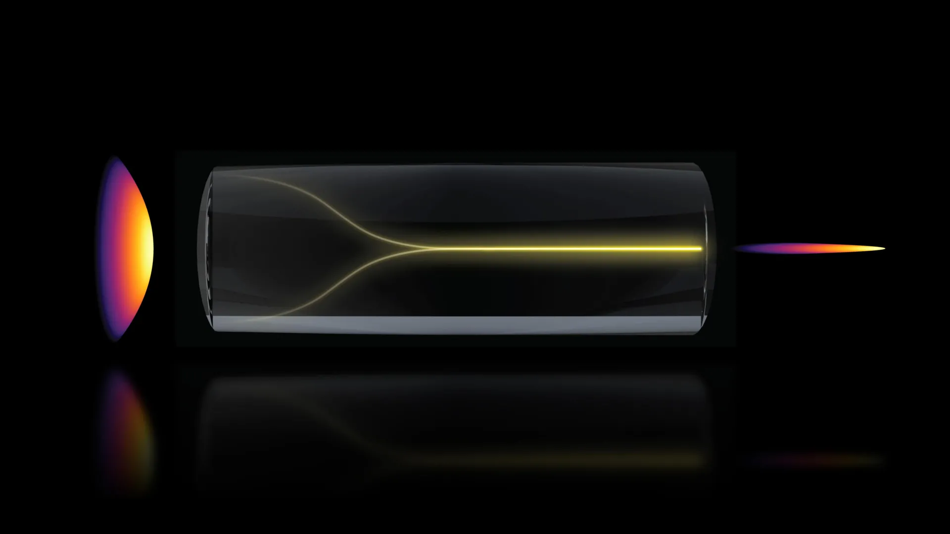
In a discovery that challenges long-standing conventions in the field of optical physics, a team of researchers at the Massachusetts Institute of Technology (MIT) has identified a phenomenon where chaotic laser signals can spontaneously reorganize into a highly focused "pencil beam." This self-organizing light effect, observed under specific high-power conditions within optical fibers, has enabled a breakthrough in biomedical imaging, allowing scientists to visualize the human blood-brain barrier in three dimensions at speeds 25 times faster than current industry standards. The research, published today in the journal Nature Methods, provides a novel pathway for observing real-time cellular interactions and drug absorption, potentially accelerating the development of treatments for neurodegenerative diseases such as Alzheimer’s and Amyotrophic Lateral Sclerosis (ALS).
The Paradigm Shift in Optical Physics
For decades, the prevailing wisdom in optical engineering has been that increasing the power of a laser traveling through a multimode optical fiber—a type of fiber capable of carrying high levels of energy—would inevitably lead to increased disorder. As light interacts with the inherent imperfections and microscopic irregularities within the glass fiber, it typically scatters, creating a "speckle pattern" that is difficult to control and unsuitable for high-precision imaging. To counteract this, researchers usually employ complex beam-shaping components or keep power levels low to avoid damaging the equipment.
However, the MIT team, led by Sixian You, an assistant professor in the Department of Electrical Engineering and Computer Science (EECS), found that the light behaves quite differently when pushed to its limits. By following the evidence of an unexpected experimental result, the team discovered that at a specific threshold of high power and precise alignment, the light undergoes a process of self-organization. Instead of disintegrating into chaos, the photons interact with the nonlinear properties of the fiber’s glass material, effectively "cleaning" the signal into a stable, narrow beam.
"The common belief in the field is that if you crank up the power in this type of laser, the light will inevitably become chaotic," says Professor You. "But we proved that this is not the case. We followed the evidence, embraced the uncertainty, and found a way to let the light organize itself into a novel solution for bioimaging."
Chronology of the Discovery: From Fiber Shapers to Self-Organization
The discovery originated from experiments involving a "fiber shaper," a device the researchers had previously developed to exert fine control over laser light as it traverses multimode fibers. Lead author Honghao Cao, an EECS graduate student, was testing the physical limits of this setup by gradually increasing the laser’s power.
In a typical scenario, the light would have become increasingly scattered, eventually risking a "catastrophic breakdown" of the fiber. Instead, as the power levels approached the threshold of potential damage, the researchers observed a sudden transition. The disordered light condensed into a single, extremely sharp "pencil beam" that remained stable even without the intervention of external beam-shaping hardware.
To replicate and validate this phenomenon, the team identified two stringent physical requirements:
- Perfect Alignment: The laser must enter the optical fiber at a zero-degree angle of incidence. This level of precision is significantly stricter than what is required for standard fiber-optic applications, where multimode fibers are chosen specifically for their ability to accept light from various angles.
- Nonlinear Interaction: The power must be high enough to trigger a "nonlinearity" in the glass. At this stage, the light does not just pass through the material; it changes the way the material responds to it. This interaction creates a balancing force that counters the intrinsic disorder of the fiber, forcing the light into a self-organized state.
"At this critical power, the nonlinearity can counter the intrinsic disorder, creating a balance that transforms the input beam into a self-organized pencil beam," Cao explained. This discovery effectively turns a traditional engineering "hassle"—disorder in fibers—into a self-correcting advantage.
Breaking the Trade-off: Speed, Resolution, and Depth
One of the most significant hurdles in optical imaging is the inherent trade-off between resolution and depth of focus. Typically, a beam that is focused very tightly to achieve high resolution will diverge quickly, resulting in a very shallow depth of focus. Conversely, a beam designed to stay narrow over a long distance (a large depth of focus) usually lacks the intensity and sharpness required for high-resolution cellular imaging.
The self-organized pencil beam defies this convention. It maintains a high resolution while simultaneously providing a large depth of focus. This allows the system to probe deep into biological tissues without the need for the mechanical "stacking" of multiple 2D images, which is the current standard for creating 3D visualizations.
By utilizing this pencil beam, the MIT team demonstrated a 25-fold increase in imaging speed compared to the "gold standard" of multi-photon microscopy. This speed is critical for "time-resolved tracking," which refers to the ability to watch biological processes as they happen in seconds or minutes rather than hours.
Real-Time Visualization of the Blood-Brain Barrier
The researchers applied this technology to one of the most challenging structures in human biology: the blood-brain barrier (BBB). The BBB is a highly selective semipermeable border of endothelial cells that prevents solutes in the circulating blood from non-selectively crossing into the extracellular fluid of the central nervous system. While this protects the brain from toxins, it also presents a massive hurdle for pharmaceutical delivery; approximately 98% of small-molecule drugs and nearly 100% of large-molecule drugs cannot cross the BBB.
Traditionally, evaluating whether a drug has crossed the barrier requires either animal models—which frequently fail to translate to human outcomes—or static, human-based engineered models that require fluorescent tagging. Fluorescent tags, while useful, can sometimes interfere with the natural behavior of the cells or the drug molecules themselves.
The MIT team’s new method is "label-free," meaning it does not require these tags. For the first time, researchers can visualize the entry of drugs into the brain in real-time, identifying the specific rates at which different cell types internalize the treatment.
Roger Kamm, the Cecil and Ida Green Distinguished Professor of Biological and Mechanical Engineering at MIT and a co-author of the study, highlighted the industrial implications: "The pharmaceutical industry is especially interested in using human-based models to screen for drugs that effectively cross the barrier… That this new method doesn’t require the cells to have a fluorescent tag is a game-changer."
Implications for Alzheimer’s and ALS Research
The ability to track drug absorption at the cellular level in 3D has immediate implications for the study of neurodegenerative diseases. In conditions like Alzheimer’s, the failure of the blood-brain barrier is often a contributing factor to the disease’s progression, while also being the primary obstacle to treatment.
By using the self-organized pencil beam, researchers can now:
- Monitor Drug Delivery: Observe if experimental antibodies or proteins are reaching the intended neurons or if they are being blocked or absorbed by the wrong cell types.
- Evaluate Tissue Engineering: Assess the health and functionality of lab-grown "brain-on-a-chip" models with unprecedented detail.
- Understand Disease Pathology: Study how the cellular architecture of the BBB changes over time in response to various stressors or therapeutic interventions.
Sarah Spitz, a postdoc and co-author, noted that the application of this technology extends beyond the brain. "This approach… enables time-resolved tracking of diverse compounds and molecular targets across engineered tissue models, providing a powerful tool for biological engineering," she said.
Technical Data and Validation
The stability of the pencil beam was a key focus of the study. Unlike conventional laser beams that often produce "sidelobes"—secondary rings of light that blur the image—the self-organized beam remained remarkably clean. Comparative tests showed that the signal-to-noise ratio was significantly improved, allowing for clearer differentiation between cell types within the dense environment of the blood-brain barrier.
The research team included a diverse group of experts from MIT’s EECS, the Research Laboratory for Electronics (RLE), Harvard University, and Beth Israel Deaconess Medical Center. The collaboration between physicists, electrical engineers, and biological engineers was essential to translating a discovery in fiber optics into a practical tool for medicine.
Future Outlook and Practical Implementation
Despite the success of the initial experiments, the team acknowledges that there is more to learn about the underlying physics. The specific "nonlinear" interactions within the glass that allow for self-organization are complex, and the team plans to further investigate these mechanisms to optimize the beam for different types of tissue.
Future goals include:
- Neuronal Imaging: Expanding the technique to image entire networks of neurons to study how they communicate in real-time.
- Clinical Portability: Developing more compact versions of the setup that could be used in clinical settings or pharmaceutical laboratories without requiring specialized expertise in optical physics.
- Broader Molecular Targets: Testing the system’s ability to track a wider range of compounds, including gene therapies and viral vectors.
"That is the charm of this method—you could do this with a normal, optical setup and without much domain expertise," Professor You remarked, suggesting that the barrier to entry for this high-speed imaging may be lower than previously thought once the initial alignment and power parameters are understood.
The work received support from several prestigious institutions, including the National Science Foundation (NSF), the Silicon Valley Community Foundation, the Diacomp Foundation, and the Harvard Digestive Disease Core. As the technology moves toward broader adoption, it stands as a testament to the value of "embracing uncertainty" in scientific research, turning what was once considered a limitation of optical fibers into a powerful new lens for looking into the human body.
