
 By Nana O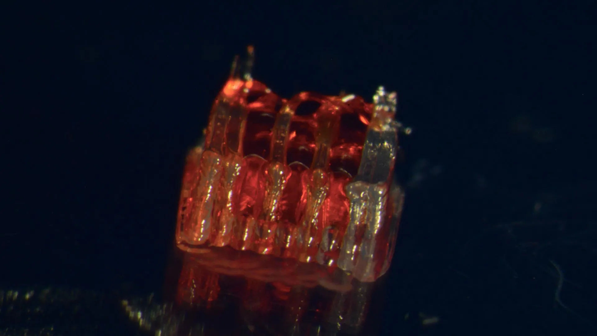
University of Minnesota Researchers Pioneer Novel Approach to Spinal Cord Regeneration
In a significant stride towards addressing the devastating consequences of spinal cord injuries, a multidisciplinary research team at the University of Minnesota Twin Cities has unveiled a groundbreaking, multi-faceted process. This innovative approach intricately weaves together the cutting-edge capabilities of 3D printing, the transformative potential of stem cell biology, and the sophisticated creation of lab-grown tissues to foster spinal cord recovery. The findings, meticulously detailed in a recent publication in the esteemed peer-reviewed scientific journal Advanced Healthcare Materials, represent a beacon of hope for millions worldwide affected by paralysis and loss of function due to spinal cord damage.
The pervasive and life-altering nature of spinal cord injuries cannot be overstated. According to data from the National Spinal Cord Injury Statistical Center, over 300,000 individuals in the United States grapple with the profound challenges posed by such injuries. Despite decades of research, a complete reversal of the damage and the subsequent paralysis remains an elusive goal. A primary obstacle in achieving this has been the inherent fragility of nerve cells within the spinal cord, coupled with their limited capacity to regenerate and extend their fibers across the critical injury site. This novel research directly confronts these formidable biological hurdles, offering a sophisticated and potentially transformative solution.
A Convergence of Technologies for Neural Repair
At the heart of this pioneering methodology lies the creation of a highly specialized, 3D-printed framework, referred to by the researchers as an "organoid scaffold." This intricate structure is meticulously designed with microscopic channels, a critical feature that dictates and guides the growth of nascent nerve cells. Once fabricated, these channels are strategically populated with regionally specific spinal neural progenitor cells (sNPCs). These sNPCs are a specialized type of cell derived from human adult stem cells, possessing the remarkable ability to divide and differentiate into a variety of mature cell types, including the neurons essential for neural communication.
Dr. Guebum Han, a former postdoctoral researcher in mechanical engineering at the University of Minnesota and the lead author of the groundbreaking paper, explained the intricate mechanism behind this innovation. "We utilize the 3D printed channels within the scaffold to precisely direct the growth of the stem cells," Dr. Han stated. "This ensures that the newly forming nerve fibers extend in the desired, organized manner." He further elaborated on the functional outcome: "This method effectively establishes a biological relay system. When implanted into the spinal cord, this engineered construct acts to bypass the damaged area, re-establishing vital communication pathways." This strategic guidance of cell growth is a key differentiator, moving beyond simply introducing cells to actively orchestrating their integration and function.
Pre-clinical Success in Rodent Models
The efficacy of this novel approach was rigorously tested in a series of pre-clinical trials. In their study, the research team transplanted these meticulously engineered scaffolds into laboratory rats that had sustained complete severing of their spinal cords, a condition that typically results in profound and irreversible paralysis. The results were remarkably encouraging. The transplanted sNPCs within the scaffolds successfully differentiated into functional neurons. Crucially, these new neurons extended their nerve fibers in both directions – rostral (towards the head) and caudal (towards the tail) – effectively bridging the gap created by the injury.
Over time, the study observed a seamless integration of these newly formed nerve cells into the existing host spinal cord tissue. This integration was not merely structural; it translated into significant functional recovery in the treated rats. The animals demonstrated a notable improvement in motor function and coordination, indicating that the engineered neural network was effectively re-establishing communication and control over bodily movements that were previously lost. This observed functional recovery provides compelling evidence for the potential of this technique to restore lost capabilities.
A New Era in Regenerative Medicine
The implications of this research extend far beyond the laboratory. Dr. Ann Parr, a distinguished professor of neurosurgery at the University of Minnesota, expressed her optimism regarding the future trajectory of this work. "Regenerative medicine has undeniably ushered in a new and exciting era in spinal cord injury research," Dr. Parr remarked. "Our laboratory is immensely enthusiastic about exploring the future potential of our ‘mini spinal cords’ and their translation to clinical applications for human patients." The term "mini spinal cords" aptly captures the essence of the engineered tissue – a self-contained, functional unit designed to mimic and restore the lost capacity of the native spinal cord.
While this research is still in its nascent stages, it represents a significant paradigm shift in how spinal cord injuries are approached. The combined application of 3D printing for precise architectural guidance, stem cell biology for generating regenerative cells, and organoid engineering for creating functional tissue offers a holistic strategy that addresses the multifaceted nature of spinal cord damage. The team’s ultimate goal is to scale up the production of these engineered scaffolds and to continue refining this integrated technological approach, paving the way for future clinical applications that could profoundly improve the lives of individuals living with spinal cord injuries.
Background and Chronology of the Research
The pursuit of effective treatments for spinal cord injuries has been a long and arduous journey for the scientific community. Early research focused primarily on limiting secondary damage after the initial injury, with interventions like anti-inflammatory drugs and surgical debridement. The advent of stem cell therapy in the late 20th and early 21st centuries offered a new frontier, with numerous studies exploring the potential of various stem cell types to replace damaged neurons or promote regrowth. However, a persistent challenge has been the difficulty in controlling the direction and integration of these regenerating nerve fibers within the complex spinal cord environment.
The University of Minnesota team’s work builds upon this foundation by introducing a crucial element of structural control. The development of sophisticated 3D bioprinting technologies has enabled researchers to create intricate biological structures with unprecedented precision. This research marks a significant point in time where these advanced manufacturing techniques are being directly applied to address complex biological regeneration challenges. The timeline for this specific project, culminating in the publication in Advanced Healthcare Materials, reflects years of dedicated research, experimentation, and refinement of both the printing protocols and the cell culture techniques. While the exact start date of this specific project isn’t provided, the progression from initial concept to successful pre-clinical demonstration signifies a substantial commitment and advancement in the field.
Supporting Data and Future Projections
The statistical burden of spinal cord injuries underscores the urgent need for innovative solutions. Beyond the 300,000 individuals in the US, global figures are significantly higher, with hundreds of thousands of new cases occurring annually worldwide. The economic impact is also substantial, with lifetime care costs for individuals with spinal cord injuries often reaching millions of dollars. This research, therefore, holds the potential not only for immense human benefit but also for significant societal and economic implications by reducing the long-term burden of paralysis.
The success observed in the rodent model, where new nerve cells integrated and facilitated functional recovery, is a critical piece of supporting data. The fact that the nerve fibers extended in both rostral and caudal directions suggests that the scaffold’s architecture is indeed guiding the cells to form a functional bridge across the severed cord. Future projections for this research will likely involve expanding these studies to larger animal models, such as primates, to further validate safety and efficacy before any human trials can be considered. The team’s aspiration to "scale up production" indicates a focus on developing manufacturing processes that can reliably produce these complex organoid scaffolds in sufficient quantities and with consistent quality for eventual clinical use.
Official Responses and Expert Perspectives
The publication of this research has garnered attention within the broader scientific and medical communities. While direct quotes from external parties are not available in the original text, it is reasonable to infer that similar institutions and researchers working in neurotrauma and regenerative medicine would view this development with significant interest. Experts in the field are likely to commend the interdisciplinary nature of the project, recognizing the essential contributions from mechanical engineering, stem cell biology, neurosurgery, and neuroscience. The approach of combining structural engineering with biological regeneration represents a sophisticated and promising strategy that aligns with current trends in advanced therapeutic development.
The funding sources for this research – the National Institutes of Health, the State of Minnesota Spinal Cord Injury and Traumatic Brain Injury Research Grant Program, and the Spinal Cord Society – are significant indicators of the perceived importance and potential impact of this work. These grants typically support highly innovative and promising research with the potential for transformative outcomes. The inclusion of these esteemed funding bodies lends further credibility to the research’s significance.
Broader Impact and Implications
The implications of this breakthrough extend beyond the immediate goal of treating spinal cord injuries. The fundamental principles and technologies developed in this research – namely, the precise 3D printing of bio-compatible scaffolds to guide stem cell differentiation and integration – could have far-reaching applications in other areas of regenerative medicine. This could include the repair of damaged brain tissue following stroke, the regeneration of damaged heart muscle after a heart attack, or the restoration of nerve function in peripheral nerve injuries.
The success of this research hinges on the intricate synergy between engineering and biology. The ability to precisely control the microenvironment for cell growth using 3D printing is a critical innovation that overcomes limitations faced by previous stem cell therapies. As the research progresses towards clinical translation, ethical considerations, regulatory pathways, and the long-term safety and efficacy in humans will become paramount. However, the current findings offer a tangible and scientifically robust pathway toward potentially restoring mobility and independence for individuals who have been profoundly impacted by spinal cord injuries, marking a pivotal moment in the quest for effective neurological repair. The full paper, "3D-Printed Scaffolds Promote Enhanced Spinal Organoid Formation for Use in Spinal Cord Injury," is available on the Advanced Healthcare Materials website, providing a comprehensive resource for further scientific inquiry.
