
 By Nana O
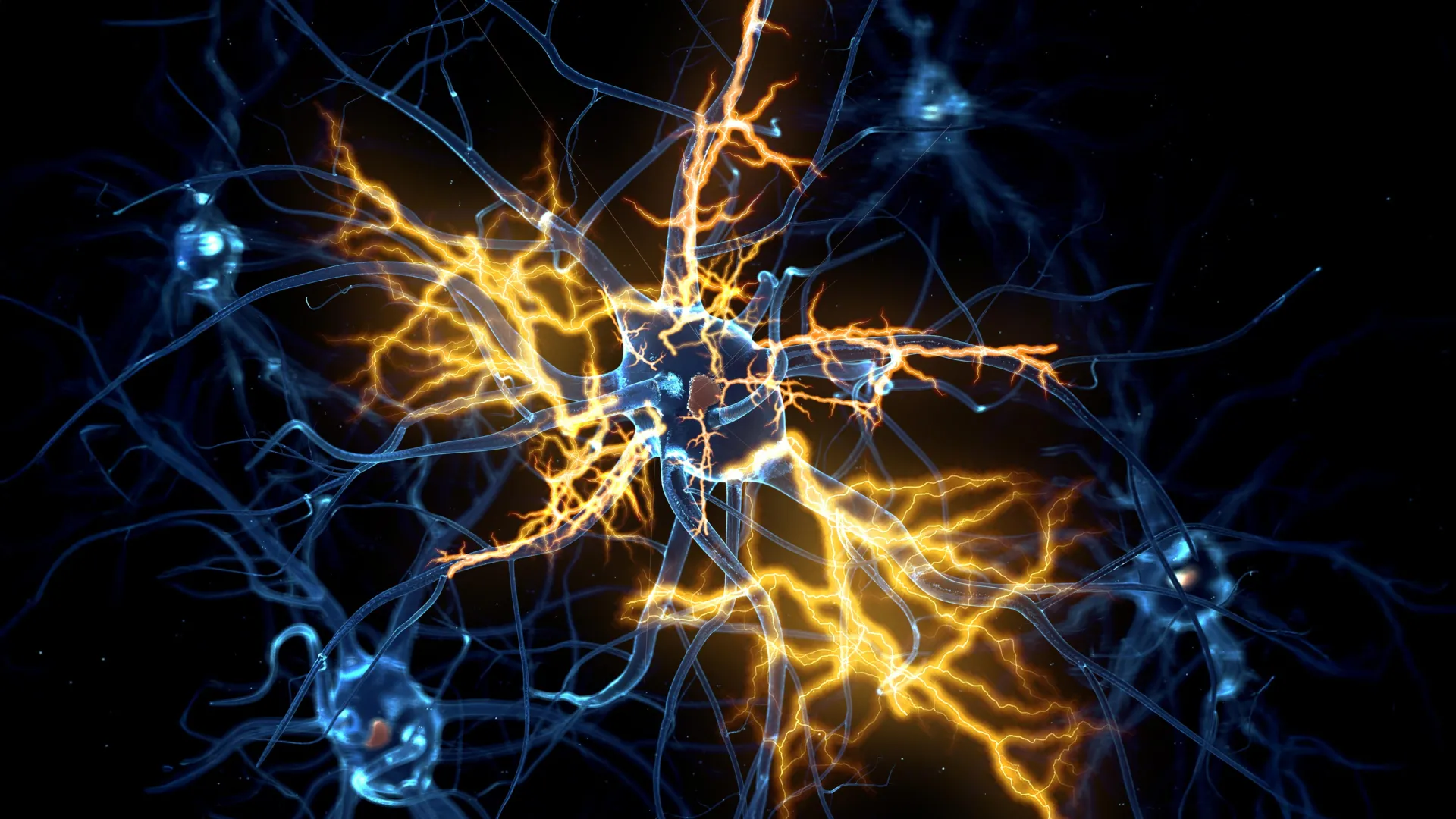
Scientists at the University of Cambridge have created tiny lab-grown brain and spinal cord systems that mimic how movement signals travel through the human nervous system. Using this model, the team discovered that nerve damage once believed to be permanent may actually be reversible under certain conditions. This groundbreaking research, published in the prestigious journal Cell Reports, offers a new glimmer of hope for individuals suffering from paralysis and other debilitating neurological conditions.
Unlocking the Secrets of Neural Regeneration
For decades, the scientific community has grappled with the devastating reality that injuries to the central nervous system, encompassing the brain and spinal cord, often result in irreversible loss of function. This is primarily due to the remarkable inability of mature neurons to regrow their axons, the crucial long nerve fibers responsible for transmitting signals that control everything from voluntary movement to sensory perception. This regenerative deficit not only impacts individuals after traumatic injuries but also plays a significant role in the progression of neurodegenerative diseases like motor neurone disease (MND) and multiple sclerosis (MS).
The intricate journey of neural development begins in the embryonic stage, where neurons form complex and precise communication networks between the brain and spinal cord. These nascent connections are characterized by robust axon growth, allowing for the establishment of functional pathways. However, as the human body matures, a fundamental shift occurs: the central nervous system largely loses this innate capacity for regeneration. This decline in regenerative potential has long been considered an inherent limitation, leading to the widespread acceptance of permanent disability following spinal cord injuries or severe brain trauma.
The Genesis of a Revolutionary Model
Building upon their earlier successes in developing miniature human brain models, Dr. András Lakatos and his team at the University of Cambridge embarked on an ambitious project: to construct a more comprehensive, interconnected system that accurately replicates the brain-spinal cord axis. In 2021, their pioneering work introduced pea-sized "brain organoids" derived from human stem cells, which mimicked aspects of the cerebral cortex. These initial models proved invaluable for studying the molecular underpinnings of conditions like motor neurone disease and for exploring potential preventative strategies.
The latest research, detailed in Cell Reports, represents a significant leap forward. The Cambridge team meticulously engineered miniature versions of the connected human brain and spinal cord systems. Recognizing the distinct yet interconnected nature of these structures, the researchers maintained the brain and spinal cord organoids in separate compartments within the laboratory. This deliberate separation allowed them to observe and analyze the natural tendency of axons from the brain tissue to extend across the intervening gap and establish connections with the spinal cord tissue. The resulting neural circuits were not merely structural; they demonstrated functional capability, remarkably triggering contractions in adjacent clusters of muscle cells. This functional demonstration provided compelling evidence that the lab-grown systems were faithfully recapitulating key aspects of human neural communication.
A Developmental Timeline of Regenerative Decline
To understand the biological mechanisms governing axon regeneration, the scientists maintained these intricate miniature systems for an extended period, exceeding one year. This prolonged observation allowed them to meticulously track the regenerative capabilities of the neurons across different developmental stages. Their findings revealed a critical developmental window: up until approximately day 150 of development, a stage roughly corresponding to the mid-pregnancy period in humans, damaged axons exhibited a significant capacity for regrowth. However, beyond this point, a dramatic decline in regenerative ability was observed.
George Gibbons, a key contributor to the study from the Department of Clinical Neurosciences at the University of Cambridge and the study’s first author, articulated the significance of this discovery: "Neurons taken from less mature organoids regrew long fibers after injury, but those from more mature organoids showed a sharp drop in their ability to regrow. In other words, poor regeneration is built into human neurons as they mature in the central nervous system." This statement underscores a pivotal insight: the loss of regenerative capacity is not an external factor but rather an intrinsic process that becomes embedded within neurons as they mature.
Unraveling the Genetic Blueprint of Regeneration Limitation
The research team then delved deeper, performing a comprehensive analysis of gene activity within the neurons responsible for connecting the brain and spinal cord. This investigation unearthed a complex network of genes that functions akin to a biological switch, actively suppressing axon growth as neurons mature and establish synaptic connections. This intricate regulatory system plays a vital role in fine-tuning neural circuits for efficient adult function but comes at the cost of diminished regenerative potential.
In a truly remarkable turn, the researchers discovered that by selectively blocking key regulators within this identified gene network, they could effectively reactivate the dormant regenerative capacity of the neurons. This intervention demonstrated that the decline in axon growth is not an immutable destiny but a biologically controlled process that can, in principle, be manipulated.
A Pharmaceutical Ally in Nerve Repair: Lynestrenol’s Potential
The pursuit of therapeutic interventions led the scientists to explore existing pharmaceutical compounds. They systematically searched a vast database of drug compounds, seeking those that might influence the newly identified gene network. Among the promising candidates identified was lynestrenol, a hormone-based drug already approved for specific menstrual disorders and contraceptive use.
When lynestrenol was administered to damaged neurons in their organoid models, it demonstrated a significant and encouraging improvement in axon regrowth. This finding is particularly noteworthy as it suggests that a drug with an established safety profile for human use could potentially be repurposed to enhance nerve repair.
The researchers acknowledged that scar tissue formation and inflammation are significant hurdles in the natural repair of nerve injuries. However, they emphasized the critical importance of understanding the neuron-specific biological mechanisms that inherently limit regeneration. Previous research has indicated that younger neurons can navigate and grow through environments that typically inhibit repair at injury sites, further supporting the notion that intrinsic neuronal factors are paramount.
Dr. András Lakatos, the senior author of the study, provided a profound perspective on the implications of their findings: "When the brain and spinal cord are damaged, the nerve fibers that carry movement signals from the brain to the spinal cord rarely grow back. That’s why paralysis is usually permanent. But we didn’t know exactly when the ability of axons to regenerate becomes limited. Our model provides a good indication that this block happens during development, and it can still be reversed after this point." He further elaborated, "Lynestrenol itself may not be the answer to spinal cord repair, but it shows us that, in principle, it should be possible to directly target human neurons and regenerate their axons. Although we still need to show that this strategy will also help to re-establish appropriate connections between the brain and spinal cord cells, this gives us hope that one day we may be able to treat conditions previously thought untreatable."
The Growing Significance of Human Organoids in Medical Research
The advent and refinement of organoid technology are revolutionizing our understanding of human biology and disease. While animal models, such as mice and rats, have historically been indispensable tools in biomedical research, crucial biological differences between species can limit the direct translatability of findings to human outcomes.
Human stem cell-derived organoids, on the other hand, offer a more faithful recapitulation of human biology. By mimicking the complexity and cellular interactions found in human tissues and organs, they serve as powerful bridges, narrowing the gap between preclinical animal studies and the clinical realities faced by patients. This is particularly relevant in neurobiology, where species-specific differences in neural circuitry and regenerative capacity are well-documented.
Dr. Lakatos further highlighted the value of these human-centric models: "Much of what we know about nerve regeneration comes from rodents, whose neurons behave differently from human neurons. Our sophisticated organoid models help bridge the knowledge gap from animal models to what we see in patients. They are also an important contribution to efforts to reduce the use of animals in research." This sentiment aligns with the growing ethical imperative in scientific research to minimize animal testing wherever viable alternatives exist.
The University of Cambridge is at the forefront of utilizing organoid technology for a diverse array of medical investigations. Beyond neuroregeneration, researchers are employing organoids to study liver repair, unravel the complexities of Crohn’s disease in children, and explore the earliest, most delicate stages of human pregnancy. The ability to model these complex biological processes in a human-relevant context promises to accelerate discoveries and improve therapeutic development across a wide spectrum of medical fields.
This pioneering research was made possible through the generous funding of UK Research and Innovation (UKRI) via the Medical Research Council and Spinal Research, underscoring the collaborative and multi-faceted support crucial for advancing such complex scientific endeavors. The findings represent a significant stride towards a future where previously untreatable neurological conditions may become amenable to effective treatment.
