
 By Gabriel Paijo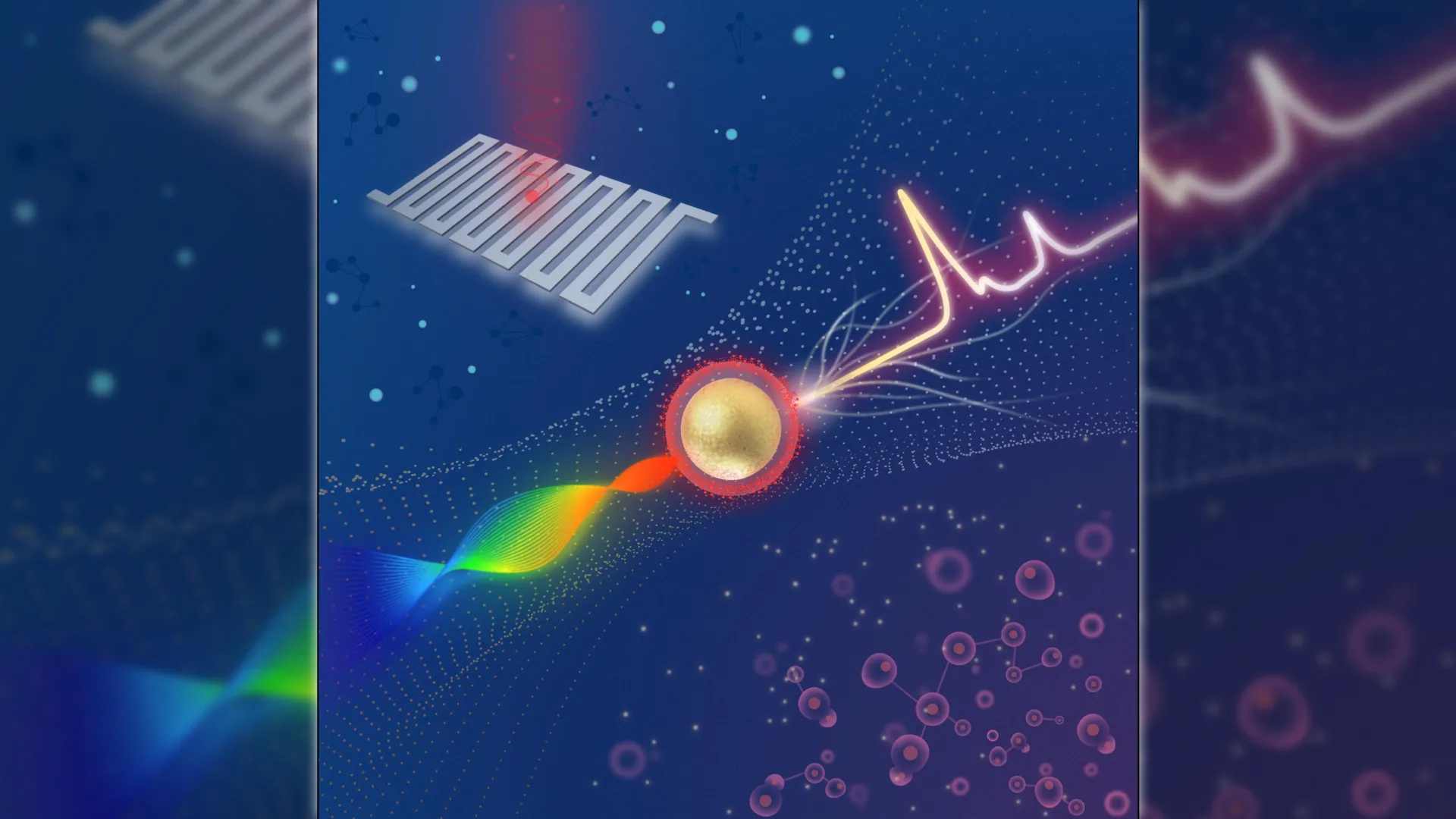
In a significant leap for optical medical imaging, researchers at Michigan State University have unveiled a compact Raman imaging system capable of distinguishing between cancerous and healthy tissue with unprecedented sensitivity. By integrating advanced superconducting nanowire single-photon detectors (SNSPDs) with a swept-source laser architecture, the team has developed a tool that addresses one of the most persistent challenges in clinical oncology: the real-time identification of tumor margins. This breakthrough, published in the journal Optica, suggests a future where the gap between laboratory-grade molecular analysis and bedside clinical practice is finally closed, potentially reducing diagnostic delays and improving surgical outcomes for millions of patients.
The Evolution of Raman Spectroscopy in Clinical Settings
For decades, Raman spectroscopy has been lauded as a "gold standard" for non-invasive chemical analysis. The technique relies on the inelastic scattering of photons, known as the Raman effect, where incident laser light interacts with molecular vibrations, resulting in a unique spectral "fingerprint." In the context of oncology, these fingerprints can reveal the specific biochemical signatures of malignancy, such as altered protein structures or increased nucleic acid concentrations.
However, the primary hurdle to widespread clinical adoption has been the inherent weakness of the Raman signal. Only about one in every ten million photons undergoes Raman scattering, making the signal difficult to detect against a background of fluorescence and noise. Traditionally, this necessitated bulky, expensive equipment, long integration times, and highly controlled laboratory environments. While Surface-Enhanced Raman Scattering (SERS)—which uses metallic nanoparticles to amplify these signals—offered a partial solution, the detection hardware remained a bottleneck.
The new system developed by the Institute for Quantitative Health Science and Engineering (IQ) at Michigan State University, led by Dr. Zhen Qiu, overcomes these limitations. By replacing traditional charge-coupled device (CCD) cameras with ultra-sensitive superconducting detectors, the researchers have managed to capture signals that are nearly four times weaker than those detectable by current state-of-the-art commercial systems.
Technical Architecture: SNSPDs and Swept-Source Lasers
The innovation of the MSU system lies in its unique combination of two high-performance technologies: superconducting nanowire single-photon detectors (SNSPDs) and a swept-source Raman architecture.
SNSPDs represent the pinnacle of light detection technology. These devices consist of a thin film of superconducting material, such as niobium nitride, patterned into a nanowire. When the wire is cooled below its critical temperature and biased with a current near its critical threshold, the absorption of even a single photon can disrupt the superconducting state, creating a measurable voltage pulse. This allows for near-unity quantum efficiency and extremely low "dark counts"—essentially eliminating the electronic noise that plagues traditional silicon-based detectors.
In most Raman systems, a fixed-wavelength laser is used, and the scattered light is dispersed across a multi-pixel camera. The MSU team inverted this design. Their system utilizes a swept-source laser that rapidly changes its wavelength over a specific range. This allows the system to use a single-pixel SNSPD rather than a bulky camera. This "fiber-coupled" configuration is not only more sensitive but also significantly more compact, facilitating the eventual miniaturization of the device into a portable unit or a handheld surgical probe.
Methodology and the Role of Targeted Nanoparticles
To validate the system’s diagnostic capabilities, the researchers employed a targeted approach using SERS nanoparticles. These nanoparticles were engineered with a gold core to provide the Raman enhancement and coated with hyaluronan acid (HA).
The choice of HA was strategic. Hyaluronan has a high affinity for CD44, a cell-surface glycoprotein that is overexpressed in a wide variety of cancers, including breast, colon, and lung carcinomas. CD44 is often associated with cancer stem cells and epithelial-mesenchymal transition, making it a reliable marker for aggressive tumor tissue.
The experimental protocol followed a rigorous progression:
- Sensitivity Testing: The system was first tested against varying concentrations of nanoparticle solutions. It achieved femtomolar sensitivity ($10^-15$ moles per liter), a level of precision that allows for the detection of minute quantities of biomarkers that would be invisible to standard imaging modalities like MRI or CT scans.
- Cellular Validation: The platform was then used to image cultured breast cancer cells. The system successfully identified the "hotspots" where nanoparticles had bound to the CD44 receptors on the cell membranes.
- Animal Model Testing: In the most critical phase, the researchers applied the system to mouse tumor models and healthy tissue samples. The results demonstrated a stark contrast: tumor samples produced robust Raman signals, while healthy tissues showed only minimal background interference.
Comparative Performance and Data Insights
According to the data published in Optica, the MSU system’s sensitivity represents a paradigm shift. In head-to-head comparisons with commercial Raman spectrometers—which typically use deep-cooled CCDs—the SNSPD-based system maintained a signal-to-noise ratio (SNR) that allowed for clear tissue differentiation even when the signal intensity was reduced by 75%.
This sensitivity gain is crucial for "intraoperative" use, where time is of the essence. In a surgical setting, a doctor cannot wait minutes for a single point of data. The high-speed detection capability of the SNSPD, combined with the efficient light collection of the swept-source architecture, paves the way for real-time "video-rate" Raman imaging.
Furthermore, the system’s ability to operate with lower laser power is a significant safety advantage. High-intensity lasers required by less sensitive systems can cause "photodamage" or localized heating of biological tissues. By requiring four times less signal, the MSU system can operate well within the safety limits for human tissue exposure while still providing high-fidelity diagnostic data.
Expert Perspectives and Clinical Implications
"Traditional methods for cancer-related diagnosis are time-consuming and labor-intensive because they require staining tissue samples and having a pathologist look for any abnormalities," noted Dr. Zhen Qiu. "While our system would not immediately replace pathology, it could serve as a rapid screening tool to accelerate diagnosis."
Pathologists currently rely on Hematoxylin and Eosin (H&E) staining, a process that can take days for a final result. During surgery, "frozen section" analysis is sometimes used, but it is prone to errors and takes 20 to 30 minutes while the patient remains under anesthesia. A compact Raman system could potentially provide an "optical biopsy" in seconds.
Industry experts have reacted positively to the integration of SNSPDs in medical tech. Quantum Opus, the collaborator that provided the superconducting devices, highlighted that this application demonstrates the "quantum advantage" in a practical, life-saving context. Historically, SNSPDs were reserved for quantum computing and deep-space communication due to the requirement for cryogenic cooling. However, recent advances in closed-cycle cryostats have made these detectors more accessible for clinical environments.
Future Research: Multiplexing and Miniaturization
The research team is already looking toward the next iteration of the technology. One of the most promising avenues is "multiplexing." Because Raman signals are so specific, it is possible to use different types of nanoparticles, each targeting a different biomarker (e.g., HER2, EGFR, and CD44) simultaneously. This would allow clinicians to create a multi-dimensional molecular map of a tumor, providing insights into its genetic makeup and potential resistance to certain therapies.
Technological refinements are also underway to increase the readout speed. The team is exploring Vertical-Cavity Surface-Emitting Lasers (VCSELs), which are cheaper and more compact than current swept-source lasers. By narrowing the sweep range to focus only on the most informative spectral peaks, the researchers believe they can further increase the frame rate of the imaging system.
Broader Impact on Healthcare Systems
The implications of this technology extend beyond the operating room. Earlier detection is the single most effective way to improve cancer survival rates. A portable Raman screening tool could be deployed in outpatient clinics or even in resource-limited settings where access to full pathology labs is restricted.
From an economic perspective, the system addresses the "positive margin" problem in oncology. In breast cancer lumpectomies, for example, up to 20-30% of patients require a second surgery because post-operative pathology reveals that tumor cells were left at the edge of the excised tissue. By providing surgeons with a real-time map of the tumor margins during the first procedure, the MSU system could significantly reduce re-operation rates, lowering healthcare costs and reducing patient trauma.
"Ultimately, such advances could enhance patient outcomes and reduce diagnostic delays, accelerating the path from detection to treatment," Dr. Qiu concluded.
As the team moves toward clinical trials, the medical community remains watchful. The transition from a controlled laboratory setting to the "noisy" environment of a human operating room is the final hurdle. However, with a detection limit that pushes the boundaries of physics and a design focused on clinical utility, this superconducting Raman system stands as a beacon of progress in the fight against cancer. By turning the "weakness" of light into a strength of diagnosis, the researchers at Michigan State University have opened a new chapter in molecular medicine.
