
 By George Ongkojoyo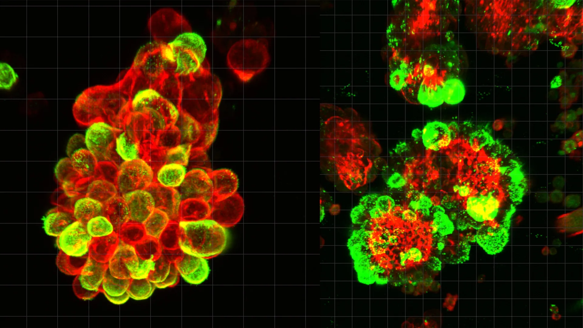
Ovarian cancer has long held a reputation as the "silent killer" of the gynecological world, characterized by its stealthy progression and devastating lethality. For decades, the medical community has struggled to understand why this specific malignancy spreads with such aggressive speed across the abdominal cavity, often evading detection until it has reached an advanced stage. A groundbreaking study led by researchers at Nagoya University in Japan has finally decoded a significant portion of this mystery. Published in the prestigious journal Science Advances, the research reveals that ovarian cancer cells do not act as solitary invaders; instead, they "hijack" healthy cells from the body’s own protective lining to facilitate their spread and shield themselves from the effects of chemotherapy.
The discovery centers on the interaction between ovarian cancer cells and mesothelial cells—the specialized cells that form the peritoneum, a protective membrane lining the abdominal cavity and covering the organs within it. The study demonstrates that these two cell types form hybrid clusters, creating a biological partnership that allows the cancer to navigate the complex environment of the abdomen with unprecedented efficiency.
The Biological Mechanism of Hybrid Spread
To uncover these findings, the research team, led by Dr. Kaname Uno of the Nagoya University Graduate School of Medicine, conducted a comprehensive analysis of abdominal fluid, known as ascites, collected from patients with ovarian cancer. Ascites often accumulates in the late stages of the disease, serving as a reservoir for floating cancer cells.
Previous oncological models suggested that cancer cells primarily drift through this fluid as individual units or small, uniform clusters. However, the Nagoya University team observed a different reality. Using advanced microscopy and single-cell RNA sequencing, they found that cancer cells frequently bond with mesothelial cells that have detached from the abdominal lining. These "hybrid spheres" were present in approximately 60% of the cancer cell clusters analyzed.
The process of recruitment is driven by a specific signaling pathway. The researchers identified that ovarian cancer cells secrete a potent signaling molecule called Transforming Growth Factor-beta 1 (TGF-β1). This protein acts as a molecular command, fundamentally altering the behavior and structure of the recruited mesothelial cells. Under the influence of TGF-β1, the mesothelial cells undergo a transition, developing sharp, needle-like protrusions known as invadopodia. These structures act as biological "drills," allowing the hybrid clusters to penetrate the surfaces of organs such as the liver, stomach, and intestines far more effectively than cancer cells could on their own.
A Departure from Conventional Metastasis
The spread of ovarian cancer represents a significant departure from the metastatic patterns seen in other common malignancies. In cancers such as breast, lung, or prostate cancer, the primary mode of spread is hematogenous or lymphatic. This means tumor cells break away from the primary site, enter the bloodstream or the lymphatic system, and travel through these defined "highways" to reach distant organs. Because these pathways are structured, clinicians can often use blood tests to monitor for circulating tumor cells or biomarkers.
Ovarian cancer, however, utilizes a method known as transperitoneal spread. When cells detach from the primary ovarian tumor, they enter the peritoneal fluid. This fluid is not static; it moves constantly, influenced by the motion of the diaphragm during breathing and the peristaltic movements of the digestive tract. This creates a "drifting" effect, where cancer cells are carried to various points within the abdominal cavity without a predictable route.
Until the publication of the Nagoya University study, the "floating phase" of ovarian cancer was poorly understood. Scientists were unsure how these cells survived the journey through the abdominal fluid or how they managed to re-attach and invade new tissues so rapidly. The discovery of the hybrid spheres provides the missing link. By recruiting mesothelial cells to do the "heavy lifting" of tissue invasion, the cancer cells remain relatively dormant and unchanged, conserving energy and maintaining their genetic integrity while their hijacked partners create the openings necessary for colonization.
The Challenge of Chemotherapy Resistance
One of the most concerning findings of the study involves the hybrid spheres’ response to medical intervention. The researchers found that these mixed cell clusters are significantly more resistant to standard chemotherapy drugs than isolated cancer cells.
This resistance appears to be a byproduct of the physical and molecular architecture of the spheres. The presence of mesothelial cells provides a protective barrier, potentially limiting the penetration of cytotoxic drugs. Furthermore, the molecular signaling between the two cell types within the sphere creates a microenvironment that favors cell survival over apoptosis (programmed cell death).
This helps explain a common clinical challenge in treating ovarian cancer: many patients initially respond well to chemotherapy, only to experience a rapid and aggressive recurrence. If these hybrid clusters remain in the abdominal fluid or tucked away in the recesses of the peritoneum, they can survive the initial chemical onslaught and later re-seed the abdomen with new tumors.
The Human Element: From Clinic to Laboratory
The impetus for this research was deeply personal for the lead author, Dr. Kaname Uno. Before transitioning into full-time research, Dr. Uno spent eight years practicing as a gynecologist. His career path was permanently altered by a specific clinical case involving a patient who had been diligent with her health screenings.
The patient had undergone a comprehensive gynecological examination that returned completely normal results. However, just three months later, she returned with severe symptoms and was diagnosed with stage IV ovarian cancer. The disease had already spread throughout her abdominal cavity, and despite aggressive treatment, she could not be saved.
"This experience haunted me," Dr. Uno stated during the announcement of the study. "Existing diagnostic tools failed to detect the disease early enough because we didn’t fully understand the mechanism of its speed. I wanted to find out why this cancer moves so fast and how it stays hidden until it’s too late."
Dr. Uno’s transition to research allowed him to apply his clinical observations to molecular biology. By using patient-derived samples and validating them through mouse models, his team was able to bridge the gap between bedside observations and laboratory breakthroughs.
Data and Methodology
The study’s conclusions are supported by a multi-faceted experimental approach:
- Patient Samples: The team analyzed ascites fluid from dozens of patients, ensuring the findings were grounded in human pathology rather than just laboratory cell lines.
- Live-Cell Imaging: Advanced microscopy allowed the scientists to watch the formation of the hybrid spheres in real-time, observing the exact moment TGF-β1 began to transform the mesothelial cells.
- Genetic Analysis: Single-cell RNA sequencing was utilized to track gene expression changes within the hybrid clusters, confirming that the cancer cells were manipulating the mesothelial cells at a genetic level.
- In Vivo Testing: Mouse models confirmed that hybrid clusters resulted in a higher "tumor take" rate and faster abdominal spread compared to the injection of pure cancer cell lines.
Implications for Future Treatment and Diagnosis
The identification of the TGF-β1/mesothelial cell axis opens several new doors for the management of ovarian cancer. Currently, the "gold standard" of treatment involves debulking surgery followed by platinum-based chemotherapy. While effective at reducing tumor mass, these treatments do not specifically target the mechanism of spread identified by the Nagoya team.
1. Targeted Therapies: Future drug development could focus on inhibitors that block the TGF-β1 signaling pathway. By preventing the cancer cells from "talking" to the mesothelial cells, doctors might be able to prevent the formation of the hybrid spheres, effectively "grounding" the cancer cells and making them easier to eliminate.
2. Liquid Biopsies of Abdominal Fluid: The study suggests that monitoring the presence and concentration of these hybrid clusters in abdominal fluid could serve as a powerful prognostic tool. Doctors could potentially use small samples of peritoneal fluid to predict how aggressively a patient’s cancer will spread or to determine if a specific chemotherapy regimen is likely to be effective.
3. Repurposing Existing Drugs: There is also the possibility of using drugs that target the formation of invadopodia. If the "drills" can be disabled, the hybrid clusters lose their ability to invade organs, potentially confining the disease to a more manageable state.
Conclusion: A New Frontier in Gynecological Oncology
Ovarian cancer remains a formidable foe, claiming the lives of more women than any other cancer of the reproductive system. However, the Nagoya University study provides a significant shift in perspective. By viewing the disease not just as a collection of rogue cells, but as a sophisticated system capable of domesticating healthy cells for its own ends, researchers have identified a critical vulnerability.
As the medical community moves toward more personalized and targeted therapies, the understanding of the "hybrid sphere" mechanism will likely play a central role in developing the next generation of treatments. For patients and clinicians alike, this research offers a glimmer of hope that the "silent killer" may one day be silenced by science.
