
 By Nana O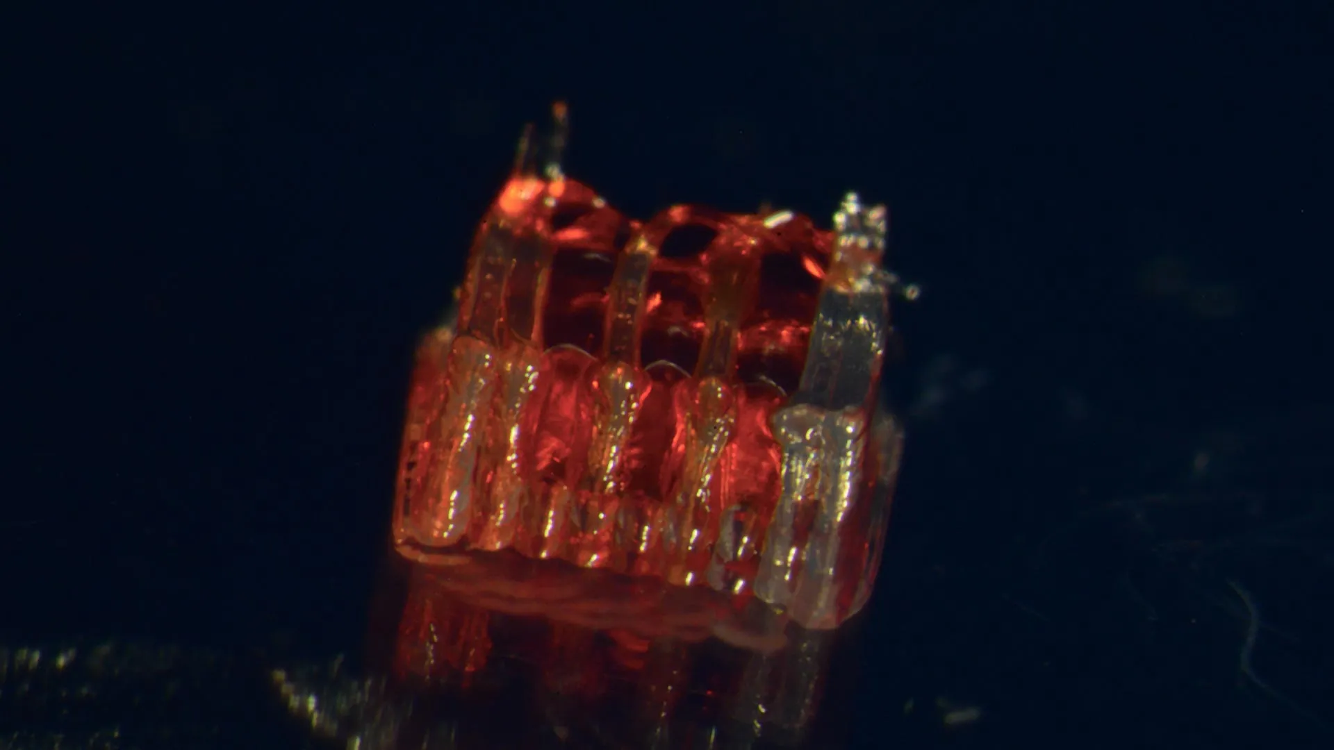
A groundbreaking fusion of 3D printing, advanced stem cell biology, and the creation of lab-grown tissues has yielded a significant breakthrough in the quest for spinal cord injury recovery, according to a study by a research team at the University of Minnesota Twin Cities. This pioneering approach offers a new paradigm for addressing the devastating consequences of spinal cord damage, a condition that affects hundreds of thousands globally with limited avenues for reversal. The findings of this transformative research were recently published in the esteemed peer-reviewed scientific journal, Advanced Healthcare Materials.
The Unmet Need: A Silent Epidemic of Spinal Cord Injury
Spinal cord injuries (SCIs) represent a profound public health challenge. In the United States alone, the National Spinal Cord Injury Statistical Center reports that more than 300,000 individuals live with the debilitating effects of these injuries. These injuries often result in paralysis, loss of sensation, and a cascade of secondary health complications, dramatically altering the lives of those affected and their families. A primary obstacle in treating SCIs has been the inherent inability of the central nervous system to effectively regenerate. Specifically, nerve cells (neurons) in the spinal cord are highly vulnerable to damage and death following trauma, and once lost, they possess a very limited capacity to regrow and re-establish connections across the injury site. This critical failure in regeneration is the root cause of persistent paralysis and functional deficits. Existing treatments largely focus on managing symptoms, preventing further damage, and rehabilitation to maximize residual function, but a true restorative therapy has remained elusive.
The Innovative Solution: Engineering a Neural Relay System
The University of Minnesota team has directly confronted this fundamental challenge by developing an innovative method that leverages the precision of 3D printing to engineer a sophisticated scaffold designed to guide neural regeneration. This scaffold, termed an "organoid scaffold," is not merely a passive support structure; it is meticulously designed with microscopic channels. These intricate pathways serve as predetermined routes for cellular growth.
The next crucial step involves populating these channels with specialized cells known as regionally specific spinal neural progenitor cells (sNPCs). These sNPCs are derived from human adult stem cells, which possess remarkable plasticity, meaning they can differentiate into various specialized cell types. In this context, the sNPCs are coaxed to develop into the specific types of neurons and supporting cells necessary for spinal cord function.
"We utilize the 3D-printed channels of the scaffold to meticulously direct the growth of the stem cells," explained Guebum Han, a former postdoctoral researcher in mechanical engineering at the University of Minnesota and the first author of the study, who now contributes his expertise at Intel Corporation. "This precise guidance ensures that the new nerve fibers grow in the desired, functional way. The resulting construct essentially acts as a biological relay system. When implanted into the spinal cord, it bypasses the damaged area, facilitating communication between the brain and the lower body."
The Experimental Journey: From Lab Bench to Animal Model
The research team’s journey involved a rigorous experimental process, culminating in a significant demonstration of their technology’s potential. After successfully fabricating the organoid scaffolds and populating them with sNPCs in vitro, the next critical phase was to test their efficacy in a living system.
The researchers selected a well-established animal model for spinal cord injury: rats with completely severed spinal cords. This severe injury model provides a stringent test of the regenerative capacity of their engineered tissue. The transplanted organoid scaffolds, containing the sNPCs guided by the 3D-printed channels, were surgically implanted into the injured spinal cords of these animals.
The results observed were remarkably encouraging. The sNPCs within the scaffold successfully differentiated into mature neurons. Crucially, these newly formed neurons extended their axons – the long, slender projections of nerve cells that transmit signals – in both directions. They grew rostrally, towards the head, and caudally, towards the tail, effectively bridging the severed gap in the spinal cord. More importantly, these axons were able to form new, functional connections with the host’s pre-existing nerve circuits. This integration is a vital step for restoring communication pathways that were lost due to the injury.
Over time, the study observed that these newly integrated nerve cells not only survived but also became an integral part of the host spinal cord tissue. This seamless integration facilitated significant functional recovery in the rats. While specific metrics of recovery were detailed in the published paper, the overall outcome indicated a tangible restoration of motor and sensory functions, a testament to the successful bridging of the injured segment by the engineered neural tissue.
A Glimpse into the Future: Regenerative Medicine’s Promise
The implications of this research extend far beyond the laboratory setting, offering a profound beacon of hope for individuals living with the devastating consequences of spinal cord injuries. Ann Parr, a professor of neurosurgery at the University of Minnesota and a key figure in the research, expressed her optimism about the future potential of their work.
"Regenerative medicine has truly ushered in a new era of possibilities for spinal cord injury research," stated Professor Parr. "Our laboratory is incredibly excited to explore the future potential of these ‘mini spinal cords’ for clinical translation. The ability to engineer functional neural tissue and guide its integration within the damaged spinal cord represents a monumental leap forward."
While acknowledging that the research is still in its nascent stages, the team is committed to advancing this technology. The immediate goals include scaling up the production of these organoid scaffolds and further refining the combination of 3D printing and stem cell technologies. The ultimate aim is to develop robust and reproducible methods for future clinical applications in human patients.
The Collaborative Engine: A Multidisciplinary Effort
This significant advancement is the product of a highly collaborative, multidisciplinary effort, bringing together expertise from various fields. The core research team included:
- From the University of Minnesota Department of Mechanical Engineering: Guebum Han and Michael McAlpine, whose expertise in 3D printing and bioengineering was instrumental in designing and fabricating the organoid scaffolds. Hyunjun Kim also contributed significantly from this department.
- From the University of Minnesota Department of Neurosurgery: Ann Parr, leading the clinical and neurosurgical aspects, alongside Nicolas S. Lavoie, Nandadevi Patil, and Olivia G. Korenfeld, who provided critical insights into spinal cord physiology and injury mechanisms.
- From the University of Minnesota Department of Neuroscience: Manuel Esguerra, contributing to the understanding of neural development and regeneration.
- From Virginia Commonwealth University, Department of Physics: Daeha Joung, whose involvement underscores the interdisciplinary nature of this cutting-edge research.
Funding the Future of Hope
The development of such complex and innovative research is made possible through dedicated funding from various sources committed to advancing medical science. This particular project received crucial support from:
- The National Institutes of Health (NIH): A primary source of funding for biomedical research in the United States, recognizing the significant potential of this work.
- The State of Minnesota Spinal Cord Injury and Traumatic Brain Injury Research Grant Program: Demonstrating state-level commitment to addressing these critical neurological conditions.
- The Spinal Cord Society: An organization dedicated to finding a cure for paralysis caused by spinal cord injury, highlighting the direct relevance and importance of this research to patient advocacy groups.
The Path Forward: Towards Clinical Realization
The publication of the full research paper, titled "3D-Printed Scaffolds Promote Enhanced Spinal Organoid Formation for Use in Spinal Cord Injury," on the Advanced Healthcare Materials website, marks a significant milestone. It provides the scientific community with detailed insights into the methodology, results, and future directions of this promising research.
The implications of this work are profound. By successfully demonstrating the ability to engineer functional neural tissue that can integrate and restore function across a completely severed spinal cord in an animal model, the University of Minnesota team has opened a new frontier in the treatment of SCIs. While human clinical trials are still a considerable undertaking, requiring extensive safety and efficacy testing, this research lays a robust foundation. The potential for developing therapies that could restore movement, sensation, and independence to individuals living with paralysis is now a more tangible prospect than ever before. The continued development of this innovative combination of 3D printing and stem cell technologies holds the promise of transforming the lives of millions worldwide.
