
 By George Ongkojoyo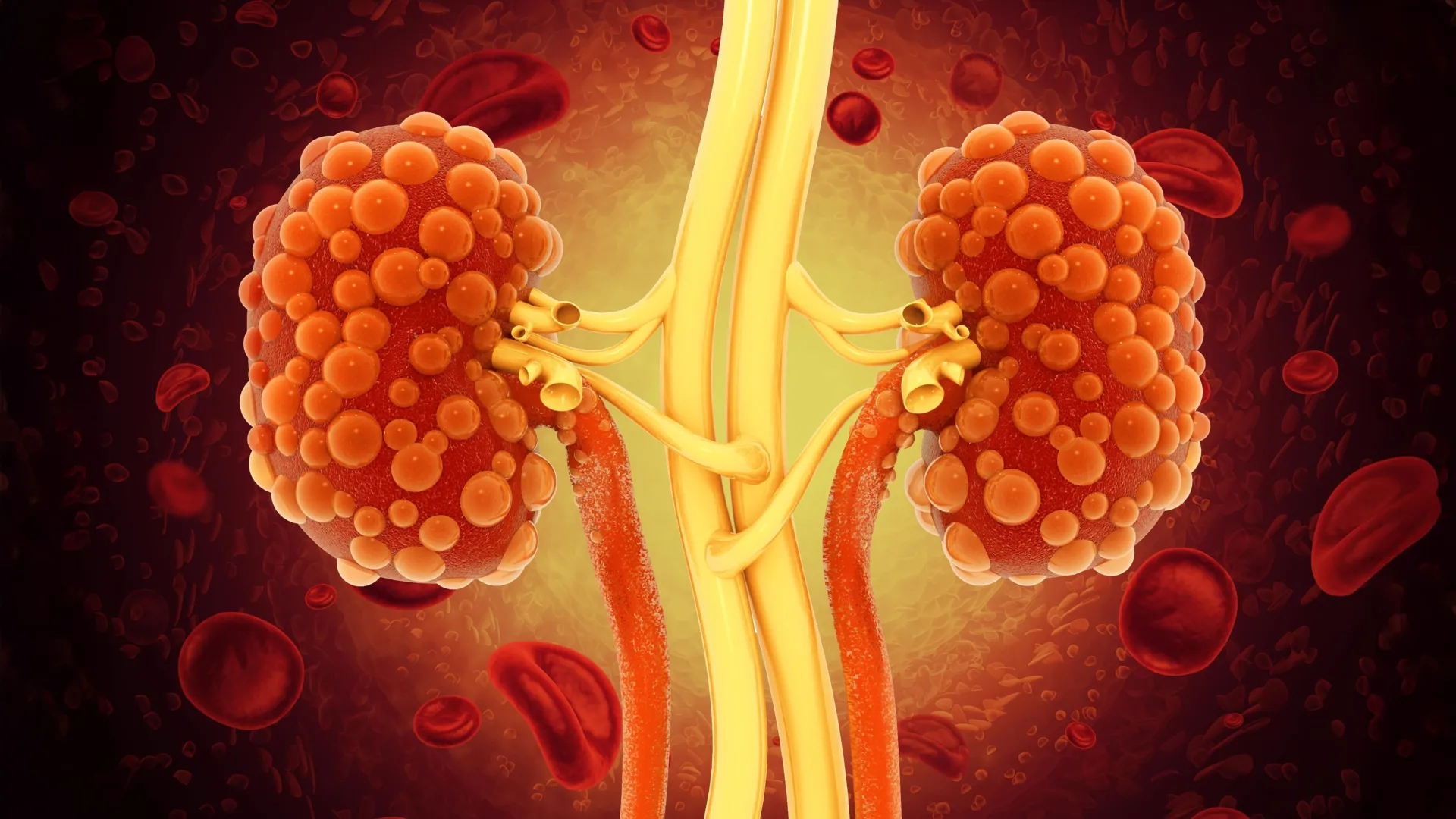
Researchers at the University of California, Santa Barbara (UCSB) have announced a significant milestone in the treatment of Polycystic Kidney Disease (PKD), a genetic disorder that has long frustrated the medical community due to its progressive nature and lack of a definitive cure. In a study published in the prestigious journal Cell Reports Medicine, a team led by biologist Thomas Weimbs detailed the development of a novel class of monoclonal antibodies designed specifically to penetrate the protective barriers of renal cysts. This breakthrough offers a potential paradigm shift from merely managing symptoms to actively disrupting the cellular mechanisms that drive the disease.
Polycystic Kidney Disease is characterized by the relentless growth of fluid-filled cysts within the kidneys. These clusters, which can number in the hundreds or thousands, gradually expand, causing the kidneys to swell to many times their original size. As the cysts grow, they compress and destroy healthy kidney tissue, eventually leading to chronic kidney disease (CKD) and, in many cases, end-stage renal disease (ESRD). For patients reaching this stage, the only remaining options are lifelong dialysis or a kidney transplant. The UCSB study suggests that by using engineered dimeric immunoglobulin A (dIgA) antibodies, clinicians may one day be able to halt this progression before the organs sustain irreversible damage.
The Pathophysiology of Polycystic Kidney Disease
To understand the significance of the UCSB research, it is essential to recognize the biological hurdles presented by PKD. The disease is primarily hereditary, resulting from mutations in the PKD1 or PKD2 genes. These mutations disrupt the normal signaling of epithelial cells lining the kidney tubules. Instead of maintaining a stable tube structure, these cells begin to proliferate uncontrollably and secrete fluid, forming "sealed chambers" or cysts.
The interior of these cysts acts as a localized microenvironment that promotes further growth. Research has shown that the cells lining the cysts secrete various growth factors into the internal fluid. These factors then bind to receptors on the same cells or neighboring cells, creating a self-sustaining autocrine and paracrine signaling loop. This "never-ending scheme," as described by Professor Weimbs, ensures that once a cyst begins to form, it possesses the internal machinery to continue expanding indefinitely, regardless of external systemic signals.
Limitations of Current Pharmacological Interventions
Currently, the therapeutic landscape for PKD is remarkably sparse. The primary FDA-approved drug for slowing the progression of Autosomal Dominant Polycystic Kidney Disease (ADPKD) is Tolvaptan, a vasopressin V2-receptor antagonist. While Tolvaptan has been shown to reduce the rate of cyst growth and the decline in kidney function, it is frequently associated with significant side effects. Patients often experience intense thirst and polyuria (excessive urination), and there are serious concerns regarding potential liver toxicity.
The challenge in developing better drugs lies in delivery. Most small-molecule drugs are filtered out by the kidneys or metabolized by the liver before they can reach therapeutic concentrations within the cyst interiors. Furthermore, standard immunotherapy approaches—which have revolutionized cancer treatment—have historically failed in the context of PKD. The most common form of lab-made protein used in immunotherapy is immunoglobulin G (IgG). While IgG is highly effective at targeting circulating antigens or surface receptors on most tissues, it is too large and lacks the specific transport mechanisms required to cross the epithelial cell layers that seal off PKD cysts. Consequently, IgG-based therapies remain "locked out" of the very areas where the disease is most active.
Engineering a "Molecular Key" with dIgA
The UCSB team’s innovation lies in the selection and modification of the antibody "backbone." Instead of using the standard IgG, the researchers turned to dimeric immunoglobulin A (dIgA). In the human body, dIgA is a critical component of the mucosal immune system. It is the primary antibody found in secretions such as tears, saliva, and the lining of the gastrointestinal and respiratory tracts.
The biological advantage of dIgA is its ability to interact with the polymeric immunoglobulin receptor (pIgR) found on the surface of epithelial cells. When dIgA binds to this receptor, it triggers a process known as transcytosis, where the cell engulfs the antibody and transports it through its interior to be released on the opposite side.
The timeline for this discovery traces back to a 2015 hypothesis proposed by the Weimbs lab. At that time, the researchers suggested that because kidney cysts are lined with epithelial cells that often overexpress pIgR, a dIgA-based drug could potentially "hitch a ride" on these receptors to gain entry into the cyst fluid. The recent study published in Cell Reports Medicine provides the empirical validation of this theory. By altering the DNA sequence of an antibody to convert its structure from IgG to dIgA, the team created a molecule capable of penetrating the previously impenetrable cyst walls.
Targeting the cMET Receptor and Inducing Selective Apoptosis
The target selected for this pilot study was the mesenchymal-epithelial transition (cMET) receptor. cMET is a tyrosine kinase receptor that, when activated by hepatocyte growth factor (HGF), triggers pathways associated with cell growth, survival, and migration. In PKD, the cMET receptor is frequently overactive, serving as a primary engine for the rapid multiplication of cyst-lining cells.
When the researchers tested their engineered dIgA antibody in mouse models of PKD, the results were highly specific. The antibody successfully navigated into the cysts and bound to the cMET receptors. This binding blocked the receptors’ activity, effectively cutting off the growth signals.
Crucially, the treatment led to a "dramatic onset of apoptosis," or programmed cell death, specifically within the cyst-lining cells. Because healthy kidney tissue does not rely on the same overactive cMET signaling pathways, the dIgA treatment left non-diseased tissue unharmed. This level of selectivity is the "holy grail" of PKD research, as it suggests a way to shrink cysts without the systemic toxicity that plagues current treatments.
Supporting Data and Research Chronology
The study represents years of interdisciplinary collaboration at UCSB. Lead author Margaret F. Schimmel, along with colleagues Bryan C. Bourgeois, Alison K. Spindt, and others, utilized advanced imaging and molecular tagging to track the movement of the dIgA antibodies in real-time.
Data highlights from the research include:
- Cyst Penetration: Fluorescence microscopy confirmed that dIgA antibodies reached the interior fluid of cysts within hours of administration, whereas IgG antibodies remained strictly in the interstitial space outside the cysts.
- Receptor Suppression: Western blot analysis of cyst tissue showed a significant reduction in phosphorylated (active) cMET levels following dIgA treatment.
- Safety Profile: Histological examination of the liver, heart, and healthy kidney segments showed no signs of inflammation or cellular damage, indicating that the dIgA backbone is well-tolerated by the biological system.
The research was supported by the National Institutes of Health (NIH) and the U.S. Department of Defense (DoD), reflecting the high level of priority given to finding solutions for PKD, which affects approximately 1 in every 400 to 1,000 people worldwide.
Broader Implications and Future Directions
While the results in animal models are promising, the transition to human clinical trials involves several hurdles. The researchers are now looking to identify partners in the biotechnology and pharmaceutical industries to scale up production and refine the antibody design for human use.
One of the most exciting implications of this research is the potential for "cocktail" therapies. Because dIgA can be engineered to target almost any protein, researchers could theoretically combine multiple antibodies into a single treatment. This would allow for the simultaneous blocking of several growth factors—such as EGFR, IGF-1, and cMET—at once, preventing the cells from developing resistance to the therapy.
Nephrologists and patient advocacy groups have reacted to the news with cautious optimism. The PKD Foundation, while not directly involved in the study, has long advocated for research into targeted therapies that reduce the reliance on dialysis. If the UCSB approach proves successful in humans, it could fundamentally change the life expectancy and quality of life for millions of patients who currently live under the shadow of eventual kidney failure.
The economic impact is also noteworthy. In the United States alone, the cost of treating end-stage renal disease exceeds $50 billion annually. A therapy that successfully halts PKD progression could save the healthcare system billions by reducing the demand for transplants and long-term dialysis sessions.
Conclusion
The work of Thomas Weimbs and his team at UC Santa Barbara represents a landmark shift in the fight against Polycystic Kidney Disease. By reimagining the architecture of monoclonal antibodies and leveraging the body’s own mucosal transport systems, they have opened a door that was previously thought to be closed. While years of rigorous testing and clinical trials remain, the ability to deliver a "payload" directly into the heart of a kidney cyst provides the first real hope for a targeted, low-toxicity treatment that could one day render PKD a manageable condition rather than a progressive death sentence for the kidneys. The next steps will involve broadening the library of target receptors and optimizing the dIgA platform to ensure it can be safely and effectively deployed in the human population.
