
 By George Ongkojoyo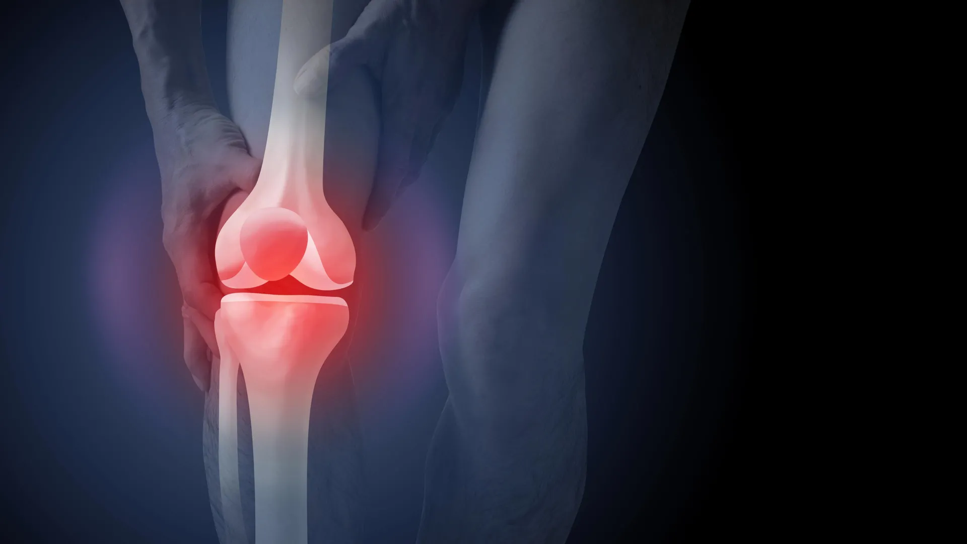
The Australian healthcare system currently faces a significant disconnect between evidence-based clinical guidelines and the practical application of diagnostic tools for knee osteoarthritis. While routine X-rays are not recommended for the primary diagnosis of the condition, recent data indicates that nearly half of all new patients presenting with knee pain to a general practitioner (GP) in Australia are referred for imaging. This trend persists despite the fact that medical history and physical symptoms provide a more accurate and less invasive diagnostic path. A new study published in PLOS Medicine highlights a concerning psychological byproduct of this over-reliance on imaging: patients who see X-ray results are significantly more likely to pursue invasive surgical interventions and develop a debilitating fear of physical activity.
The financial implications of this diagnostic trend are staggering. According to the Australian Institute of Health and Welfare (AIHW), imaging for osteoarthritis costs the national health system approximately A$104.7 million annually. When the scope is widened to include the downstream effects of these diagnoses—most notably joint replacement surgeries—the costs escalate to $3.7 billion. Beyond the fiscal burden, the reliance on X-rays is fostering a "wear and tear" narrative that experts say is fundamentally flawed and psychologically damaging to patients.
Understanding Osteoarthritis: A Dynamic Repair Process
To understand why X-rays can be misleading, it is necessary to redefine osteoarthritis (OA) through the lens of modern pathology. For decades, the condition was described as a passive "wearing out" of the joint, similar to the tread on a tire. However, current medical understanding views osteoarthritis as an active, albeit sometimes unsuccessful, repair process. It is a condition that affects the entire joint ecosystem, including the subchondral bone, cartilage, ligaments, and surrounding muscles.
The joint undergoes structural changes as it attempts to adapt to various stressors, such as high body weight, a history of traumatic injury, or the natural biological shifts associated with aging. These changes are most prevalent in older adults, but they do not always correlate with clinical symptoms. Many individuals with significant "bone-on-bone" appearances on an X-ray report little to no pain, while others with minimal structural changes experience debilitating discomfort. This discrepancy is the primary reason why clinical guidelines, such as those from the Royal Australian College of General Practitioners (RACGP) and the National Safety and Quality Health Service (NSQHS), advise against routine imaging.
The PLOS Medicine Study: Methodology and Chronology
The new research aimed to quantify exactly how the presentation of X-ray evidence influences patient psychology. Researchers recruited 617 participants from across Australia to participate in a controlled study. The cohort was randomly assigned to watch one of three scripted video scenarios involving a consultation with a GP regarding persistent knee pain.
In the first scenario, the patient received a "clinical diagnosis." The GP explained that based on the patient’s age (over 45) and symptoms—specifically pain during activity and morning stiffness lasting less than 30 minutes—the diagnosis of knee osteoarthritis was clear without the need for an X-ray.
In the second and third scenarios, the patients were sent for X-rays. In one of these groups, the doctor merely discussed the results; in the other, the doctor physically showed the X-ray images to the patient, pointing out the structural changes. Following the videos, all 617 participants were surveyed to assess their beliefs regarding the severity of their condition, their fear of movement, and their perceived need for surgery.
Key Findings: The "Structural Fixation" Effect
The results of the study were stark. Participants who were shown their X-ray images reported a 36% higher perceived need for knee replacement surgery compared to the group that received a clinical diagnosis alone. This suggests that the visual evidence of joint changes creates a "structural fixation" in the patient’s mind, leading them to believe that the only solution is a mechanical replacement of the "damaged" part.
Furthermore, the study found that the X-ray groups were significantly more likely to view exercise and physical activity as potentially harmful. This phenomenon, known as kinesiophobia, is particularly detrimental in the context of osteoarthritis, where movement is the primary evidence-based treatment. Patients who saw their X-rays were more worried about their condition worsening and expressed greater fear that walking or climbing stairs would accelerate the "wear and tear" of their joints.
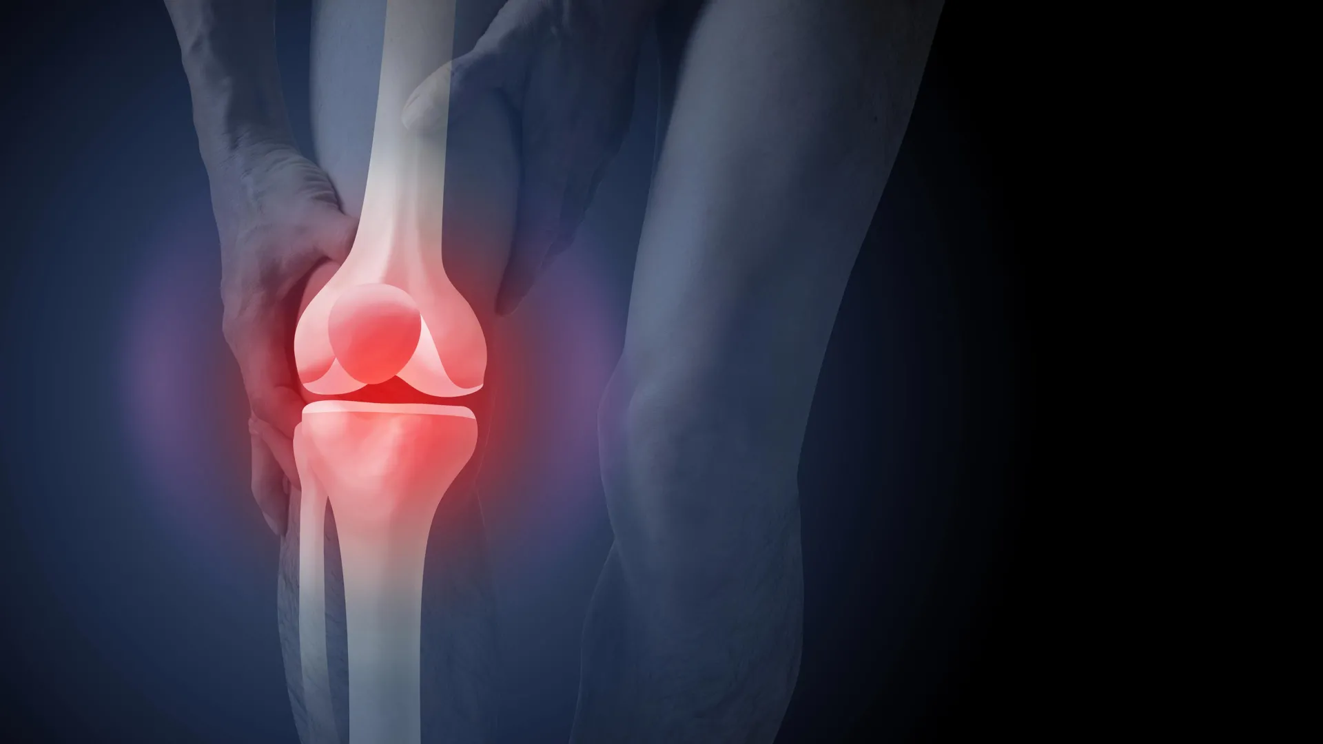
Paradoxically, the researchers found that patients were slightly more satisfied with a diagnosis that included an X-ray than one that did not. This highlights a deep-seated cultural expectation that medical technology is inherently superior to a doctor’s clinical judgment. Patients often feel that their pain is not being taken seriously unless it can be "seen" on a screen, even if that visual data does not change the course of treatment.
The Economic and Clinical Burden of Surgery
The push toward surgery is a growing concern for health administrators. In the 2021–22 period, more than 53,000 Australians underwent knee replacement surgery due to osteoarthritis. While these procedures can be life-changing for patients with end-stage symptoms who have exhausted all other options, they are not without risk.
Joint replacement is a major surgery that carries the potential for serious adverse events, including deep vein thrombosis (blood clots), pulmonary embolisms, and surgical site infections. Furthermore, clinical data suggests that up to 20% of patients who undergo knee replacement surgery continue to experience chronic pain or are dissatisfied with the outcome.
The $3.7 billion spent on hospital services for osteoarthritis in 2020–21 represents a massive allocation of resources toward a "last resort" treatment. Experts argue that if even a fraction of this funding were redirected toward early-intervention non-surgical programs, the long-term health of the population would improve significantly.
Non-Surgical Management: The Gold Standard
Medical guidelines are unanimous in their recommendation of three core pillars for managing knee osteoarthritis:
- Education: Understanding that the joint is not "wearing out" but is in a state of active repair helps reduce anxiety and empowers patients to take control of their health.
- Exercise: Strengthening the muscles around the knee (specifically the quadriceps and hamstrings) provides better support for the joint and reduces the load on the bone and cartilage.
- Weight Management: For those carrying excess weight, even a modest reduction (5–10% of body weight) can significantly decrease the mechanical pressure on the knee joints and systemic inflammation.
When patients are diagnosed via X-ray, they are less likely to adhere to these three pillars because they believe their joints are too "fragile" for exercise. In contrast, a clinical diagnosis focuses on function and symptom management, which naturally aligns with active rehabilitation.
Broader Implications and the Path Forward
The findings of the PLOS Medicine study mirror similar trends in other areas of musculoskeletal health. Research into lower back pain and shoulder pain has consistently shown that early imaging leads to worse clinical outcomes, higher costs, and more invasive treatments without providing any benefit in terms of pain relief or mobility.
The challenge for the Australian healthcare system lies in changing both practitioner behavior and patient expectations. GPs are often under immense pressure from patients to provide a "definitive" scan. To combat this, public health initiatives must focus on "de-prescribing" unnecessary imaging and educating the public on the validity of clinical diagnoses.
Reducing the number of unnecessary X-rays would have a multi-faceted benefit: it would lower radiation exposure for the population, save over $100 million in direct imaging costs, and—most importantly—prevent thousands of patients from entering a psychological downward spiral of fear and surgical dependency.
As the Australian population ages and the prevalence of osteoarthritis rises, the sustainability of the healthcare system will depend on a shift toward these high-value, low-cost management strategies. For the individual patient, the message is clear: an X-ray is a snapshot of anatomy, not a roadmap of your future mobility. Avoiding the scan may be the first step toward a more active and less painful life.
