
 By Billy Candra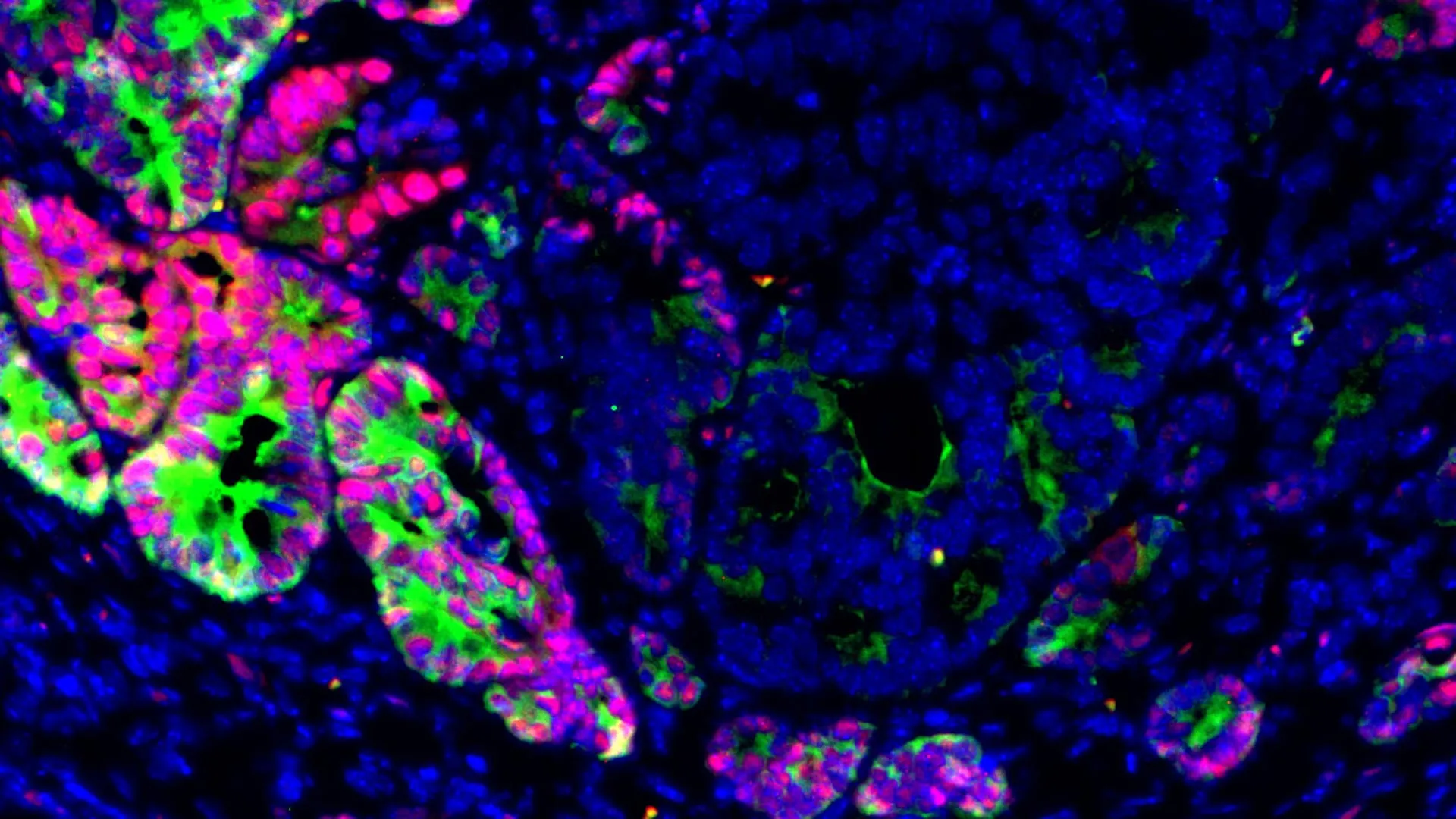
Researchers at Weill Cornell Medicine and the Massachusetts Institute of Technology have identified a key factor that may help colorectal cancer spread to the liver. Their findings suggest that losing GATA6, a transcription factor that helps control which genes are turned on or off, can push cancer cells into a more primitive and adaptable state that makes metastasis possible. Understanding how this transformation occurs could lead to new strategies for preventing one of the deadliest aspects of colorectal cancer. This groundbreaking study, published June 22 in Cell Stem Cell, marks a significant advancement in the understanding of metastatic disease, shifting focus from purely genetic mutations to the critical role of epigenetic changes in cancer progression.
The Deadly Challenge of Colorectal Cancer Metastasis
Colorectal cancer (CRC) stands as a formidable global health challenge. It is the third most commonly diagnosed cancer and the second leading cause of cancer-related deaths worldwide, with an estimated 1.9 million new cases and 935,000 deaths annually, according to the World Health Organization. In the United States alone, the American Cancer Society projects over 153,000 new cases and more than 52,000 deaths in 2023. While early-stage colorectal cancer is highly curable, with a five-year survival rate exceeding 90% for localized disease, this prognosis dramatically declines once the cancer metastasizes, or spreads, to distant organs. For patients with metastatic colorectal cancer, the five-year survival rate plummets to approximately 15-20%.
The liver is the most common site for colorectal cancer metastasis, affecting up to 70% of patients during the course of their disease. This predilection is due to the liver’s extensive blood supply, which filters blood from the intestines, making it a fertile ground for circulating tumor cells. Once colorectal cancer spreads to the liver, treatment becomes far more challenging, often involving complex surgeries, chemotherapy, and targeted therapies with limited success. Metastasis, therefore, remains the leading cause of death from the disease, underscoring the urgent need for a deeper understanding of its mechanisms and the development of effective preventative or interventional strategies. For years, scientists have intensively searched for specific genetic mutations that might trigger liver metastasis, but despite extensive research, no clear, consistent driver mutations have emerged that fully explain this deadly process. This new study points to a fundamentally different mechanism, offering a fresh perspective on a long-standing mystery in oncology.
Unveiling the Role of GATA6: An "Identity Keeper" Dethroned
The core of the recent discovery lies in the transcription factor GATA6. Transcription factors are proteins that bind to specific DNA sequences, thereby controlling the rate at which genetic information is transcribed from DNA to messenger RNA, ultimately regulating gene expression. In essence, they act as master switches, turning genes on or off, and orchestrating a cell’s identity and function. GATA6 normally serves as a molecular "identity keeper" in the specialized epithelial cells that line the intestine, playing a crucial role in helping them maintain their differentiated functions and ensuring proper tissue architecture. This steady state is vital for the intestine’s complex tasks of digestion and nutrient absorption.
However, the study found a stark deviation from this norm in metastatic colorectal cancer. GATA6 levels were observed to be much lower in liver metastases from both mouse models and human patients with colorectal cancer. This reduction was not merely an incidental finding; the researchers also established a compelling correlation between reduced GATA6 expression and poorer patient outcomes, including decreased overall survival and increased disease progression. This observation strongly suggested that GATA6 loss was not just a symptom of advanced cancer but potentially a critical driver. "We discovered that GATA6 loss acts as a critical switch that can change cancer cells in the primary tumor from non-metastatic to pro-metastatic," said Dr. Norihiro Goto, assistant professor of medicine in the Division of Gastroenterology & Hepatology at Weill Cornell, who co-led the research. This statement underscores the pivotal nature of GATA6 in dictating cellular behavior and metastatic potential.
A Paradigm Shift: Epigenetics Over Pure Genetics
The significance of GATA6 loss extends beyond a mere correlation; it represents a paradigm shift in understanding metastatic mechanisms. Unlike genetic mutations, which involve permanent alterations to the DNA sequence itself (e.g., a change in a single base pair or a deletion/insertion), epigenetic changes influence which genes are active or inactive without altering the underlying DNA code. These changes can include DNA methylation, histone modification, and non-coding RNA mechanisms, all of which affect chromatin structure and gene accessibility. Consequently, epigenetic modifications determine which proteins cells produce and, ultimately, their phenotype and behavior.
The finding that GATA6 loss, an epigenetic event, can act as a "critical switch" for metastasis is profound. It suggests that the metastatic cascade may be driven less by the accumulation of specific DNA mutations within cancer cells and more by dynamic, reversible changes in gene expression programs that allow cells to adapt and survive in foreign environments. This offers a new avenue for therapeutic intervention, as epigenetic changes are, in principle, more malleable and reversible than genetic mutations. Dr. Norihiro Goto emphasized this point, stating, "Our findings suggest that epigenetic changes may be more important for promoting liver metastasis." Dr. Saori Goto, an instructor in medicine at Weill Cornell, served as the first author of the study, meticulously detailing the molecular mechanisms involved. Dr. Omer H. Yilmaz, associate professor of biology at the Massachusetts Institute of Technology, also co-led the impactful work, bringing his expertise in stem cell biology and cancer.
Innovative Research: Organoid Models Illuminate Early Metastatic Events
To truly unravel the intricate process of metastasis, researchers faced a significant hurdle: established liver metastases, whether from patient samples or traditional animal models, often present a snapshot of the end stage of the process, obscuring the crucial early events that initiate spread. "When researchers analyze patient samples from liver metastases, we fail to capture the important signals occurring in the early stages of the metastatic process," Dr. Norihiro Goto explained. This limitation has long hampered efforts to identify precise windows for intervention.
To overcome this, the research team employed a cutting-edge laboratory model: organoids derived from liver metastases. Organoids are miniature, three-dimensional cellular structures grown in vitro that closely mimic the architecture, cellular composition, and functionality of real organs or tumors. These sophisticated models allow scientists to study complex biological processes in a controlled environment that more accurately reflects in vivo conditions than traditional 2D cell cultures.
The scientists developed organoids from liver metastases and then iteratively implanted these into the colons of mice. This innovative approach allowed them to observe how the transplanted cells formed increasingly aggressive primary tumors that subsequently spread to the liver. By repeating this process several times, the team could meticulously track the evolutionary trajectory of cancer cells, witnessing their gradual acquisition of metastatic abilities over successive generations. This iterative modeling provided an unprecedented opportunity to dissect the molecular and cellular changes occurring as cancer cells transition from a non-metastatic to a pro-metastatic state, offering critical insights into the early, often elusive, stages of the metastatic cascade.
The Mechanism of Metastasis: Lineage Plasticity and Fetal-like States
The meticulously designed organoid experiments yielded crucial insights into the mechanism by which GATA6 loss promotes metastasis. Their findings revealed that the absence of GATA6 promotes a phenomenon known as lineage plasticity. Lineage plasticity refers to the remarkable ability of cells to alter their identity, developmental trajectory, and specialized functions in response to various cues. This cellular shapeshifting is a fundamental process in normal embryonic development, tissue regeneration, and wound repair, allowing cells to adapt to changing environmental demands.
In the context of cancer, however, this inherent flexibility can be hijacked for nefarious purposes. When GATA6 was absent, colorectal cancer cells activated alternative genetic programs, essentially "reprogramming" themselves. They adopted a flexible, primitive, and often fetal-like state, characterized by a less differentiated phenotype. These transformed cells, shedding their original specialized intestinal identity, became remarkably more adaptable and resilient. This altered state endowed them with enhanced capabilities crucial for metastasis: they were better equipped to detach from the primary tumor, survive the arduous journey through the bloodstream, evade immune surveillance, and ultimately establish viable tumors in distant organs like the liver.
This cellular reprogramming was further evidenced by the appearance of cells lacking LGR5, a well-established marker commonly found in highly proliferative intestinal stem cells. Earlier research had indicated that LGR5-negative cells possess an increased capacity to initiate liver metastases. The new study powerfully demonstrated that shutting down GATA6 caused a distinct cellular transition: cancer cells shifted from an LGR5-positive state to an LGR5-negative, fetal-like state. These LGR5-negative cells not only displayed characteristics reminiscent of early development but also exhibited a pronounced ability to disseminate and form secondary tumors. Conversely, when the researchers genetically restored GATA6 activity, or activated related signaling pathways, the metastatic potential of colorectal cancer cells was significantly reduced, providing strong evidence for GATA6’s suppressive role in metastasis.
Beyond Tumor Size: Metastasis as a State Transition
One of the most compelling aspects of the study’s findings challenges a long-held assumption in oncology: that metastasis is primarily driven by the aggressive growth and sheer mass of the primary tumor. The researchers observed that genetically deleting GATA6 in their mouse models significantly increased both the frequency and burden of liver metastases, yet it had little discernible effect on the growth rate or size of the primary tumor. This crucial distinction suggests that the capacity for metastasis is not solely a function of primary tumor growth kinetics.
"When we genetically delete GATA6, the frequency and burden of liver metastases in mouse models significantly increase, while having little effect on primary tumor growth," reiterated Dr. Norihiro Goto, who is also a member of the Jill Roberts Institute for Research in Inflammatory Bowel Disease and the Sandra and Edward Meyer Cancer Center, both at Weill Cornell. This observation indicates that metastasis may depend more on specific transitions between cellular states – a qualitative change in cell identity and behavior – rather than merely on the quantitative aspects of how quickly a primary tumor grows or how large it becomes. This conceptual shift opens new avenues for therapeutic intervention, suggesting that therapies might need to target these specific cellular transformations rather than just focusing on reducing primary tumor bulk.
Translational Potential: Biomarker and Therapeutic Target
The profound implications of this research extend directly to clinical practice and future therapeutic development. The findings raise the exciting possibility that GATA6 could serve as a valuable biomarker for metastatic risk in colorectal cancer patients. Tumors exhibiting low levels of GATA6 expression may be more likely to harbor cells capable of undergoing the crucial epigenetic reprogramming that leads to a metastasis-promoting state. Such information, readily obtainable through tumor biopsy analysis, could empower clinicians to better stratify patients, identify those at higher risk of developing liver metastases, and tailor treatment strategies accordingly. Patients identified with low GATA6 levels might benefit from closer surveillance, more aggressive systemic therapies, or prophylactic interventions aimed at preventing metastatic spread.
Furthermore, the study points toward a promising new therapeutic strategy centered on maintaining cellular identity or actively preventing cancer cells from entering these highly flexible, pro-metastatic states. If GATA6 loss is indeed a "critical switch," then therapeutic approaches could be designed to either restore GATA6 function, activate its downstream pathways, or block the alternative genetic programs that emerge in its absence. However, Dr. Norihiro Goto cautioned that researchers will need to carefully navigate the inherent complexities of such interventions. The challenge lies in finding ways to specifically target these cancer-driven processes of cellular plasticity without inadvertently interfering with normal tissue repair and regeneration, which rely on similar biological programs and the body’s natural ability to adapt.
The Road Ahead: Future Research Directions
This study represents a significant milestone, yet it also lays the groundwork for a rich landscape of future research. The immediate focus will be on identifying specific vulnerabilities unique to GATA6-deficient cancer cells. By understanding the distinct metabolic or signaling dependencies of these reprogrammed cells, scientists could uncover novel targets for drug development that selectively incapacitate metastatic cells while sparing healthy tissues.
The research team also plans to investigate the intricate interplay between the tumor and its microenvironment. The tumor microenvironment, a complex ecosystem comprising immune cells, fibroblasts, blood vessels, and various signaling molecules, plays a crucial role in cancer progression and metastasis. Understanding how specific components of the liver microenvironment, including liver-specific signals and resident immune cells, influence these GATA6-driven cellular transitions in preclinical models will be vital for developing more effective therapies. This holistic approach acknowledges that cancer cells do not operate in isolation but are profoundly influenced by their surroundings.
Ultimately, the broader goal of this research is to translate these fundamental scientific discoveries into tangible clinical benefits. "In addition to treating primary tumors, we need to find strategies to target the mechanism of liver metastasis," Dr. Norihiro Goto stated. "Our study is a step toward developing therapies that block the spread of cancer at the earliest stages." This commitment reflects the urgent need for new interventions that can tackle metastasis head-on, offering renewed hope for patients battling colorectal cancer.
This pivotal research was supported by a consortium of dedicated organizations, including the Astellas Foundation; Research Abroad from Japan Society for the Promotion of Science; the National Institutes of Health (grants R00AG076987, 01CA254314, 5U01CA25055, R01CA258523, R01CA25723, R01DK133919, R01DK140310, R01CA299955, and 3OT2CA297570); Pew-Stewart Trust; AFAR and Glenn Foundation for Medical Research Breakthroughs in Gerontology; Kenneth Rainin Foundation; Crohn’s & Colitis Foundation and Mark Foundation for Cancer Research. The collaborative effort and substantial funding underscore the critical importance of this work in the ongoing fight against cancer.
