
 By Budi Oetomo Narendra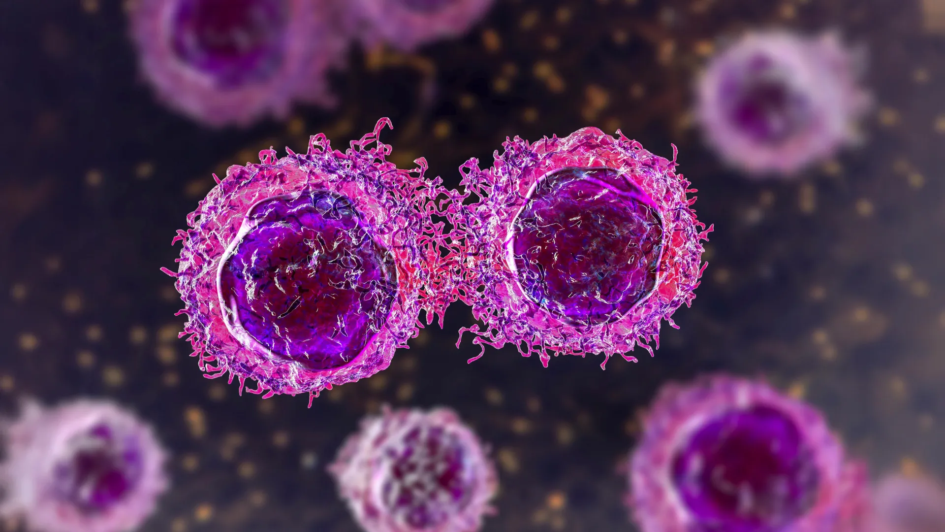
Groundbreaking research from Sanford Burnham Prebys Medical Discovery Institute, in collaboration with institutions across North America, has pinpointed specific environmental conditions within the tumor microenvironment that enable cancer cells to construct this formidable protective layer. Published on August 7, 2026, in the esteemed journal Science Advances, these findings not only shed light on a crucial mechanism of immune evasion but also suggest a promising strategy to dismantle this sugar-rich shield, potentially rendering cancer cells more vulnerable to the body’s natural defenses and advanced immunotherapies.
The study represents a significant stride in understanding the intricate interplay between a tumor’s surroundings and its metabolic adaptability, particularly how cancer cells exploit their environment to cloak themselves from the immune system. For years, scientists have grappled with the mystery of why certain cancers are stubbornly resistant to immune attack. While various factors contribute, the identification of this sugar-derived coating, known as the glycocalyx, as a key camouflage mechanism has opened new avenues for therapeutic intervention.
The Immune Evasion Challenge: A Sugar Shield
The human immune system is a sophisticated defense network, constantly patrolling the body for abnormal or foreign cells. Cancer cells, however, are notoriously adept at developing strategies to bypass this surveillance. One such strategy involves altering their cell surface, often by modifying their glycocalyx. This dense, fuzzy layer of carbohydrates attached to proteins and lipids on the cell surface serves various normal physiological functions, from cell-cell recognition to protection against mechanical stress. In cancer, however, it can be weaponized.
When the glycocalyx becomes overly thick or its composition changes, it can physically block immune cells from accessing critical recognition markers on the cancer cell surface. It can also send inhibitory signals to immune cells, effectively telling them to stand down. This "sugar shield" makes it exceedingly difficult for T-cells and other immune components to identify and eliminate malignant cells, contributing to tumor growth and metastasis. Understanding the factors that influence the formation and composition of this glycocalyx is therefore paramount in developing more effective cancer treatments.
The Tumour Microenvironment: A Complex Arena
Central to this new discovery is the concept of the tumor microenvironment (TME). Far from being isolated entities, cancer cells are embedded within a dynamic and heterogeneous TME, a complex ecosystem comprising various cell types—including immune cells, fibroblasts, and endothelial cells—along with extracellular matrix components, growth factors, and metabolites. This environment is highly influential in shaping tumor behavior, dictating everything from growth and spread to therapeutic response.
Researchers have increasingly recognized that the TME is not merely a passive backdrop but an active participant in cancer progression. Its biophysical properties, such as stiffness, and its biochemical composition, including nutrient availability, can profoundly impact how cancer cells function. The TME in primary tumors is often significantly stiffer than surrounding healthy tissue due to altered collagen deposition and increased cellular density. This mechanical stress, alongside metabolic shifts, creates a unique set of challenges and opportunities for cancer cells to adapt and thrive. The Sanford Burnham Prebys study delved into how these specific characteristics of the TME could be exploited by cancer cells to enhance their protective glycocalyx.
Unraveling the Metabolic Mystery: Dr. Tharp’s Approach
The genesis of this groundbreaking research lies in the prior work of lead and corresponding author Dr. Kevin Tharp, an assistant professor in the Cancer Metabolism and Microenvironment Program at the Sanford Burnham Prebys NCI-Designated Cancer Center. Dr. Tharp had previously investigated how physical pressure on cells could induce unexpected alterations in mitochondrial function, the cell’s powerhouses. This line of inquiry naturally led him to consider tumors as an ideal biological model, given that cancer cells within them are frequently subjected to substantial mechanical forces.
"Primary tumors are typically stiffer than their surrounding tissue," Dr. Tharp explained, setting the stage for his hypothesis. "This led me to hypothesize that the biophysical properties of cells influence the altered metabolic programs that everyone observes in tumors." This insight was crucial, as it linked physical aspects of the TME to cellular metabolism, a field known to be profoundly dysregulated in cancer. One well-documented metabolic change in tumors is a reduced oxidative metabolism of glucose, often referred to as the Warburg effect. Previous studies had hinted that this metabolic shift might not be an inherent, immutable trait of cancer cells but rather a context-dependent adaptation, influenced by the availability of nutrients in their immediate surroundings. Dr. Tharp’s team aimed to experimentally validate this context dependency and understand its implications.
Experimental Rigor: Mimicking Physiological Conditions
To rigorously test their hypothesis, Dr. Tharp and his colleagues designed a series of meticulous experiments. They exposed cells to abundant glucose, a common nutrient for cancer cells, while manipulating several environmental parameters:
-
Biophysical Environment:
- Stiff Environments: Cells were grown on substrates engineered to mimic the increased stiffness characteristic of primary tumors.
- Softer Environments: Other cells were cultured in softer conditions, analogous to normal, healthy tissue. This allowed the researchers to directly assess the impact of mechanical forces on cellular behavior.
-
Culture Medium Composition:
- Standard Laboratory Medium: A conventional cell culture medium, widely used in research but often not fully representative of the human body’s complex nutrient milieu.
- Physiological Medium: A specially formulated medium designed to more closely match the diverse nutrient composition found within the human body. This distinction proved to be critically important, highlighting the need for physiologically relevant experimental models.
-
Glucose Levels: Both types of culture media were tested under normal glucose concentrations and under elevated glucose conditions, allowing the researchers to simulate hyperglycemia, a state of high blood sugar prevalent in conditions like diabetes.
These carefully controlled experimental conditions enabled the researchers to isolate and study the individual and combined effects of mechanical stress, nutrient availability, and glucose levels on cancer cell metabolism and surface characteristics.
Key Discoveries: Glycocalyx Thickness and Glucose
The environmental manipulations yielded profound and notable changes in the cells. The researchers observed significant differences in the proteins produced by the cells, the concentrations of intracellular metabolites, and, most importantly, the thickness of the sugar-derived layer covering their surfaces – the glycocalyx.
A critical finding emerged: excess glucose only led to an increase in glycocalyx thickness when the cells were grown in the physiological medium, the one that more closely mirrored conditions inside the human body. This observation underscored the importance of using relevant experimental models and suggested that findings from conventional laboratory settings might not always translate directly to the complex biological reality of a human body. "We observed that changing the physiological media composition and changing the available metabolites for those tumor cells reveals distinct biology for normal and tumor cell metabolism," Dr. Tharp noted, emphasizing the nuanced nature of cellular responses to environmental cues.
The glycocalyx is constructed from glycoconjugates, which are carbohydrates attached to proteins or lipids. Given that glucose is a primary raw material for producing these glycoconjugates, the team hypothesized that changes in glucose metabolism or the presence of hyperglycemia might directly alter how this protective layer is assembled. Indeed, their investigations confirmed this suspicion. "We found stark separation between the glycoconjugates of cells cultured in conventional medium versus those cultured in a medium that better reflects the nutrient composition of the human body," Dr. Tharp elaborated. Furthermore, they discovered that hyperglycemia specifically changed the composition of glycoconjugates produced by the cells, suggesting a qualitative as well as quantitative alteration of the sugar shield.
HSF1: The Unsuspected Orchestrator
To delve deeper into the molecular mechanisms driving these changes, the researchers sought to identify which proteins became more abundant when cells were exposed to hyperglycemia. Their experiments quickly highlighted Heat Shock Factor 1 (HSF1). HSF1 is a transcription factor best known for its role in helping cells survive high temperatures and other forms of cellular stress by regulating the expression of heat shock proteins. However, previous research had also implicated HSF1 in various aspects of cancer biology, including the progression and metastasis of breast cancer, indicating its broader significance beyond stress response.
The team’s findings unequivocally demonstrated that the presence or absence of HSF1 directly influenced the composition of glycoconjugates produced by the cells. This established HSF1 as a key player in the assembly of the glycocalyx under hyperglycemic conditions. The researchers then conducted a series of experiments to investigate the intricate relationship among hyperglycemia, HSF1, the tumor microenvironment, and the immune system’s capacity to recognize and attack cancer cells.
Their results were compelling: hyperglycemia enhanced cancer cells’ ability to evade immune detection only when HSF1 was present and when the cells were cultured under conditions that mimicked the tumor microenvironment. This discovery solidified HSF1’s central role as a mediator, linking environmental stressors and metabolic imbalances to immune evasion.
A New Therapeutic Avenue: Targeting HSF1
The identification of HSF1 as a crucial nexus in this immune evasion pathway presents a significant therapeutic opportunity. The findings strongly suggest that drugs designed to specifically target HSF1 could potentially reduce the thickness and alter the composition of the glycocalyx, effectively stripping away some of the protective camouflage that allows cancer cells to avoid immune surveillance. Such a strategy could revolutionize current cancer treatments, particularly immunotherapies.
Immunotherapy, while highly effective for certain patients and cancer types, still faces challenges, with many patients not responding or developing resistance. By making cancer cells more visible to the immune system through glycocalyx modification, HSF1 inhibitors could act as sensitizers, enhancing the efficacy of existing immunotherapies like checkpoint inhibitors. This approach could broaden the patient population that benefits from these advanced treatments and potentially overcome some forms of resistance.
Bridging the Gap: Hyperglycemia, Metabolism, and Cancer Outcomes
"Our findings indicate that changes in mitochondrial function lead to the synthesis of cell surface sugar-derived molecules that make it difficult for the immune system to recognize and kill cancer cells," Dr. Tharp articulated, summarizing the core mechanism. "Now that we know this, this creates an enormous drug discovery opportunity to take away the surface coating that protects them from immune surveillance. And we think this will be a really effective strategy to attack metastatic disease and improve immunotherapy responses."
This research holds particular relevance in the context of a global health crisis: the escalating prevalence of metabolic syndrome and type 2 diabetes. Both conditions are characterized by chronic hyperglycemia. Dr. Tharp highlighted that high blood sugar is increasingly recognized as an important risk factor for individuals with cancer. A substantial body of epidemiological and clinical research has consistently linked high blood sugar levels to both an increased risk of developing various cancers and worse outcomes following cancer treatment. However, the precise biological mechanisms underpinning this relationship have remained largely elusive, representing a significant gap in cancer research.
The new findings from Sanford Burnham Prebys offer one of the most plausible and detailed explanations to date. By demonstrating how elevated glucose levels, particularly within the specific context of the tumor microenvironment and mediated by HSF1, can directly assist tumors in escaping immune attack, the study provides a concrete mechanistic link. "What we found is a plausible mechanism by which hyperglycemia directly contributes to immune evasion," Dr. Tharp affirmed. "And potentially a way to take away a pro-tumor advantage from hyperglycemia caused by metabolic syndrome and modern diets." This insight could also inform dietary and lifestyle recommendations for cancer patients, emphasizing the importance of metabolic control.
Future Prospects and Public Health Implications
The implications of this study extend beyond the immediate discovery. The identification of HSF1 as a druggable target opens the door for pharmaceutical companies to develop novel compounds that specifically interfere with this pathway. Subsequent research will likely involve preclinical studies in animal models to validate the efficacy and safety of HSF1 inhibitors, followed by human clinical trials if promising results are observed. Exploring whether HSF1 inhibition could be combined with other therapeutic modalities, such as chemotherapy or radiation, also presents a fertile area for future investigation.
From a public health perspective, this research reinforces the critical connection between metabolic health and cancer. As rates of obesity, type 2 diabetes, and metabolic syndrome continue to climb globally, understanding their mechanistic links to cancer progression becomes ever more urgent. This study underscores that managing metabolic health, including maintaining healthy blood glucose levels, could be a vital component of both cancer prevention and treatment strategies. It also calls for greater integration of metabolic health assessments into oncology care, potentially leading to personalized treatment plans that consider a patient’s metabolic profile.
The work of Dr. Tharp and his collaborators represents a significant step forward in the ongoing battle against cancer. By illuminating how cancer cells masterfully adapt to their environment and exploit metabolic vulnerabilities to evade immune detection, the research not only enhances our fundamental understanding of tumor biology but also paves the way for innovative, targeted therapies that could disarm cancer’s protective sugar shield and significantly improve patient outcomes, particularly in an era where metabolic diseases are increasingly intertwined with cancer.
Study Support and Collaborators
The comprehensive study was made possible through the generous support of several key funding bodies, including the National Institutes of Health, the National Cancer Institute, the National Foundation for Cancer Research, the Canada Excellence Research Chair in Glycomics, and the Ovarian Cancer Research Alliance. The collaborative efforts across multiple institutions in North America were instrumental in bringing this complex and impactful research to fruition.
