
 By Gabriel Paijo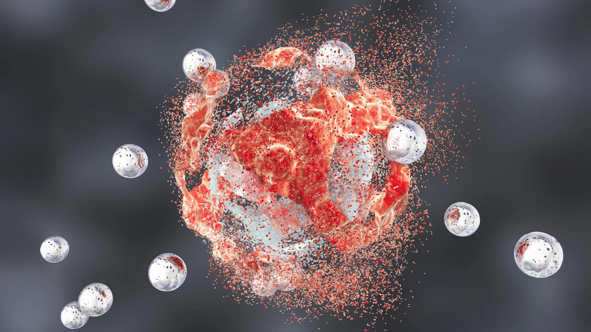
In a significant advancement for the field of nanomedicine, researchers from Weill Cornell Medicine and the Cornell Duffield College of Engineering have announced the development of a novel therapeutic platform utilizing tiny silica nanoparticles. These engineered particles, which were originally conceived for diagnostic imaging, have demonstrated a remarkable ability to directly eradicate prostate tumors while simultaneously stimulating a robust immune response. The findings, published in the peer-reviewed journal Cancer Research, suggest a potential paradigm shift in the treatment of aggressive, treatment-resistant cancers through a dual-action mechanism that combines physical cell destruction with immunological "reawakening."
The study, conducted primarily in preclinical mouse models of aggressive prostate cancer, revealed that these nanoparticles—known as Cornell Prime dots or "C’ dots"—can induce complete tumor remission in a significant percentage of subjects when used in conjunction with existing immunotherapies. This discovery marks a critical milestone in a long-running collaborative effort between materials scientists and clinical oncologists to harness the unique properties of amorphous silica for therapeutic intervention.
The Evolution of C’ Dots: From Medical Imaging to Cancer Therapy
The journey of the C’ dot began not as a weapon against cancer, but as a lens to see it. Developed over the last two decades in the laboratory of Dr. Ulrich Wiesner at Cornell University, these ultrasmall fluorescent core-shell silica nanoparticles were engineered to improve the precision of medical imaging. Because of their minute size—typically less than 10 nanometers—and their biocompatible silica composition, they are capable of circulating through the bloodstream and penetrating deep into tissues before being safely cleared through the kidneys.
Silica is a naturally occurring substance found in various foods, including leafy green vegetables and cereal grains, as well as in the fossilized remains of microscopic organisms. This inherent biological compatibility allowed the C’ dots to progress into late-stage clinical trials for image-guided surgery, where they serve as tracers to help surgeons identify the margins of tumors and the location of sentinel lymph nodes.
However, during the course of these clinical and preclinical investigations, researchers observed an unexpected phenomenon: the particles were not merely passive observers. In certain environments, they appeared to exert a biological pressure on cancer cells. This led the research team, headed by Dr. Michelle Bradbury and Dr. Wiesner, to investigate whether the particles could be optimized to actively destroy malignant cells.
A Novel Mechanism of Destruction: Harnessing Ferroptosis
One of the most profound revelations of the new study is the nanoparticles’ ability to trigger ferroptosis. Unlike apoptosis, which is a common form of programmed cell death that many cancer cells learn to evade, ferroptosis is a specialized process driven by iron-dependent lipid peroxidation.
The researchers found that the silica particles, when engineered with specific surface properties, act as vehicles for iron transport. Evidence suggests that the particles collect positively charged iron ions from the systemic circulation and concentrate them within the tumor microenvironment. Once inside the acidic and metabolically volatile environment of a cancer cell, these iron ions catalyze the production of reactive oxygen species.
This internal "oxidative stress" leads to the catastrophic damage of fatty molecules (lipids) within the cell membranes. As the membranes lose their structural integrity, the cancer cell essentially collapses and dies. Because ferroptosis is a relatively recently discovered pathway, many tumors that have developed resistance to traditional chemotherapy and radiation remain highly vulnerable to this form of attack.
Transforming the Tumor Microenvironment: From Cold to Hot
While the direct killing of cancer cells via ferroptosis is a significant achievement, the C’ dots provide a second, perhaps more critical, benefit: they alter the "immune landscape" of the tumor.
In the context of oncology, prostate tumors are often characterized as "cold." This means they are immunologically quiet, often surrounded by a protective barrier of immunosuppressive cells that prevent the body’s T-cells from recognizing or attacking the malignancy. This "cold" state is a primary reason why many prostate cancer patients do not respond to modern immunotherapy drugs, such as checkpoint inhibitors.
The study demonstrated that the introduction of C’ dots effectively "heats up" the tumor. By inducing ferroptosis and disrupting the metabolic processes of the cells within the tumor microenvironment, the nanoparticles signal the immune system to mobilize. The researchers observed that T-cells and macrophages—the primary soldiers of the immune system—shifted from an inactive or suppressed state into an aggressive, cancer-fighting state.
Dr. Jedd Wolchok, a world-renowned oncologist and co-author of the study, noted that this "immune remodeling" is essential for long-term success. By breaking down the tumor’s defenses, the C’ dots allow immunotherapy drugs to finally reach their targets, creating a synergistic effect that is far more powerful than either treatment used in isolation.
Analysis of Study Data and Survival Rates
The preclinical trials involved mouse models specifically designed to mimic the most aggressive and treatment-resistant forms of human prostate cancer. The research team compared several different treatment regimens to determine the efficacy of the C’ dots.
- Monotherapy Results: When used alone, both the C’ dots and standard immunotherapy drugs showed only modest effects on tumor growth and survival rates.
- Dual Combination Therapy: When the C’ dots were combined with an immune checkpoint blockade (a standard form of immunotherapy), the results improved dramatically. In this group, four out of ten mice achieved complete or nearly complete remission, surviving indefinitely.
- Triple Combination Therapy: To further enhance the response, the team added a third component: a CSF-1R blockade. This drug specifically targets tumor-associated macrophages that often protect the cancer. With this triple-threat approach, the rate of complete remission rose to five out of ten mice (50%).
These figures are particularly striking given the aggressive nature of the cancer models used. In the control groups, which received no treatment or only partial treatments, tumor growth was rapid and lethal. The ability to achieve 50% total remission in a model of "cold" prostate cancer is considered a major milestone in preclinical oncology.
Precision Targeting and Safety Profiles
To ensure that the silica particles did not cause collateral damage to healthy organs, the team utilized a targeting molecule that recognizes PSMA (Prostate-Specific Membrane Antigen). PSMA is a protein that is overexpressed on the surface of nearly all prostate cancer cells but is largely absent in healthy tissue.
By coating the C’ dots with PSMA-targeting ligands, the researchers ensured that the particles would bypass healthy cells and latch onto the tumors. While the study noted that some particles were temporarily filtered through the spleen—a common occurrence for any nanoparticle—no signs of systemic toxicity or organ damage were found. The researchers attributed this safety profile to the "ultrasmall" nature of the particles, which allows for rapid renal clearance of any dots that do not find their way to a tumor.
Dr. Wiesner remarked on the "unreal" nature of the findings, questioning whether the long-standing presence of silica in the natural environment and human diet has created a unique biological pathway that scientists are only now beginning to understand.
Institutional Support and Future Directions
The research was a massive undertaking involving multiple laboratories and institutions. Significant contributions were made by co-first authors Drs. Nabil Siddiqui, Li Zhang, and Gabriel DeLeon, alongside graduate students from the Wiesner lab. The project received funding from the Department of Defense, the National Cancer Institute (NCI), and the Parker Institute for Cancer Immunotherapy at Weill Cornell Medicine.
The collaboration between Weill Cornell Medicine and the Cornell Duffield College of Engineering exemplifies the modern "bench-to-bedside" approach, where materials science and clinical medicine intersect to solve complex biological problems.
The next phase for the research team involves refining the synthesis of the nanoparticles for large-scale production and preparing for human clinical trials. Because the C’ dots have already cleared several regulatory hurdles for imaging purposes, the path to therapeutic trials may be shorter than that of entirely new drug compounds.
Broader Implications for Oncology
The success of this study in prostate cancer has broad implications for other types of "cold" tumors, such as pancreatic cancer and certain types of brain tumors, which have historically been resistant to immunotherapy. If the C’ dots can successfully "remodel" the immune environment of these cancers, they could unlock the potential of immunotherapy for millions of patients who currently have few options.
Furthermore, the discovery of ferroptosis as a viable clinical target opens new avenues for drug development. The ability to trigger a specific, iron-driven form of cell death through a non-toxic nanoparticle carrier represents a sophisticated evolution in cancer therapy—one that moves away from the "carpet-bombing" approach of traditional chemotherapy toward a more nuanced, targeted, and biologically integrated strategy.
As the scientific community looks toward the future of oncology, the integration of nanotechnology and immunology stands out as one of the most promising frontiers. The Cornell study provides a compelling blueprint for how engineered materials can work in harmony with the body’s own defenses to overcome the most resilient forms of disease.
