
 By Gabriel Paijo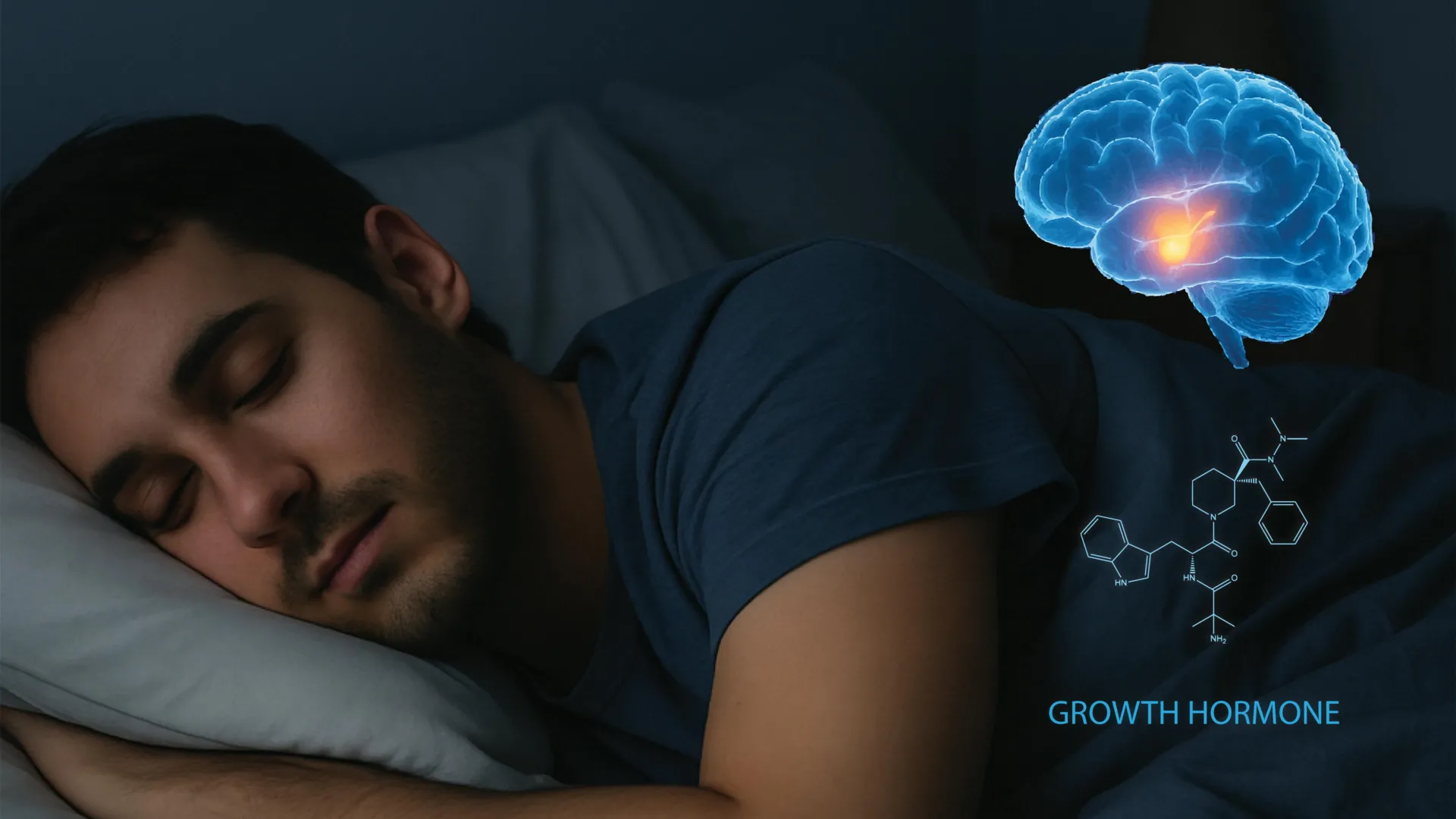
The intricate relationship between the human sleep cycle and the endocrine system has long been a focal point of biological research, yet the specific neurological "switchboard" that governs these interactions has remained largely elusive. In a landmark study published in the journal Cell, a team of neuroscientists at the University of California, Berkeley, has successfully mapped the brain circuitry responsible for the surge of growth hormone (GH) during sleep. This discovery not only clarifies how the brain orchestrates tissue repair and metabolic regulation during rest but also identifies a novel feedback loop that maintains the delicate equilibrium between wakefulness and slumber. The findings carry profound implications for the treatment of metabolic disorders, such as type 2 diabetes, and neurodegenerative conditions, including Alzheimer’s and Parkinson’s disease.
Growth hormone, secreted by the pituitary gland, is a fundamental pillar of human physiology. While its role in childhood development is well-documented, its importance in adulthood is equally vital, serving as a primary driver for muscle synthesis, bone density maintenance, and the regulation of glucose and lipid metabolism. For decades, clinicians have observed that GH levels peak during the deepest stages of sleep, particularly during the slow-wave oscillations of non-rapid eye movement (non-REM) sleep. However, the precise neural architecture that signals the pituitary gland to release these pulses at specific intervals during the night was a mystery until now.
Mapping the Hypothalamic Control Center
The research, led by Dr. Yang Dan, a professor of neuroscience and molecular and cell biology at UC Berkeley, focused on the hypothalamus—a region of the brain often described as the "master regulator" of the autonomic nervous system and endocrine functions. Within this region, the team identified a specific set of nerve cells that coordinate the release of GH. These include growth hormone-releasing hormone (GHRH) neurons and two distinct types of somatostatin neurons.
In the biological "tug-of-war" that regulates hormone levels, GHRH acts as the accelerator, stimulating the pituitary gland to produce GH, while somatostatin acts as the brake, inhibiting its release. Using advanced optogenetic techniques—where light is used to control neurons that have been genetically sensitized to it—the researchers were able to observe how these neurons fluctuate in activity across different sleep stages.
The study utilized murine models, which are essential in neuroendocrinology due to the highly conserved nature of the hypothalamic-pituitary axis across mammalian species. Because mice sleep in short, frequent bursts throughout a 24-hour cycle, the researchers were able to gather a vast dataset of hormone fluctuations across hundreds of sleep-wake transitions. This provided a high-resolution view of how GHRH and somatostatin neurons shift their behavior as the brain moves from light sleep to deep non-REM sleep and eventually into REM sleep.
The Dual Role of Sleep Stages in Hormone Secretion
One of the most significant findings of the UC Berkeley study is the nuance with which different sleep stages handle hormone regulation. Traditionally, it was believed that GH release was almost exclusively tied to non-REM sleep. However, the researchers discovered that the dynamics are more complex.
During REM sleep—the stage associated with vivid dreaming and high brain activity—both GHRH and somatostatin levels increase. This simultaneous rise leads to a significant and controlled release of growth hormone. Conversely, during non-REM sleep, somatostatin levels (the "brake") fall significantly while GHRH levels (the "accelerator") rise only moderately. This suggests that the body uses different neural pathways to achieve growth hormone peaks depending on the specific brain state.
This discovery challenges the monolithic view of sleep-hormone interaction and suggests that the quality of both REM and non-REM sleep is essential for metabolic health. If either stage is truncated—due to sleep apnea, insomnia, or lifestyle-induced sleep deprivation—the body’s ability to repair tissue and manage fat storage is compromised.
A Novel Feedback Loop: The Locus Coeruleus Connection
The study’s most surprising revelation involves a feedback mechanism involving the locus coeruleus (LC), a small nucleus in the brainstem. The LC is the brain’s primary source of norepinephrine and is central to maintaining alertness, attention, and the "fight or flight" response.
The UC Berkeley team found that as growth hormone builds up in the system during sleep, it directly activates neurons in the locus coeruleus. Initially, this activation promotes wakefulness, acting as a signal that the body has completed a cycle of repair. However, the researchers identified a self-regulating threshold: if the activity in the LC becomes excessively high, it triggers a compensatory mechanism that actually promotes sleepiness.
"This suggests that sleep and growth hormone form a tightly balanced system," noted Daniel Silverman, a postdoctoral fellow and study co-author. "Too little sleep reduces growth hormone release, and too much growth hormone can in turn push the brain toward wakefulness. This balance is essential for growth, repair, and metabolic health."
This feedback loop serves as a biological "safety valve," ensuring that the brain does not remain in a state of hyper-arousal or permanent lethargy. It also explains why individuals with disrupted GH levels often report significant cognitive "fog" or chronic fatigue, as the locus coeruleus is unable to properly regulate the transitions between sleep and wakefulness.
Clinical Implications for Metabolic and Neurodegenerative Diseases
The implications of mapping this circuit extend far beyond basic biology. By identifying the specific neurons that control GH, the researchers have opened the door for targeted therapies for a variety of conditions.
1. Metabolic Disorders and Obesity
Growth hormone is a potent regulator of lipolysis (the breakdown of fats) and insulin sensitivity. Chronic sleep deprivation is a known risk factor for obesity and type 2 diabetes. According to data from the Centers for Disease Control and Prevention (CDC), more than one-third of American adults do not get enough sleep, which correlates with the rising national rates of metabolic syndrome. By understanding the GHRH/somatostatin circuit, pharmacologists may be able to develop treatments that mimic the hormonal benefits of deep sleep for patients who are unable to achieve it naturally, potentially mitigating the risk of cardiovascular disease.
2. Neurodegenerative Conditions
The locus coeruleus is one of the first areas of the brain to show signs of degeneration in Alzheimer’s and Parkinson’s disease. These conditions are also characterized by severe sleep disturbances. The UC Berkeley study suggests that the LC’s role in growth hormone regulation may be a critical link in the progression of these diseases. If growth hormone signaling is disrupted, the LC may lose its ability to regulate the sleep-wake cycle, leading to the accumulation of toxic proteins (like amyloid-beta) that are typically cleared during deep sleep.
3. Precision Gene Therapy
The researchers highlighted the potential for experimental gene therapies that target specific cell types within the hypothalamus. "This circuit could be a novel handle to try to dial back the excitability of the locus coeruleus," Silverman explained. Such precision medicine could allow doctors to "tune" the brain’s hormonal output without the systemic side effects associated with traditional hormone replacement therapy.
Chronology of the Research and Future Directions
The study represents the culmination of years of research within the lab of Dr. Yang Dan. The team’s journey began with the observation that sleep-deprived mice exhibited metabolic markers similar to aging, specifically a decline in muscle mass and an increase in adipose tissue. This led the researchers to investigate the "master clock" of the hypothalamus.
Following the initial identification of GHRH and somatostatin involvement, the team spent the last year refining the circuit tracing. By using viral vectors to map the connections between the hypothalamus and the locus coeruleus, they were able to confirm that the feedback loop was a direct neural pathway rather than a secondary chemical response.
The research was a collaborative effort involving experts from UC Berkeley’s Department of Neuroscience and the Helen Wills Neuroscience Institute, with additional contributions from Stanford University. The project received significant support from the Howard Hughes Medical Institute (HHMI) and the Pivotal Life Sciences Chancellor’s Chair fund.
Looking ahead, the team plans to investigate how this circuit changes with age. As humans grow older, the amount of deep sleep they experience decreases, and GH levels naturally decline—a process known as somatopause. Understanding whether the degradation of the GHRH circuit causes aging, or is a result of it, could be the key to extending "healthspan" in the elderly.
Conclusion
The work of Ding, Silverman, Dan, and their colleagues provides a definitive map of one of the most important regulatory systems in the mammalian brain. By proving that growth hormone is not just a passive byproduct of sleep, but an active participant in regulating our states of consciousness, the study elevates the importance of sleep hygiene to a matter of metabolic and neurological necessity. As the scientific community continues to explore the "glymphatic system" and other waste-clearing functions of the brain during rest, the UC Berkeley study adds a vital piece to the puzzle: a blueprint of the neural machinery that ensures we wake up not just rested, but biologically renewed.
