
 By George Ongkojoyo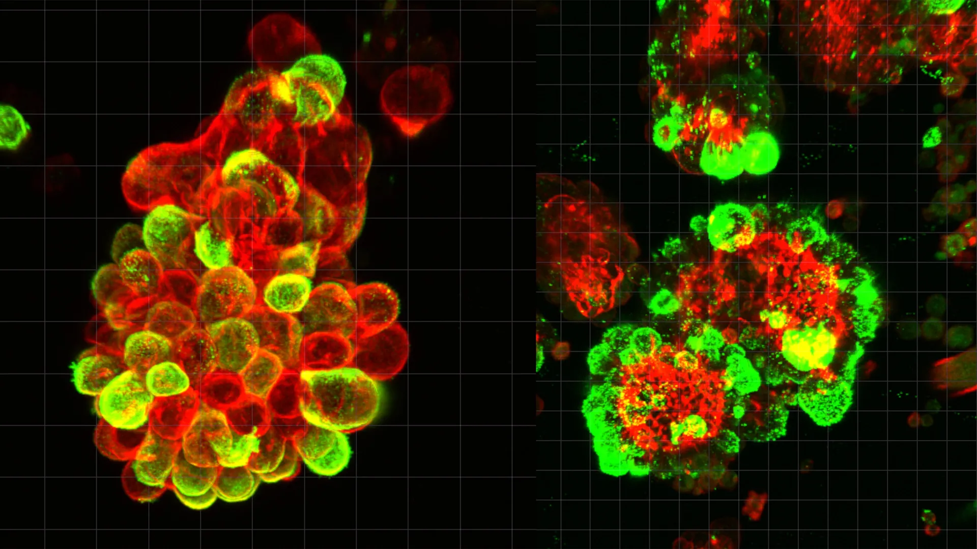
A landmark study led by researchers at Nagoya University in Japan has unveiled a sophisticated biological mechanism that explains why ovarian cancer is uniquely aggressive and difficult to detect in its early stages. The research, recently published in the prestigious journal Science Advances, identifies a "cooperative" relationship between malignant cancer cells and the healthy mesothelial cells that line the abdominal cavity. This discovery provides a long-awaited answer to why ovarian cancer spreads with such devastating speed and why it so frequently resists conventional chemotherapy treatments.
Ovarian cancer has earned a reputation as the "silent killer" of gynecological oncology. Because the symptoms are often vague—ranging from bloating to minor abdominal discomfort—most patients do not seek medical attention until the disease has reached an advanced stage. By the time of diagnosis, the cancer has typically moved beyond the ovaries to colonize the peritoneum and other abdominal organs. While researchers have long observed the rapid progression of these tumors, the specific cellular interactions driving this speed remained a mystery until now.
The Mechanism of Hybrid Cell Clusters
The research team, spearheaded by Dr. Kaname Uno, discovered that ovarian cancer cells do not operate as isolated units when spreading through the body. Instead, they actively recruit mesothelial cells—specialized cells that normally provide a smooth, protective, and friction-reducing lubricant for the internal organs.
Through a detailed analysis of abdominal fluid (ascites) taken from ovarian cancer patients, the Nagoya University team found that cancer cells frequently bind to these mesothelial cells to form compact, spherical "hybrid clusters." The study estimated that approximately 60% of the cancer cell spheres found in patient fluid samples were actually these mixed-cell entities.
The transformation begins when the primary tumor releases a signaling protein known as Transforming Growth Factor-beta 1 (TGF-β1). This molecule acts as a biochemical command, hijacking the neighboring mesothelial cells and altering their fundamental structure. Under the influence of TGF-β1, the once-protective mesothelial cells undergo a transition that causes them to develop "invadopodia"—sharp, spike-like protrusions. These structures act as biological drills, allowing the hybrid clusters to penetrate the linings of other organs with far greater efficiency than cancer cells could achieve on their own.
A Departure from Traditional Metastasis
The study highlights a critical distinction between ovarian cancer and other common malignancies like breast or lung cancer. In most cancers, metastasis occurs through the hematogenous route, where tumor cells invade blood vessels and are transported via the circulatory system to distant sites. Because the blood follows a predictable and enclosed network of vessels, clinicians can sometimes detect circulating tumor cells (CTCs) or biomarkers through routine blood tests.
Ovarian cancer, however, primarily spreads through "peritoneal seeding." In this process, cancer cells detach from the primary tumor and enter the peritoneal fluid, the liquid that naturally fills the space between abdominal organs. This fluid is in a state of constant, unpredictable motion, driven by the rhythmic movements of breathing, digestion, and physical activity.
This "floating phase" was previously thought to be a passive period where cancer cells were simply drifting. The Nagoya University study proves otherwise. It reveals that the floating phase is a period of intense biological activity where cancer cells "outsource" the labor of invasion to recruited mesothelial cells. By using these hijacked cells to create pathways, the cancer cells can remain relatively stable at a molecular level, focusing their energy on survival and proliferation rather than the complex genetic changes usually required for tissue invasion.
Resilience Against Chemotherapy
One of the most alarming findings of the research is the increased resistance of these hybrid clusters to standard treatments. When cancer cells are bundled with mesothelial cells, they exhibit a significantly higher survival rate when exposed to chemotherapy drugs compared to solitary cancer cells.
The mesothelial cells appear to provide a protective shield, potentially through both physical barrier effects and biochemical signaling that prevents the induction of apoptosis (programmed cell death) in the cancer cells. This discovery helps explain why many ovarian cancer patients experience a recurrence of the disease. Even if chemotherapy successfully eliminates individual cancer cells, the more resilient hybrid clusters may survive, eventually attaching to new surfaces and forming secondary tumors.
Methodology and Real-Time Observation
To validate their findings, the research team utilized a multi-disciplinary approach. They employed advanced live-cell imaging and microscopy to observe the formation of the hybrid spheres in real-time using fluid samples from human patients. These observations were then replicated in mouse models, where the researchers could track the movement of the clusters across the abdominal cavity.
Furthermore, the team performed single-cell RNA sequencing to analyze gene activity within the clusters. This allowed them to pinpoint the exact moment the TGF-β1 signal began to alter the mesothelial cells. The genetic data confirmed that the cancer cells themselves undergo minimal changes; they effectively remain "passengers" while the mesothelial cells do the "heavy lifting" of drilling into healthy tissue.
The Human Element: A Physician’s Motivation
The study’s lead author, Dr. Kaname Uno, brought a unique perspective to the laboratory. Before becoming a Visiting Researcher at Nagoya University’s Graduate School of Medicine, Dr. Uno spent eight years practicing as a gynecologist. His transition into full-time research was fueled by the clinical frustrations of treating ovarian cancer.
Dr. Uno recounted the story of a specific patient who had undergone a regular gynecological screening and received a clean bill of health. Only three months later, she returned with advanced, Stage IV ovarian cancer that had already spread throughout her abdomen. The speed of the progression and the failure of existing diagnostic tools to catch the disease in that short window profoundly impacted Dr. Uno.
"The inability of current medical technology to save patients who were vigilant about their health was a driving force for this study," Dr. Uno stated. "We needed to understand the ‘why’ behind the speed of this disease to develop better ‘hows’ for treatment."
Statistical Background and Clinical Urgency
The implications of this study are underscored by the sobering statistics surrounding ovarian cancer. According to global health data, ovarian cancer has the lowest five-year survival rate of all gynecological cancers, hovering around 30% to 49% depending on the region and the specific subtype.
In the United States alone, the American Cancer Society estimates that over 19,000 women will receive a new diagnosis of ovarian cancer annually, with more than 13,000 deaths. The high mortality rate is directly linked to the fact that approximately 75% of cases are diagnosed at Stage III or IV, when the cancer has already disseminated through the peritoneum—the exact process explained by the Nagoya University research.
Future Implications for Treatment and Diagnosis
The discovery of the hybrid cell clusters opens several new avenues for clinical intervention. Current oncological strategies for ovarian cancer are largely "cancer-centric," focusing on cytotoxic drugs designed to kill rapidly dividing malignant cells. However, this study suggests that the "microenvironment"—specifically the mesothelial cells—is a critical "accomplice" that must be addressed.
- Targeted Signaling Inhibition: Future therapies could focus on blocking the TGF-β1 signaling pathway. By preventing the cancer cells from communicating with mesothelial cells, doctors might be able to stop the formation of the hybrid clusters, effectively "disarming" the cancer and preventing it from invading new organs.
- Enhanced Diagnostics: The presence of these hybrid clusters in abdominal fluid could serve as a new diagnostic biomarker. Developing a "liquid biopsy" for peritoneal fluid could allow doctors to monitor the progression of the disease or the effectiveness of a specific chemotherapy regimen with much higher precision.
- Preventing Attachment: If researchers can identify the specific proteins that allow cancer cells to "grab" onto mesothelial cells, they could develop drugs that prevent this adhesion, leaving the cancer cells in a solitary, more vulnerable state.
Conclusion
The research from Nagoya University represents a paradigm shift in how scientists view ovarian cancer metastasis. By demonstrating that the disease’s rapid spread is a result of a "hijacking" of the body’s own protective mechanisms, the study provides a roadmap for more sophisticated and effective treatments. While ovarian cancer remains a formidable challenge, understanding the secret partnership between cancer and mesothelial cells brings the medical community one step closer to turning the "silent killer" into a manageable, and perhaps even curable, condition.
