
 By Budi Oetomo Narendra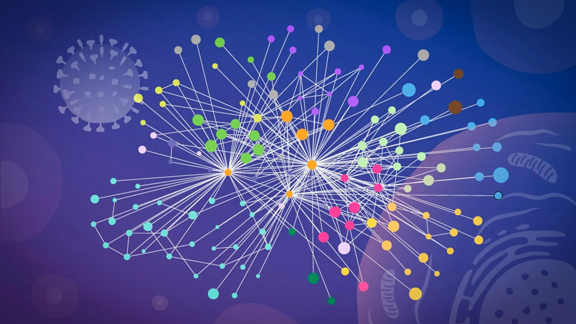
This groundbreaking research, published in Nature Microbiology, marks a significant leap in understanding the intricate molecular warfare waged by the influenza A virus inside human cells. By developing a customized workflow that allowed them to observe protein interactions directly inside intact cells – a departure from traditional methods that often rely on fragmented cellular samples – the scientific team has provided an unprecedented "snapshot" of the virus’s takeover strategy. This detailed mapping of influenza’s interaction with host proteins at a structural level within their native environment offers crucial insights that could accelerate the development of more effective flu vaccines and antiviral drugs, addressing a persistent global health challenge.
The Enduring Threat of Influenza A: A Global Health Imperative
Influenza, commonly known as the flu, remains a formidable public health concern worldwide. Seasonal influenza strains are responsible for an estimated 3 to 5 million cases of severe illness and between 290,000 and 650,000 respiratory deaths globally each year, according to the World Health Organization (WHO). Among the various types of influenza viruses, influenza A is particularly notorious for its capacity to cause widespread epidemics and pandemics. Its history is punctuated by devastating outbreaks, most notably the 1918 Spanish Flu pandemic, caused by an H1N1 strain, which infected an estimated 500 million people and claimed tens of millions of lives globally. More recently, influenza A strains like H1N1 (2009 swine flu pandemic) and highly pathogenic avian influenza A (HPAI) H5N1 and H7N9 continue to pose a significant pandemic threat due to their potential for mutation and transmission to humans.
The modus operandi of the influenza virus is a masterclass in cellular hijacking. Once it successfully enters a human cell, the virus releases its RNA genetic material, which contains the blueprints for a small but potent set of viral proteins. These proteins then rapidly disseminate throughout the host cell, systematically redirecting its complex molecular machinery. In essence, the infected cell is transformed into a highly efficient factory dedicated to the production of new virus particles, ensuring the propagation of the infection. Understanding precisely how these viral proteins interact with, and manipulate, human proteins is paramount to disrupting this cycle. Scientists need to identify the specific viral proteins involved, the human proteins they target, the exact locations of these interactions within the cell, and the mechanisms by which the virus exploits these connections for its own replication advantage.
Overcoming Methodological Hurdles: Mapping in Native Context
For decades, studying these delicate and dynamic protein-protein interactions during an active viral infection has presented immense experimental challenges. Many earlier studies relied on biochemical techniques that necessitated the physical disruption of cells before measuring protein contacts. This approach, while providing valuable data, inherently carries a significant drawback: the process of breaking cells open can profoundly distort the biological reality of the living cell. Internal cellular compartments, which naturally segregate proteins, are destroyed, potentially leading to spurious interactions between proteins that would never normally meet in vivo. Conversely, weak, transient, or highly localized interactions – often the most crucial for viral processes – can be lost entirely in the chaotic environment of a cell lysate. Consequently, researchers often struggled to definitively ascertain which protein connections truly existed and were functionally relevant during the actual infection process.
The breakthrough in the current study emerged from a novel methodological advancement. "This is when we learned that our collaborators, Boris Bogdanow and Fan Liu, at FMP Berlin had developed a specialized version of cross-linking mass spectrometry (XL-MS), a long-established technique for mapping protein contacts, tailored specifically to virus-infected cells," stated Jan Kosinski, Group Leader at EMBL Hamburg and Centre for Structural Systems Biology (CSSB). This specialized XL-MS method provided the critical technological leap the team needed. It allowed for the capture of protein-protein interactions directly within intact infected cells, preserving the native cellular context. Crucially, it could detect interactions that occur only briefly or within specific, confined regions of a cell – precisely the types of interactions often overlooked by conventional methods.
Boris Bogdanow, now a Junior Research Group Leader at the Institute of Virology, Charité — Universitätsmedizin Berlin, further elaborated on the power of this technique: "XL-MS allows us to capture protein-protein interactions directly in infected intact cells, while also providing structural information about how these interactions are happening. This gives us insight into the interface between the virus and the human cell and may, through structural modeling, help identify actionable targets for future pharmaceutical interventions." The ability to glean structural information about how these proteins fit together represents a significant step forward, moving beyond simply identifying interactors to understanding the mechanics of their binding.
The AI Advantage: Integrating Experimental Data with AlphaFold
To transform the raw interaction data from XL-MS into actionable structural insights, the research team employed a sophisticated computational approach. They paired their XL-MS results with advanced structural modeling techniques, allowing them not only to identify the viral and human proteins that interact but also to estimate the precise positioning of these proteins when they connect. This synergistic approach marries experimental observation with computational prediction, offering a more complete picture than either method could achieve alone.
At the heart of their computational strategy was a modified version of AlphaFold, the revolutionary protein structure prediction algorithm developed by DeepMind, which earned its creators a Nobel Prize in Chemistry (indirectly, through its impact on structural biology) for its profound ability to predict the three-dimensional structures of proteins with unprecedented accuracy. The EMBL Hamburg team ingeniously adapted this powerful tool. "The key advantage of the modified AlphaFold approach is that it allowed us to feed our experimental cross-linking data directly into the structural modeling," explained Kosinski. This direct integration meant that the model was guided by empirical evidence, effectively "telling" the algorithm which parts of the viral and host proteins were in close proximity within the infected cells. This directed approach was particularly beneficial for predicting the structures of virus-host complexes, which are notoriously challenging to model reliably due to their dynamic nature and the lack of extensive prior structural data. By grounding the computational predictions in real-world experimental observations, the researchers significantly enhanced the accuracy and reliability of their structural models, providing a detailed blueprint of the viral-host interface.
Unveiling Influenza’s Cellular Hijack Tactics: Two Key Discoveries
The meticulous work, published in Nature Microbiology, yielded two particularly notable discoveries regarding the strategies influenza A employs to seize control of human cells. These findings illuminate specific molecular vulnerabilities that could be exploited for therapeutic intervention.
The first strategy revolves around hemagglutinin, a critical protein found on the surface of the influenza virus. Hemagglutinin plays a dual role: it is responsible for the virus’s attachment to human host cells and facilitates its entry into them. The researchers meticulously tracked the journey of hemagglutinin as it navigated through the cell’s intricate internal transport and processing network. This network comprises various subcellular compartments, including the endoplasmic reticulum and Golgi apparatus, which are essential for the proper folding, modification, and ultimate dispatch of proteins to their correct cellular destinations. The detailed analysis revealed that several human proteins actively participate in the correct folding and modification of hemagglutinin during the infection process. Intriguingly, some of these host proteins had previously poorly understood or undefined functions, highlighting new potential targets for antiviral therapies that could disrupt the maturation of this vital viral surface protein.
The second significant discovery centered on paraspeckles, fascinating small, droplet-like compartments located within the cell nucleus. These dynamic structures are known to play roles in RNA regulation, cellular stress responses, and antiviral defense mechanisms. The team observed a striking phenomenon: influenza A infection consistently caused these paraspeckles to dissolve. When the paraspeckles broke apart, they released a pool of RNA-binding proteins that had been sequestered within them. The researchers hypothesize that the virus then capitalizes on these newly liberated host proteins, co-opting them to support its own replication machinery.
Iuliia Kotova, a former predoctoral fellow at the Kosinski Group at EMBL Hamburg and first author of the publication, now at ETH Zurich, expressed her surprise: "What surprised us most was the paraspeckles. Watching these tiny organelles in the nucleus dissolve, consistently across every cell line and every flu strain we tested, told us this isn’t a side effect of infection — it might be a strategy." This consistency across different experimental conditions strongly suggested a deliberate viral mechanism rather than a collateral effect. Furthermore, Jan Kosinski added a crucial layer to this understanding: "There may also be a second benefit for the virus: some evidence suggests paraspeckles contribute to cellular stress responses and antiviral gene regulation, so disrupting them could also weaken parts of the cell’s defense response." This dual advantage – gaining access to host proteins for replication and simultaneously suppressing host antiviral immunity – underscores the sophisticated nature of influenza’s cellular manipulation.
A Collaborative Endeavor Across Leading Institutions
This complex and multidisciplinary project was a testament to the power of collaborative science, leveraging shared technology and diverse expertise from three prominent European institutions. The specialized cross-linking mass spectrometry (XL-MS) work, critical for capturing the protein interactions in their native context, was expertly carried out at Charité – Universitätsmedizin Berlin. The sophisticated glycoproteomics analyses, essential for understanding protein modifications, were completed at the EMBL Proteomics Core Facility, a hub of advanced biochemical analysis.
The computational backbone of the study, including the modified AlphaFold modeling, relied heavily on the robust infrastructure of the EMBL Compute Cluster, demonstrating the indispensable role of high-performance computing in modern structural biology. Complementing these molecular and computational approaches, the critical microscopy imaging that provided visual confirmation and context for the observed cellular changes took place at CSSB’s Advanced Light and Fluorescence Microscopy (ALFM) Facility. This seamless integration of expertise and resources across EMBL Hamburg, FMP, Charité, and CSSB highlights a model for tackling grand challenges in infectious disease research.
Broader Implications: A New Paradigm for Pandemic Preparedness
The findings of this study extend far beyond influenza A, demonstrating a powerful new paradigm for understanding how viruses interact with and commandeer human cellular machinery. By meticulously mapping molecular contacts directly inside intact infected cells, the research offers a clearer picture of both where and how a virus exerts control. This "mapping in context" approach holds immense promise for deciphering the operational strategies of other pathogenic viruses.
"While the exact host factors and mechanisms often differ from virus to virus, we think our overall approach — combining in-cell cross-linking, structural modeling, and targeted cell-biology follow-up to map native virus-host interactions at specific stages of infection — remains broadly applicable," Kosinski asserted. This statement underscores the potential for this methodology to become a standard tool in virology and infectious disease research.
Although the current study focused on a laboratory-adapted strain of influenza, the researchers are optimistic about the broader applicability of their strategy. Boris Bogdanow concurs: "Although this study has focused on a lab-adapted strain, this study lays the groundwork to apply the methodology to viruses of potential pandemic relevance, such as H5N1, and for uncovering the interaction networks that support their multiplication in human cells." The ability to rapidly map the detailed host-pathogen interaction networks of emerging or highly virulent strains could significantly accelerate the identification of novel drug targets, facilitate the design of next-generation vaccines, and enhance global preparedness against future pandemics.
This innovative research not only deepens our understanding of influenza’s cunning strategies but also provides a powerful methodological framework for future investigations into the complex world of host-pathogen interactions. By revealing the precise molecular dance between virus and host, scientists are better equipped to develop targeted interventions that could save lives and mitigate the impact of infectious diseases worldwide. The next steps will involve applying this robust methodology to investigate different stages of infection, diverse viral strains, and other viruses, further solidifying its role as a cornerstone in the fight against viral threats.
