
 By Budi Oetomo Narendra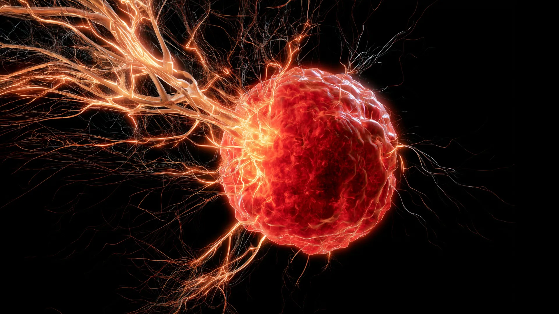
New findings from the University of Oklahoma (OU) have illuminated a previously underappreciated mechanism by which an aggressive form of breast cancer, triple-negative breast cancer (TNBC), manipulates the body’s immune system to facilitate its growth and potential resistance to treatment. Published in the esteemed journal Cell Death & Differentiation, this groundbreaking study details how cancer cells co-opt specific immune cells, known as macrophages, to draw nerve fibers into tumors, creating a microenvironment conducive to disease advancement. This discovery not only resolves a long-standing question in oncology regarding tumor innervation but also paves the way for innovative therapeutic strategies, potentially leveraging existing pharmacological agents.
The Enigma of Tumor Innervation: A Critical Knowledge Gap
For many years, scientists have observed that solid tumors are not merely collections of aberrant cells but complex ecosystems teeming with various cell types, including an intricate network of nerves. The presence of these nerve networks within tumors has been consistently associated with worse prognoses, increased pain, and enhanced metastatic potential across various cancer types. However, the precise mechanisms by which these nerves infiltrate and establish themselves within tumor masses have remained largely elusive. Understanding this critical interaction is vital, as nerves are increasingly recognized as active participants in the tumor microenvironment, influencing everything from cell proliferation and angiogenesis (new blood vessel formation) to immune suppression and drug resistance. The new research from the University of Oklahoma specifically addresses this gap, providing a clear explanation for this process in the context of triple-negative breast cancer, a disease notorious for its aggressive nature and limited targeted treatment options.
Triple-Negative Breast Cancer: A Formidable Foe in Oncology
Triple-negative breast cancer represents approximately 10-15% of all breast cancers, yet it accounts for a disproportionately high number of breast cancer deaths. It is defined by the absence of three key receptors commonly found in other breast cancer types: estrogen receptor (ER), progesterone receptor (PR), and human epidermal growth factor receptor 2 (HER2). The lack of these receptors means that TNBC does not respond to hormone therapies or HER2-targeted drugs, which are highly effective treatments for other breast cancer subtypes. Consequently, treatment options for TNBC have historically been limited primarily to chemotherapy, radiation, and surgery. While advancements in immunotherapy have shown promise for a subset of TNBC patients, the disease remains highly aggressive, characterized by rapid growth, a higher likelihood of recurrence, and a greater propensity for distant metastasis, often affecting younger women and women of African American descent more frequently. The urgent need for novel therapeutic targets and a deeper understanding of its biology drives much of the research in this field.
The OU Breakthrough: Macrophages as Master Manipulators
The core of the OU study’s revelation lies in the discovery of how tumors actively recruit and reprogram macrophages, a type of immune cell typically tasked with fighting infections and repairing damaged tissues. These versatile cells, part of the innate immune system, are usually beneficial to the body. However, within the tumor microenvironment, they can be "corrupted" by cancer cells, transforming into tumor-associated macrophages (TAMs) that, paradoxically, promote tumor growth and progression.
The researchers, led by Dr. Maureen Cox, an assistant professor in the Department of Microbiology and Immunology at the OU College of Medicine and a research member of OU Health Stephenson Cancer Center, found that once inside the tumor, these co-opted macrophages begin to secrete a specific protein: brain-derived neurotrophic factor (BDNF). BDNF is a well-known neurotrophin, a family of proteins that support the growth, survival, and differentiation of neurons. While its most widely recognized role is in the central nervous system, where it is crucial for brain development, learning, and memory, the OU team demonstrated its sinister role in breast cancer. By releasing BDNF, these tumor-associated macrophages effectively send out a powerful chemical signal that encourages nearby nerves to sprout, grow, and ultimately infiltrate the cancerous tissue. This process of nerve recruitment into the tumor mass is now understood as a critical, actively driven mechanism rather than a passive infiltration.
Decoding BDNF and Macrophages: A Dual-Edged Sword of Biology
The intricate biological dance between macrophages, BDNF, and nerve growth highlights the complex interplay within the tumor microenvironment. Macrophages are incredibly plastic, meaning they can differentiate into various phenotypes depending on the signals they receive from their surroundings. In a healthy context, macrophages might adopt an M1 phenotype, initiating inflammatory responses and directly attacking pathogens or abnormal cells. However, in the context of cancer, they are often polarized towards an M2 phenotype, which is associated with immunosuppression, tissue repair, angiogenesis, and, as this study shows, nerve growth promotion. This "re-education" of macrophages by the tumor is a hallmark of cancer’s ability to evade and exploit the host immune system.
BDNF, in its normal physiological role, is indispensable for the health and function of the nervous system. It binds to its receptor, TrkB (tropomyosin receptor kinase B), on nerve cells, triggering signaling pathways that promote neuronal survival, differentiation, and synaptic plasticity. The University of Oklahoma research reveals a sinister repurposing of this essential biological pathway by aggressive breast cancer. The tumor effectively weaponizes BDNF, transforming a vital neurotrophic factor into a pro-tumorigenic signal that fosters nerve infiltration. This nerve growth, in turn, is hypothesized to contribute to various aspects of cancer progression, including increased tumor proliferation, enhanced metastatic potential, and potentially conferring resistance to conventional therapies.
Experimental Validation: From Lab to Living Models
To validate their hypothesis, Dr. Cox and her colleagues embarked on a series of rigorous experiments. Their investigation began with in vitro studies, observing the interactions between macrophages, cancer cells, and nerve cells in controlled laboratory settings. These initial observations provided compelling evidence that macrophages, when stimulated by tumor factors, indeed secreted BDNF, which subsequently induced neurite outgrowth (the extension of nerve fibers) from neuronal cells.
The research then progressed to in vivo models, employing mice engineered with triple-negative breast cancer. This crucial step allowed the team to study the complex interactions within a living organism. In these mouse models, the researchers confirmed that nerves actively grew into the tumors, correlating with the presence of macrophages and elevated BDNF levels. The pivotal moment came when they tested a therapeutic intervention: they administered a drug known to block BDNF signaling. The results were striking and highly promising. In mice treated with this BDNF-blocking drug, the infiltration of nerves into the tumors was significantly curtailed, and critically, tumor growth itself was substantially reduced. This direct causal link between BDNF-mediated nerve growth and tumor progression provided robust evidence for the therapeutic potential of targeting this pathway.
Clinical Relevance: Insights from Patient Data
Moving beyond preclinical models, the research team sought to determine the clinical relevance of their findings to human patients. They analyzed data from individuals diagnosed with triple-negative breast cancer, examining tumor samples for the presence of macrophages and BDNF levels. Their analysis revealed a compelling correlation: patients whose tumors exhibited higher levels of both macrophages and BDNF experienced poorer survival outcomes. This crucial piece of evidence suggests that the mechanism observed in mice is highly pertinent to human disease, underscoring the potential translational impact of their discovery. The presence of elevated macrophage infiltration and BDNF signaling could serve as prognostic markers, identifying patients at higher risk of aggressive disease progression and potentially informing treatment decisions.
A New Therapeutic Horizon: Repurposing Existing Drugs
The implications of this discovery for cancer treatment are profound. Instead of solely focusing on cytotoxic therapies designed to destroy cancer cells, future strategies could involve interrupting the intricate communication network that supports tumor growth. The finding that a drug already on the market can effectively block BDNF signaling and reduce tumor growth in mice is particularly exciting. This opens the door to drug repurposing, a strategy that accelerates the development of new treatments by finding new uses for existing, approved drugs. Repurposing can significantly cut down the time and cost associated with drug development, as the safety profile and pharmacokinetics of such drugs are already well-established. Dr. Cox expressed optimism about this prospect, stating, "It looks really promising that we can use this drug, which is already on the market, to target BDNF." This suggests that a new class of anti-cancer therapies, focused on modulating the tumor microenvironment rather than directly killing cancer cells, could soon emerge, either as standalone treatments or in combination with existing modalities.
Boosting the Body’s Defenses: Implications for Immunotherapy
Beyond directly inhibiting tumor growth, the researchers hypothesize that blocking nerve infiltration could have another significant benefit: boosting the body’s natural immune response against cancer. "We believe that the nerves are immunosuppressive, so if we can stop the nerves from growing in the first place, maybe we can boost the immune response to help fight the cancer," Dr. Cox explained. This insight is particularly relevant in the era of immunotherapy, where treatments like immune checkpoint inhibitors aim to unleash the patient’s own immune system to attack cancer cells. If nerves within tumors actively suppress immune cells, their removal or prevention of growth could sensitize tumors to immunotherapy, making these powerful treatments effective for a broader range of patients. This could represent a synergistic approach, combining nerve-targeting therapies with existing immunotherapies to achieve more robust and durable anti-tumor effects, especially in immune-cold tumors that typically do not respond well to immunotherapy.
Beyond Breast Cancer: A Broader Impact
The potential impact of this research extends beyond triple-negative breast cancer. The mechanisms uncovered – immune cell manipulation, neurotrophic factor signaling, and nerve infiltration – are likely not unique to TNBC. Many other aggressive cancers, including high-grade ovarian cancer, pancreatic cancer, and prostate cancer, are known to be heavily innervated and are notoriously difficult to treat. Dr. Cox and her team are already planning to test the same intervention in high-grade ovarian cancer, another aggressive malignancy with significant unmet therapeutic needs. If similar mechanisms are at play in these other cancers, the therapeutic strategy of targeting BDNF-mediated nerve growth could have widespread applicability, offering new hope for patients facing a variety of challenging diagnoses.
The Road Ahead: Unraveling Nerve-Tumor Interactions
While the current study provides a critical piece of the puzzle, many questions remain. Dr. Cox and her team are now focused on delving deeper into exactly how nerves contribute to tumor growth and progression. Several hypotheses are being explored. Some evidence suggests that nerves may play a role in stimulating angiogenesis, the formation of new blood vessels, which are essential for supplying tumors with oxygen and nutrients. Without a robust blood supply, tumors cannot grow beyond a minimal size. Other research indicates that cancer cells may exploit nerve fibers as conduits, using them as "highways" to migrate away from the primary tumor site and metastasize to distant organs, a process known as perineural invasion, which is often linked to aggressive disease. Understanding these downstream effects of nerve infiltration will be crucial for developing comprehensive treatment strategies. Ultimately, the overarching goal of this research is to "turn the anti-tumor immunity back on in cancer patients so their own immune systems can reject the tumors," a vision that could revolutionize cancer care.
Supporting Groundbreaking Science
This pioneering research was made possible through significant support from various institutions. The National Institute of General Medical Sciences of the NIH (award numbers P20GM103447 and P20GM103639) provided essential funding, underscoring the national importance of this line of inquiry. Additionally, Oklahoma’s Tobacco Settlement Endowment Trust (TSET), a primary funder of the Stephenson Cancer Center and TSET Health Promotion Research Center at the University of Oklahoma, played a crucial role. Further support came from the Oklahoma Shared Clinical and Translational Resources through an Institutional Development Award from the National Institute of General Medical Sciences (grant no. U54GM104938). Such foundational funding is indispensable for advancing our understanding of complex diseases like cancer and for translating laboratory discoveries into tangible benefits for patients. The collaborative efforts and sustained support highlight the commitment to pushing the boundaries of medical science and ultimately improving patient outcomes in the fight against cancer.
