
 By George Ongkojoyo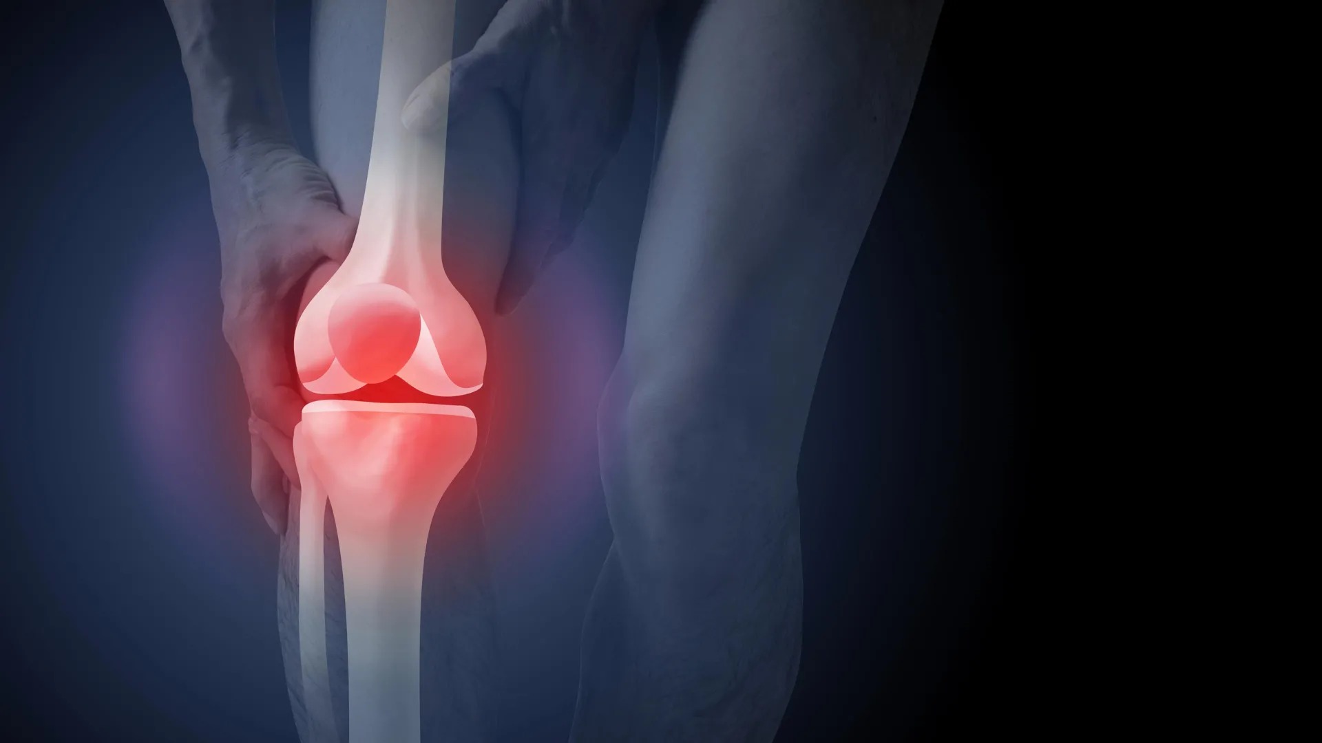
The diagnostic pathway for knee osteoarthritis in Australia is facing intense scrutiny following the release of a comprehensive study indicating that routine X-rays may do more harm than good for patient outcomes. Despite clear clinical guidelines stating that imaging is unnecessary for a standard diagnosis, nearly half of all new patients presenting with knee pain are still referred for radiological scans. This disconnect between evidence-based guidelines and clinical practice is not only costing the Australian healthcare system hundreds of millions of dollars annually but is also fundamentally altering how patients perceive their own health, often leading them toward invasive and potentially unnecessary surgical interventions.
The Disconnect Between Guidelines and Clinical Practice
Knee osteoarthritis is a prevalent chronic condition characterized by changes in the joint structure, where the body works to repair itself in response to various stressors. While it is often colloquially referred to as "wear and tear," medical professionals emphasize that it is a complex biological process affecting the entire joint, including bones, cartilage, ligaments, and muscles. Current clinical standards, including those set by the Australian Commission on Safety and Quality in Health Care, specify that a diagnosis should be made based on a patient’s medical history and physical symptoms rather than imaging.
Specifically, a clinical diagnosis is recommended for individuals aged 45 and over who experience activity-related joint pain and have either no morning stiffness or stiffness that resolves within 30 minutes. Despite these straightforward criteria, data from the Australian Institute of Health and Welfare (AIHW) and recent research published in PLOS Medicine highlight a persistent reliance on X-rays. In Australia, approximately 45% of patients visiting a GP for new knee pain are referred for imaging, contributing to an annual diagnostic imaging cost of A$104.7 million for osteoarthritis alone.
Analyzing the PLOS Medicine Study: Methodology and Results
A new study led by researchers in Australia sought to quantify the psychological and behavioral impact of using X-rays for diagnosis. The study recruited 617 participants from across the country to investigate whether seeing a physical "image" of joint degradation changed a patient’s outlook on their recovery and treatment options.
The researchers employed a randomized controlled design where participants were assigned to watch one of three hypothetical consultation videos between a GP and a patient complaining of knee pain:
- Group A (Clinical Diagnosis): The GP provided a diagnosis based solely on age and symptoms, explaining that no X-ray was required.
- Group B (X-ray Diagnosis – Image Shown): The GP used an X-ray to diagnose the condition and showed the participant the images of the joint.
- Group C (X-ray Diagnosis – Image Not Shown): The GP used an X-ray for the diagnosis but did not show the physical images to the patient.
The findings were stark. Participants in Group B—those who saw their X-ray images—reported a 36% higher perceived need for knee replacement surgery compared to those in the clinical diagnosis group. Furthermore, these participants expressed significantly higher levels of anxiety regarding the future of their condition. They were more likely to view exercise and physical activity as potentially harmful to their joints and exhibited a higher degree of "kinesiophobia," or fear of movement.
Interestingly, the study also found that patients were slightly more satisfied with a diagnosis that included an X-ray. This suggests a deep-seated cultural belief that "seeing is believing" and that medical technology is inherently superior to clinical judgment. This satisfaction, however, appears to be a double-edged sword, as it reinforces misconceptions that lead to worse long-term health choices.
The Economic and Physical Toll of Knee Replacement Surgery
The push toward surgery is a significant concern for health economists and practitioners alike. In the 2020–21 financial year, hospital services for osteoarthritis, driven primarily by joint replacement procedures, cost the Australian economy approximately A$3.7 billion. During the 2021–22 period, more than 53,000 Australians underwent knee replacement surgery due to osteoarthritis.
While total knee arthroplasty (TKA) can be life-changing for patients with end-stage disease who have exhausted all other options, it is not without risk. Experts warn that surgery should only be considered a last resort. Complications can include deep vein thrombosis (blood clots), surgical site infections, and chronic post-operative pain. Furthermore, a significant percentage of patients—estimated in some studies to be as high as 20%—report dissatisfaction or a lack of full recovery following the procedure.
The financial burden extends beyond the surgery itself. The "over-diagnosis" via imaging leads to a cascade of specialist referrals and follow-up scans that inflate healthcare spending without necessarily improving patient mobility or pain levels.
Debunking the "Wear and Tear" Myth
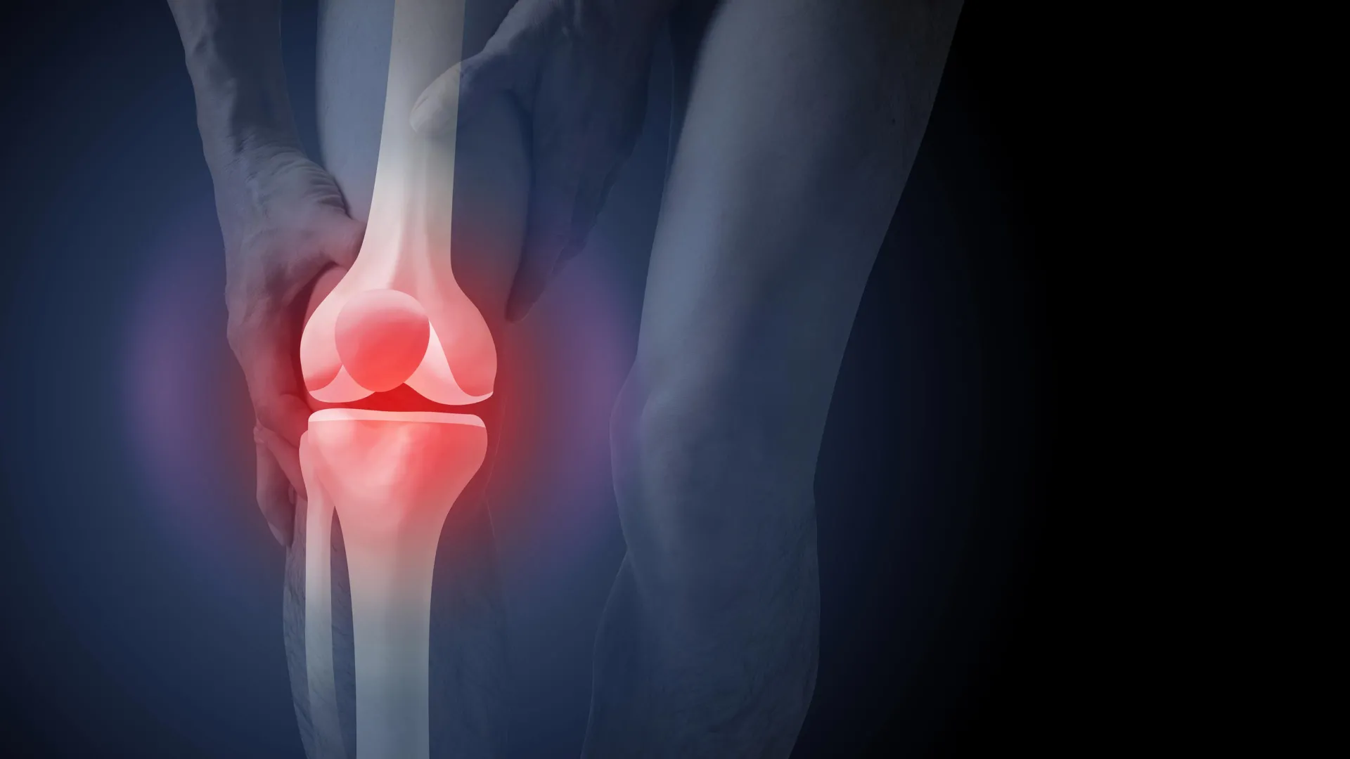
One of the primary drivers of the demand for X-rays is the persistent myth that osteoarthritis is a mechanical failure of the joint—a simple matter of "wearing out" the cartilage. However, medical research has long shown a poor correlation between structural changes seen on an X-ray and the actual pain experienced by a patient.
Many individuals with significant "bone-on-bone" changes visible on an X-ray report little to no pain and maintain high levels of activity. Conversely, many patients experiencing debilitating pain show only minimal changes on an image. Because X-rays do not capture the inflammatory environment of the joint or the functional strength of the surrounding muscles, they are often a poor guide for treatment.
When patients are shown X-rays that depict narrowed joint spaces or bone spurs, they often internalize the idea that their joints are "crumbling" or "damaged." This "nocebo effect"—where negative expectations lead to worse symptoms—can cause patients to stop exercising, which ironically accelerates the progression of the disease by allowing the supporting muscles to weaken.
First-Line Treatments: The Evidence-Based Alternative
The consensus among global health organizations, including the Royal Australian College of General Practitioners (RACGP), is that the management of knee osteoarthritis should focus on non-surgical, conservative care. The three pillars of effective management are:
- Education: Understanding that the joint is not "wearing out" and that movement is actually "lotion" for the joint.
- Exercise: Targeted strength training and aerobic activity to support the joint and reduce inflammation.
- Weight Management: Reducing the mechanical load on the knee and decreasing systemic inflammation associated with adipose tissue.
Programs such as GLA:D (Good Life with Arthritis: Denmark), which has been implemented widely in Australia, have demonstrated that structured exercise and education can reduce pain by up to 33% and significantly decrease the desire for surgery.
Chronology of Clinical Guidelines and Shifting Paradigms
The move away from routine imaging is not a new recommendation, but it has gained momentum over the last decade as the costs of over-treatment have escalated:
- 2014: The National Institute for Health and Care Excellence (NICE) in the UK strengthened its stance against routine imaging for OA diagnosis.
- 2017: The Australian Commission on Safety and Quality in Health Care released the Osteoarthritis of the Knee Clinical Care Standard, explicitly advising against X-rays for routine diagnosis.
- 2018: The RACGP updated its clinical guidelines to emphasize conservative management as the first-line treatment for all patients.
- 2024: The latest study in PLOS Medicine provides the first robust evidence of the psychological harm caused by showing patients their X-rays, providing a new impetus for changing GP behavior.
Implications for the Future of Healthcare
The implications of this research are twofold. First, there is a clear need for a cultural shift in how doctors communicate with patients. Rather than using X-rays as a "shortcut" to validate a patient’s pain, GPs are being encouraged to spend more time explaining the clinical diagnosis and the benefits of conservative care.
Second, there is a systemic need to address the incentives that lead to over-imaging. Reducing unnecessary X-rays would not only save the government over A$100 million annually but also reduce the cumulative radiation exposure of the population.
Health advocates argue that if patients understand that an X-ray might actually make them feel worse and lead them toward a risky surgery they might not need, they may be less likely to demand one. The goal is to move toward a "value-based" healthcare model where the focus is on functional outcomes—walking further, climbing stairs with less pain, and maintaining independence—rather than "fixing" an image on a screen.
As Australia’s population ages, the prevalence of osteoarthritis is expected to rise. Addressing the "imaging epidemic" now is seen as a critical step in ensuring the sustainability of the healthcare system and the physical well-being of millions of Australians. For the individual patient, the message from the latest research is clear: your symptoms matter more than your scans, and staying active is the most effective way to protect your joints for the long term.
