
 By George Ongkojoyo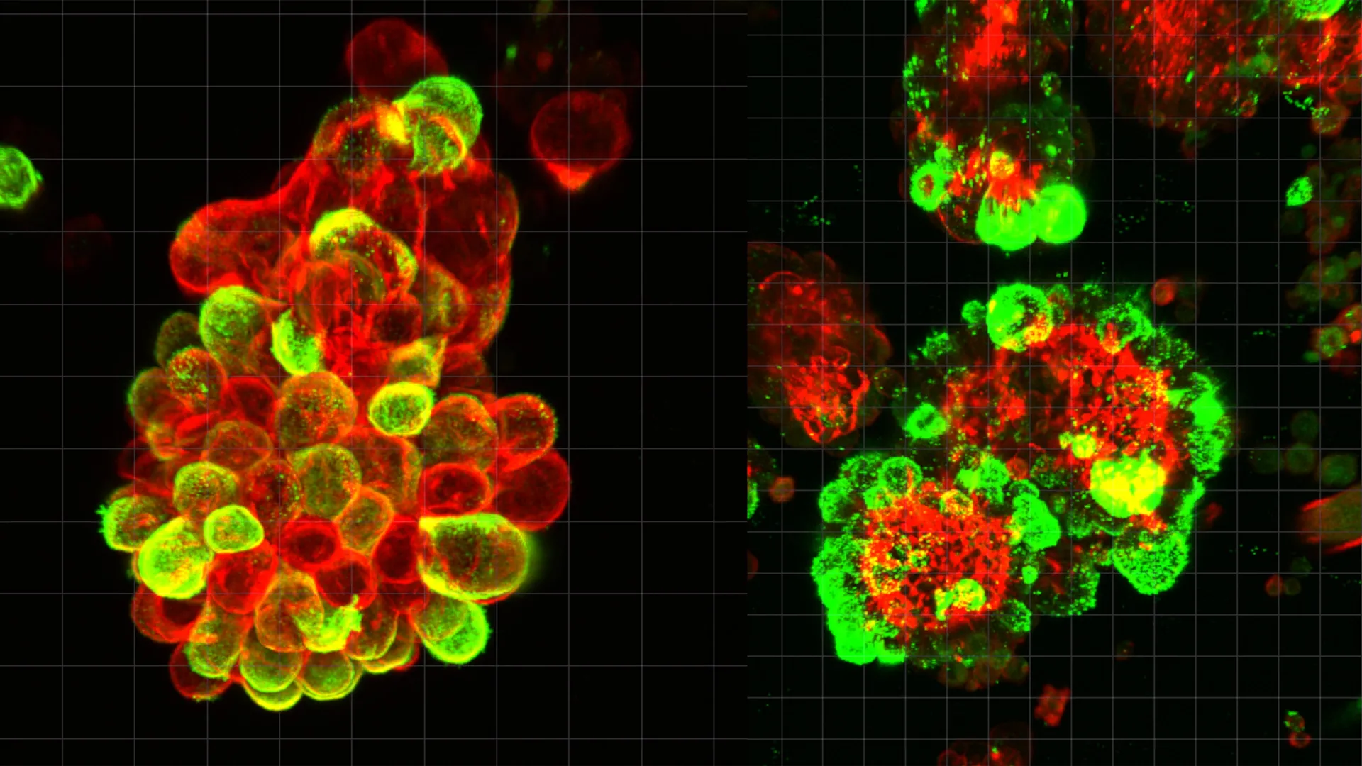
The study, recently published in the prestigious journal Science Advances, details how ovarian cancer cells do not operate as isolated entities during the metastatic process. Instead, they actively coerce mesothelial cells—the protective cells that line the peritoneal cavity—into forming hybrid clusters. These clusters act as highly efficient, mobile invasion units that are significantly more resilient to conventional chemotherapy than independent cancer cells. By uncovering this cooperative relationship, the research provides a new roadmap for developing targeted therapies that could potentially halt the spread of the disease in its tracks.
The Biological Architecture of Ovarian Cancer Spread
To understand the significance of this discovery, it is necessary to examine the unique environment in which ovarian cancer thrives. Unlike many other forms of cancer, such as breast or lung cancer, which primarily utilize the circulatory or lymphatic systems to reach distant organs, ovarian cancer utilizes the peritoneal fluid. This fluid, which naturally lubricates the abdominal organs, serves as a medium for "transperitoneal" spread.
When an ovarian tumor reaches a certain stage, cells detach from the primary site and enter the fluid-filled space of the abdomen. Historically, scientists believed these floating cells were passive travelers, drifting until they happened to land on an organ surface. However, the Nagoya University team, led by Dr. Kaname Uno, found that the process is far more calculated. Upon entering the peritoneal fluid, cancer cells immediately begin seeking out mesothelial cells that have shed from the abdominal lining.
Through advanced single-cell RNA sequencing and high-resolution microscopy, the researchers observed that approximately 60% of the cancer cell aggregates found in patient abdominal fluid were actually hybrid spheres. These spheres are composed of both malignant cancer cells and recruited mesothelial cells. This partnership is initiated by the cancer cells through the secretion of a potent signaling protein known as Transforming Growth Factor-beta 1 (TGF-β1).
The Role of Invadopodia: "Contracting" the Dirty Work
The most striking finding of the study involves how these hybrid spheres interact with healthy tissue. Once the TGF-β1 signal is received, the mesothelial cells undergo a radical transformation. Normally smooth and protective, these cells begin to develop "invadopodia"—sharp, spike-like protrusions rich in proteolytic enzymes.
In a remarkable display of biological manipulation, the cancer cells themselves remain relatively passive during the initial phase of tissue penetration. Instead, they utilize the transformed mesothelial cells as a sort of "biological drill." The mesothelial cells use their invadopodia to puncture the basement membrane of abdominal organs, creating physical openings. The cancer cells then simply follow these pre-cleared paths to establish new colonies.
Dr. Kaname Uno, a Visiting Researcher at Nagoya University’s Graduate School of Medicine, noted that this strategy allows the cancer cells to conserve energy and avoid the complex genetic mutations typically required for a cell to become invasive on its own. "They manipulate mesothelial cells to do the tissue invasion work," Dr. Uno explained. "The cancer cells undergo minimal genetic and molecular changes and just migrate through the openings that mesothelial cells create."
This "outsourcing" of labor explains why ovarian cancer can spread so rapidly. By leveraging the existing machinery of healthy cells, the tumor bypasses many of the biological hurdles that usually slow down the metastatic process in other types of cancer.
Clinical Context: The Challenge of Early Detection and Treatment
The implications of this research are particularly profound given the current landscape of ovarian cancer statistics. Globally, ovarian cancer is the eighth most common cause of cancer-related death in women. Because the symptoms—such as bloating, pelvic pain, and feeling full quickly—are often vague and mimic less serious conditions, nearly 70% to 80% of patients are diagnosed at Stage III or IV.
At these advanced stages, the cancer has already moved beyond the pelvis and into the peritoneal cavity. The five-year survival rate for localized ovarian cancer is approximately 93%, but this drops precipitously to about 30% once the disease has spread to distant abdominal sites. The discovery of hybrid cell clusters helps explain not only why the disease spreads so fast but also why it is so difficult to treat once it has done so.
The Nagoya University study found that these hybrid spheres are significantly more resistant to chemotherapy than clusters consisting solely of cancer cells. The presence of mesothelial cells appears to provide a protective "shield" or an altered metabolic environment that allows the cancer cells to survive doses of medication that would otherwise be lethal. This finding may explain why many ovarian cancer patients experience a recurrence of the disease even after undergoing aggressive chemotherapy and surgery.
Methodology and the Path to Discovery
The research team utilized a comprehensive "bench-to-bedside" approach to validate their findings. The study began with the analysis of ascites—the fluid that accumulates in the abdomen of cancer patients. Using advanced microscopy, the scientists were able to watch the formation of these hybrid spheres in real-time using samples taken directly from human patients.
To confirm the role of TGF-β1 and invadopodia, the team employed mouse models. They observed that when the TGF-β1 signaling pathway was inhibited, the formation of hybrid spheres was disrupted, and the ability of the cancer to invade abdominal organs was significantly reduced. Furthermore, single-cell genetic analysis allowed the researchers to map the specific gene expressions that occur during the "hijacking" process, providing a molecular blueprint of the cancer-mesothelial interaction.
The impetus for this research was deeply personal for the lead author. Before entering the realm of molecular research, Dr. Kaname Uno spent eight years as a practicing gynecologist. He recalls a specific patient who had undergone a routine screening that showed no abnormalities, only to be diagnosed with terminal, advanced-stage ovarian cancer just three months later. The speed at which the disease had progressed defied the medical tools available at the time. This experience drove Dr. Uno to transition into research to find the "missing link" in ovarian cancer progression.
Future Implications for Oncology and Drug Development
The discovery of the mesothelial hijacking mechanism opens several new avenues for the treatment and monitoring of ovarian cancer. Currently, the standard of care involves "debulking" surgery followed by platinum-based chemotherapy. While effective at killing rapidly dividing cancer cells, these treatments do not specifically target the cooperative biological structures identified by the Nagoya team.
1. Targeted Signal Interruption:
Future therapeutic strategies could focus on blocking the TGF-β1 signal. By preventing the cancer cells from communicating with mesothelial cells, doctors might be able to prevent the formation of the hybrid spheres entirely, effectively "trapping" the cancer cells in the peritoneal fluid where they are more vulnerable to treatment.
2. Inhibiting Invadopodia:
Drugs designed to inhibit the formation of invadopodia could prevent the "drilling" process. If the mesothelial cells cannot create pathways into the organs, the cancer cells would remain suspended in the fluid, unable to take root and form secondary tumors.
3. Liquid Biopsy and Monitoring:
The study suggests that monitoring the presence and concentration of these hybrid cell clusters in the abdominal fluid could serve as a powerful diagnostic and prognostic tool. By analyzing the composition of these spheres, oncologists could better predict how aggressively a patient’s cancer is likely to spread and tailor their treatment plans accordingly.
4. Redefining Chemotherapy Resistance:
Understanding that resistance is partially driven by "accomplice" cells allows researchers to develop sensitizing agents. These agents could break down the protective environment created by the mesothelial cells, making the underlying cancer cells susceptible to standard chemotherapy once again.
Conclusion: A New Paradigm in Gynecological Oncology
The work of the Nagoya University team marks a paradigm shift in how scientists view ovarian cancer metastasis. For years, the focus was almost entirely on the intrinsic mutations of the cancer cell itself. This new data suggests that the "microenvironment"—the neighborhood of healthy cells surrounding the tumor—is just as critical to the disease’s success as the tumor’s own genetic makeup.
By identifying the mesothelial cell as an unwilling but essential partner in the spread of ovarian cancer, the research moves the medical community closer to a future where "silent killers" are no longer able to operate in the shadows. While further clinical trials are necessary to translate these laboratory findings into bedside treatments, the roadmap is clearer than ever before. For the millions of women at risk of ovarian cancer, this discovery offers not just a better understanding of the disease, but the genuine hope of more effective, life-saving interventions.
