
 By Billy Candra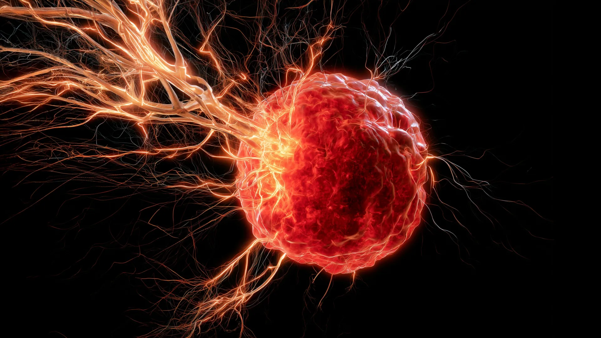
For years, scientists have recognized the presence of extensive nerve networks within many solid tumors. While the observation was clear, the precise mechanisms by which these nerves penetrate the tumor microenvironment remained largely elusive. The University of Oklahoma team, led by Dr. Maureen Cox, an assistant professor in the Department of Microbiology and Immunology at the OU College of Medicine and a research member of OU Health Stephenson Cancer Center, has now elucidated a critical pathway involving immune cells that are typically viewed as beneficial to the body. Their findings suggest a sophisticated manipulation by cancer cells, turning the body’s own defense mechanisms into unwitting accomplices in tumor growth and resistance to therapy.
The Enigma of Tumor Innervation: A Long-Observed Phenomenon, Newly Understood
The presence of nerves within tumors, often termed "tumor innervation," has been a subject of increasing scientific interest over the past two decades. Early observations, dating back to the late 19th and early 20th centuries, noted the occasional presence of nerve fibers within cancerous growths. However, it was largely dismissed as an incidental finding, without clear functional significance. Modern research, aided by advanced imaging and molecular techniques, has begun to reveal that tumor innervation is far from incidental. Instead, it is a complex and dynamic process, now understood to play a critical role in various aspects of cancer biology, including initiation, progression, metastasis, and resistance to treatment.
Nerves within the tumor microenvironment can influence cancer cells through various mechanisms. They can release neurotransmitters and neurotrophic factors that directly stimulate cancer cell proliferation, survival, and migration. Conversely, cancer cells can also secrete factors that promote nerve growth, creating a reciprocal feedback loop. This intricate interplay between the nervous system and cancer has led to the emerging field of "neuro-oncology," which seeks to understand and exploit these connections for therapeutic gain. However, a fundamental question persisted: how do these nerves get there in the first place? The OU research addresses this critical gap, focusing on a specific and aggressive cancer type.
Triple-Negative Breast Cancer: A Formidable Foe
Triple-negative breast cancer (TNBC) represents about 10-15% of all breast cancers, but its impact is disproportionately severe. It is characterized by the absence of estrogen receptors (ER), progesterone receptors (PR), and human epidermal growth factor receptor 2 (HER2) overexpression. This "triple-negative" status means that TNBC does not respond to hormone therapy or HER2-targeted drugs, which are highly effective treatments for other types of breast cancer. Consequently, treatment options for TNBC are limited primarily to chemotherapy, radiation therapy, and more recently, immunotherapy and PARP inhibitors for specific genetic mutations.
Patients with TNBC often face a more aggressive disease course, higher rates of recurrence, and poorer prognosis compared to those with other breast cancer subtypes. It tends to affect younger women and women of African American or Hispanic descent more frequently. The challenges in treating TNBC underscore the urgent need for novel therapeutic targets and a deeper understanding of its unique biology. The University of Oklahoma’s discovery offers a fresh perspective on a previously overlooked aspect of TNBC’s aggressiveness, suggesting that its ability to manipulate the immune system to recruit nerves may be a key factor in its rapid progression and resistance to conventional treatments.
Unraveling the Mechanism: Macrophages as Orchestrators
The OU research team’s breakthrough centers on the unexpected role of macrophages, a type of immune cell traditionally recognized for its beneficial functions in fighting infections and repairing damaged tissues. These versatile cells are part of the innate immune system and act as phagocytes, engulfing cellular debris, foreign substances, and pathogens. They also play a crucial role in orchestrating immune responses by presenting antigens and secreting cytokines. However, within the complex and often paradoxical environment of a tumor, macrophages can be "re-educated" or "polarized" by cancer cells to adopt pro-tumorigenic roles. These tumor-associated macrophages (TAMs) are known to promote angiogenesis (blood vessel formation), suppress anti-tumor immunity, and facilitate metastasis.
In this study, Dr. Cox and her colleagues discovered a new pro-tumorigenic function for TAMs in TNBC. They found that tumors actively attract macrophages to their vicinity. Once inside the tumor microenvironment, these macrophages do not perform their typical anti-cancer duties. Instead, they become instrumental in the tumor’s strategy to recruit nerves. The key player in this process is brain-derived neurotrophic factor (BDNF), a protein released by the macrophages.
Brain-Derived Neurotrophic Factor: A Signal Hijacked
BDNF is widely recognized for its vital role in the central and peripheral nervous systems. It is a member of the neurotrophin family, proteins that support the survival, growth, and differentiation of neurons. In healthy physiological contexts, BDNF is essential for brain development, synaptic plasticity, memory formation, and the maintenance of neuronal health. Its receptor, TrkB (tropomyosin receptor kinase B), is expressed on the surface of neurons, and BDNF binding activates signaling pathways crucial for nerve cell function and survival.
However, the OU researchers found that TNBC tumors exploit this very same biological signal. By prompting macrophages to secrete BDNF within the tumor microenvironment, the cancer effectively creates a chemical beacon, encouraging nearby nerves to grow towards and infiltrate the cancerous mass. This "hijacking" of a fundamental neurodevelopmental pathway for pathological purposes represents a sophisticated mechanism by which cancer fosters its own growth. This nerve infiltration, driven by macrophage-derived BDNF, is believed to contribute significantly to cancer progression, potentially by providing direct trophic support to cancer cells, influencing the tumor microenvironment, and contributing to treatment resistance.
Dr. Cox emphasized the critical, yet paradoxical, role of macrophages in this scenario: "Macrophages are the critical source for drawing nerves into the tumor. Although macrophages typically play a positive role in the body, they are facilitating a negative function in this scenario of breast cancer." This statement highlights the intricate and often contradictory roles immune cells can play within the tumor microenvironment, underscoring the need for highly specific interventions that can re-educate or target these cells without compromising their essential functions elsewhere in the body.
Experimental Validation: Promising Preclinical Results
The discovery of the macrophage-BDNF-nerve axis in TNBC provided a clear therapeutic target. The research team moved from identifying the mechanism to testing interventions aimed at disrupting it. They hypothesized that blocking the BDNF signaling pathway could prevent nerve infiltration and, consequently, slow tumor growth. To test this strategy, Dr. Cox and her colleagues conducted preclinical experiments using mouse models of TNBC.
They administered a drug designed to block BDNF signaling, specifically by inhibiting its receptor, TrkB. The results were highly encouraging: in the mice treated with the BDNF-blocking drug, nerves no longer grew into the tumors. More importantly, the inhibition of nerve infiltration correlated with a significant reduction in tumor growth. This direct correlation provides strong evidence that the recruited nerves are not merely bystanders but active contributors to tumor expansion.
The potential for drug repurposing is a particularly exciting aspect of these findings. Dr. Cox noted, "It looks really promising that we can use this drug, which is already on the market, to target BDNF." This statement refers to TrkB inhibitors, several of which have been developed for neurological conditions or other cancer types. The ability to repurpose an existing, FDA-approved drug could drastically accelerate the timeline for bringing this novel therapeutic strategy to patients, bypassing many of the lengthy and costly stages of de novo drug development. Such a strategy would also likely benefit from an already established safety profile, reducing risks in clinical trials.
Translational Insights: Evidence from Human Patients
To determine the clinical relevance of their findings, the researchers extended their investigation beyond preclinical models. They examined data from human patients diagnosed with triple-negative breast cancer, seeking to ascertain whether the same biological pattern observed in mice was evident in human disease. This translational step is crucial for validating laboratory discoveries and determining their potential impact on patient care.
Their analysis of patient data revealed a compelling correlation: tumors from individuals with higher levels of both macrophages and BDNF were significantly linked with poorer survival outcomes. This association provides robust evidence that the macrophage-BDNF-nerve mechanism is not confined to laboratory models but is indeed relevant to the progression and prognosis of TNBC in human patients. The convergence of preclinical and clinical data strengthens the validity of their hypothesis and underscores the potential for this discovery to translate into tangible clinical benefits. This finding also suggests that quantifying macrophage and BDNF levels in TNBC tumors could serve as prognostic biomarkers, helping to identify patients at higher risk of aggressive disease and informing treatment decisions.
Broader Implications and Future Directions
The University of Oklahoma’s research marks a significant step forward in understanding cancer biology and developing new treatment modalities. The implications of this discovery extend beyond triple-negative breast cancer and could reshape how scientists and clinicians approach cancer therapy.
Shifting Paradigms in Cancer Treatment
Historically, cancer treatment has largely focused on directly eliminating cancer cells through chemotherapy, radiation, or targeted therapies. More recently, immunotherapy has emerged as a powerful approach by harnessing the patient’s own immune system to fight cancer. The OU study introduces another critical layer to this complex picture: the nervous system’s involvement in tumor progression. By identifying a mechanism where immune cells inadvertently facilitate nerve growth, the research suggests that future therapies might not only target cancer cells or immune checkpoints but also interrupt the signaling pathways between immune cells and nerves that appear to support tumor growth. This "neuro-immune" approach represents a novel paradigm in oncology.
The Promise of Immunotherapy Enhancement
One of the most exciting implications of the research, as highlighted by Dr. Cox, is the potential to enhance the effectiveness of existing immunotherapies. She stated, "We believe that the nerves are immunosuppressive, so if we can stop the nerves from growing in the first place, maybe we can boost the immune response to help fight the cancer." This hypothesis aligns with a growing body of evidence suggesting that tumor innervation can contribute to an immunosuppressive tumor microenvironment, making it harder for the immune system to recognize and attack cancer cells. By blocking nerve infiltration, it may be possible to "re-awaken" or augment the anti-tumor immune response, potentially making patients more responsive to immunotherapeutic agents like checkpoint inhibitors. This could be particularly impactful for TNBC, where immunotherapy has shown promise but often faces challenges with patient response rates.
Unanswered Questions and Next Frontiers
While the study provides a critical explanation for nerve infiltration, it also opens up new avenues for further research. Dr. Cox and her team are now focused on elucidating the exact mechanisms by which these nerves contribute to tumor growth. Several hypotheses are under investigation:
- Angiogenesis Stimulation: Nerves may stimulate the formation of new blood vessels (angiogenesis), which are crucial for supplying tumors with oxygen and nutrients, thereby fueling their rapid growth.
- Metastasis Facilitation: Some evidence suggests that cancer cells may utilize nerve fibers as "highways" to migrate away from the primary tumor, facilitating metastasis to distant sites. This concept of "perineural invasion" is well-established in certain cancers, such as pancreatic and head and neck cancers, and its role in breast cancer, particularly TNBC, warrants further investigation.
- Direct Trophic Support: Nerves could release factors that directly promote the proliferation and survival of cancer cells.
- Modulation of the Immune Microenvironment: Beyond immunosuppression, nerves might influence other aspects of the tumor microenvironment, impacting the behavior of other immune cells, stromal cells, and the extracellular matrix.
The researchers also plan to extend their investigations to other aggressive cancers that are difficult to treat, such as high-grade ovarian cancer. This demonstrates the potential generalizability of their findings and the hope that targeting the macrophage-BDNF-nerve axis could benefit patients across various cancer types. "Ultimately, we want to turn the anti-tumor immunity back on in cancer patients so their own immune systems can reject the tumors," Dr. Cox articulated, encapsulating the overarching goal of their research program.
Powering Breakthroughs: The Role of Research Funding
This pioneering research was made possible through significant financial support from several key institutions. The National Institute of General Medical Sciences of the NIH provided crucial funding through award numbers P20GM103447 and P20GM103639. Additionally, Oklahoma’s Tobacco Settlement Endowment Trust (TSET), a primary funder of the Stephenson Cancer Center and TSET Health Promotion Research Center at the University of Oklahoma, played a vital role. Further support came from the Oklahoma Shared Clinical and Translational Resources through an Institutional Development Award from the National Institute of General Medical Sciences (grant no. U54GM104938). These investments underscore the importance of sustained public and institutional funding in driving fundamental scientific discoveries that have the potential to revolutionize patient care and combat aggressive diseases like triple-negative breast cancer. The collaborative efforts between academic institutions, research centers, and funding bodies are indispensable for advancing the frontiers of biomedical science and translating laboratory breakthroughs into clinical realities.
