
 By Nana O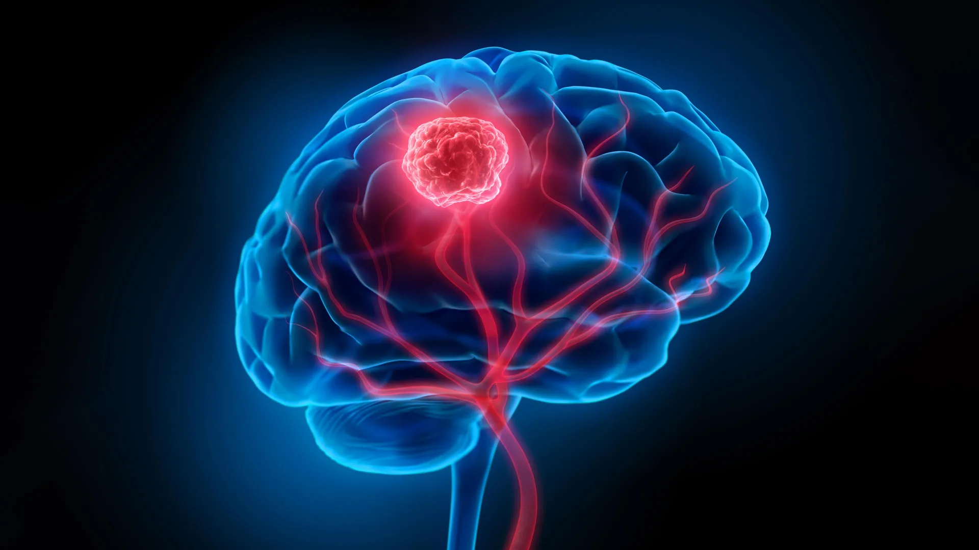
Glioblastoma, the most aggressive form of brain cancer, presents a formidable challenge to medical science due to its insidious nature and resistance to conventional treatments. Its diffuse growth pattern, where cancer cells infiltrate surrounding healthy brain tissue, complicates surgical intervention, making complete tumor eradication an exceptionally difficult feat. Surgeons are compelled to operate with extreme precision, balancing the removal of cancerous cells with the preservation of vital neurological functions. Compounding this surgical hurdle is the blood-brain barrier, a highly selective physiological shield that significantly impedes the delivery of therapeutic agents, including chemotherapy drugs and radiotherapy, to the tumor site. These combined obstacles contribute to the grim reality of glioblastoma, where the five-year survival rate hovers at a mere 7 percent, underscoring the urgent need for innovative treatment strategies.
In a significant stride towards overcoming these deeply entrenched challenges, researchers from the University of Technology Sydney (UTS), in collaboration with Harvard University and Henan University, have unveiled a groundbreaking ‘double-punch’ nanozyme platform. This sophisticated system of "smart" nanoparticles is engineered to address both the surgical and post-surgical treatment limitations of glioblastoma, employing a single, integrated technological solution. The pioneering findings of this research were recently published in the esteemed scientific journal, Science Translational Medicine, signaling a potential paradigm shift in the therapeutic landscape for this devastating disease.
A Two-Pronged Attack: Imaging and Targeted Therapy
The core innovation of this new platform lies in its ability to perform two distinct, yet complementary, functions sequentially, all activated by the same near-infrared light. This dual-action capability was articulated by Dr. Bingyang Shi, Chair Professor of Nanomedicine at UTS’s School of Electrical, Mechanical and Biomedical Engineering, who stated, "We’ve engineered a single material that does two jobs in sequence. It’s a precise guide for the surgeon during the operation, and then a targeted clean-up treatment afterward." This sequential approach promises to enhance the efficacy of treatment by improving both the identification of cancerous tissue during surgery and the elimination of residual microscopic disease.
At the heart of this revolutionary platform is an exceptionally thin, two-dimensional sheet composed of individual atoms meticulously arranged. This atomic-level precision in construction, adapted from advanced semiconductor manufacturing techniques, imbues the material with its remarkable versatility. This unique structure allows the nanoparticle to seamlessly transition between its two critical roles: serving as a highly sensitive imaging agent during surgical procedures and subsequently acting as a targeted therapeutic agent for phototherapy after the tumor has been surgically excised. The synchronized activation by near-infrared light simplifies the treatment protocol, ensuring that both functions can be deployed efficiently within the same operative and post-operative timeframe.
Illuminating the Invisible: Enhanced Surgical Guidance
During surgical operations, the nanoparticle platform functions as a sophisticated imaging agent, providing surgeons with unprecedented visualization capabilities. "During surgery, it functions as a highly sensitive imaging agent," explained Professor Shi. "A fluorescent dye engineered onto the sheet glows under a near-infrared wavelength invisible to the naked eye, allowing surgeons to see individual tumor cell clusters as small as 44 micrometers, a resolution beyond current clinical imaging tools." This remarkable resolution means that even minuscule clusters of glioblastoma cells, which often evade detection by standard imaging modalities, can be precisely identified.
Furthermore, the nanoparticle is equipped with a targeting molecule that facilitates its passage across the formidable blood-brain barrier. This targeted approach ensures that the nanoparticles preferentially accumulate within glioma cells, minimizing off-target effects and maximizing the concentration of the imaging agent at the tumor site. By enabling surgeons to visualize and resect these previously undetectable microscopic foci of cancer, the platform aims to significantly reduce the likelihood of leaving behind cancerous remnants that could lead to tumor recurrence. This enhanced intraoperative visualization is a critical advancement, directly addressing one of the most significant limitations in current glioblastoma surgery.
Eradicating Residual Disease: Post-Operative Phototherapy
Following the removal of the visible tumor, the same nanoparticle material can be reintroduced into the surgical cavity. Once in place, it is reactivated by the same wavelength of near-infrared light. This activation triggers the therapeutic phase of the platform, designed to eliminate any microscopic cancer cells that may have escaped surgical removal. "After the visible tumor is removed, the same material is administered into the surgical cavity and reactivated with the same wavelength of light for postoperative phototherapy," Professor Shi elaborated.
The therapeutic mechanism involves the platinum atoms embedded within the nanoparticle. These atoms play a crucial role by converting the tumor’s inherent hydrogen peroxide into oxygen. This conversion is particularly significant because glioblastoma tumors often create a hypoxic (low-oxygen) microenvironment, which typically shields cancer cells from treatment and promotes their survival and proliferation. By increasing oxygen levels, the nanoparticles disrupt this protective shield. Simultaneously, the near-infrared light triggers the generation of heat and reactive oxygen species (ROS), potent molecules that are highly destructive to cancer cells. This combination of oxygen generation, localized heat, and ROS production effectively targets and eradicates microscopic cancer cells that eluded surgical resection, thereby addressing the persistent threat of tumor recurrence.
Addressing the Relapse: Targeting Post-Surgical Remnants
The insidious nature of glioblastoma is particularly evident in its propensity for recurrence. Even after meticulous surgical removal of the macroscopic tumor, microscopic cancer cells often remain disseminated within the brain parenchyma. These residual cells serve as the seeds for future tumor growth, leading to the high recurrence rates associated with glioblastoma. The dual-function nanoparticle platform is specifically engineered to confront this critical challenge. By providing both enhanced visualization for more complete initial resection and a targeted post-operative therapy, it aims to significantly reduce the chances of the cancer returning.
The efficacy of this approach has been demonstrated in preclinical studies conducted on mouse models of glioblastoma. In these studies, mice treated with the nanoparticle platform following surgery exhibited a marked reduction in tumor recurrence. Notably, all treated mice survived for 60 days post-surgery, a significant improvement compared to the control group that received surgery alone, where the median survival was 42 days. Furthermore, follow-up neurological and motor assessments revealed no detectable impairments associated with the nanoparticle treatment, suggesting a favorable safety profile in these animal models. These results are highly encouraging and provide strong preclinical validation for the platform’s therapeutic potential.
A Glimmer of Hope, Grounded in Early Research
While the research findings are undeniably promising, the scientists involved are keen to emphasize the early stage of this development. Professor Shi cautioned, "The results are very encouraging, but this is still early-stage research carried out in mouse models, not in people – and that distinction is important." The transition from animal studies to human clinical trials is a rigorous and lengthy process, involving extensive safety and efficacy testing. The performance of the nanoparticle platform in terms of both its imaging and therapeutic capabilities will need to be rigorously evaluated at the scale and complexity of a human brain.
The research team is optimistic about the future potential of this technology. "If this continues to hold up through that process, the hope is that surgeons could one day see more of the tumor during an operation and treat more of what’s left behind afterward," Professor Shi stated. "It’s a meaningful step towards reducing recurrence, which remains one of the biggest challenges for people with glioblastoma." The ultimate goal is to translate these laboratory findings into a tangible clinical benefit for patients, offering a more effective strategy to manage and potentially overcome this devastating cancer.
Broader Implications and Future Directions
The development of this dual-function nanozyme platform represents a significant advancement in nanomedicine and its application to oncology. The ability to integrate diagnostic and therapeutic capabilities into a single, targeted system holds immense promise for a range of complex diseases. For glioblastoma, the implications are profound. By enhancing surgical precision and effectively clearing residual disease, this technology could potentially extend survival times, improve quality of life for patients, and ultimately contribute to a more optimistic prognosis.
The success of this platform could pave the way for similar dual-function nanotechnologies in treating other challenging cancers that are characterized by diffuse spread and resistance to therapy. The modular nature of the nanoparticle design, which allows for the incorporation of different targeting molecules and therapeutic payloads, suggests a flexible framework for future innovation.
However, significant hurdles remain. The scalability of manufacturing these atomically precise nanoparticles, ensuring their long-term stability and biocompatibility in humans, and navigating the complex regulatory pathways for novel medical devices and therapies are all critical considerations. Nevertheless, the research published in Science Translational Medicine marks a pivotal moment, providing a compelling proof-of-concept that could fundamentally alter the approach to treating glioblastoma and inspire further research into the synergistic application of imaging and therapy at the nanoscale. The journey from laboratory bench to patient bedside is long, but this innovative nanoparticle platform offers a beacon of hope in the ongoing fight against one of the most aggressive forms of cancer.
