
 By Nana O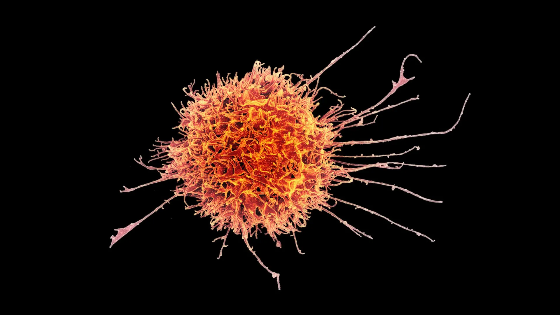
Cellular immunotherapies, a revolutionary approach that leverages the body’s own immune system to fight disease, have profoundly transformed the treatment landscape for certain hematological malignancies and lymphatic cancers. These "living drugs" have demonstrated remarkable efficacy, offering new hope and durable remissions for patients previously facing limited options. However, the battle against solid tumors—which constitute the vast majority of cancer diagnoses—remains a formidable challenge. Solid tumors present a complex biological fortress, characterized by dense cellular structures that impede the infiltration of immune cells and microenvironments that actively suppress immune responses. This inherent resistance has largely shielded them from the potent effects seen with blood cancers.
Now, a significant breakthrough from researchers at Stanford Medicine, in collaboration with scientists from Ohio State University and Washington University School of Medicine, offers a compelling new strategy to overcome these long-standing obstacles. Published last month in the prestigious journal Science Translational Medicine, their pioneering work details the successful development of a method to engineer natural killer (NK) cells into a specialized, tissue-resident form. These modified NK cells are designed to possess enhanced capabilities to penetrate solid tumors and mount a potent destructive attack against cancer cells, potentially paving the way for a new generation of broadly accessible cancer therapies.
Engineering the Next Generation of Cancer Fighters
The core of this innovation lies in transforming conventional NK cells, a crucial component of the innate immune system known for its rapid response to abnormal cells, into a specialized variant that can effectively reside within and navigate the challenging terrain of solid tumors. John Sunwoo, MD, the Edward C. and Amy H. Sewall Professor in the School of Medicine and senior author of the study, highlighted the remarkable effectiveness of this engineered approach. "We show that these tissue-resident natural killer cells infiltrate into the solid tumors much better than conventional natural killer cells," Dr. Sunwoo stated. "It was very reproducible, very striking and very clear."
The study’s co-lead authors, Nina Horowitz, PhD, a former doctoral student in otolaryngology; Imran Mohammad, PhD, a postdoctoral fellow in the Sunwoo lab; and June Ho Shin, PhD, a senior scientist in the Sunwoo lab, were instrumental in unraveling the complexities of NK cell differentiation and function. Their meticulous research efforts have culminated in a strategy that not only enhances anti-tumor activity but also addresses a critical logistical challenge in cell therapy development.
Promising Preclinical Results Against Solid Tumors
Initial testing of this experimental therapy in preclinical mouse models has yielded highly encouraging results. The modified NK cells demonstrated a significant ability to slow the growth of a variety of solid tumor types. This effect was further amplified when the engineered NK cells were administered in conjunction with an antibody treatment. This combination approach leverages monoclonal antibodies, such as cetuximab, which act as a molecular beacon, helping to guide NK cells directly to cancer cells and enhancing their targeting precision.
The therapeutic potential of these modified NK cells extends beyond their direct anti-tumor activity. A key practical advantage identified by the research team is their potential for allogeneic transplantation, meaning they could be derived from healthy donors rather than requiring individual extraction and modification from each patient. Unlike most current immune cell therapies, which necessitate personalized manufacturing processes—a time-consuming and costly endeavor—a treatment based on these engineered NK cells could potentially be produced in large batches, cryopreserved, and made readily available. This "off-the-shelf" model could dramatically reduce manufacturing bottlenecks and accelerate patient access to advanced cellular therapies.
"It would be almost an off-the-shelf drug," Dr. Sunwoo elaborated. "It could make cell therapy much more accessible to a wider variety of patients." This vision addresses a significant disparity in cancer care, where cutting-edge treatments are often limited by manufacturing capacity and cost.
The Significance of Tissue-Resident Immune Cells
Natural killer (NK) cells, first identified in the 1970s, are a cornerstone of the innate immune system. Their name aptly describes their primary function: to rapidly identify and eliminate abnormal cells, including those that have become cancerous or infected by viruses. A key distinguishing feature of NK cells, compared to other lymphocytes like B cells and T cells, is their pre-programmed ability to recognize and act against target cells without prior sensitization or antigen presentation. This intrinsic readiness allows for immediate responses to threats.
Historically, immunological research has predominantly focused on immune cells circulating within the bloodstream, as these cells are readily accessible for study and are known to patrol the body for signs of infection and disease. However, a growing body of evidence has underscored the critical importance of immune cells that take up residence within specific tissues. These "tissue-resident" immune cells adapt their functions to the unique microenvironment of their host tissue, playing specialized roles in local immunity, tissue homeostasis, and repair.
"For a long time, the study of immunology and disease in humans was concentrated on the blood immune cells," Dr. Sunwoo explained. "With the advancement of tools and bioinformatics, we are now starting to look more at what’s going on in tissue. For most immune cells, the tissue is where the action is." This paradigm shift in understanding highlights the critical role of tissue-resident immune cells in maintaining health and fighting disease.
Tissue-resident NK (TRNK) cells are found in various tissues, including the skin, mucosal linings of organs like the lungs and gastrointestinal tract, and the liver. Their precise functions have been a subject of ongoing scientific inquiry, with early studies yielding somewhat conflicting results. Some research suggested these cells might possess weaker cytotoxic capabilities or even play a role in immune suppression, while others indicated potent tumor-killing activity. The Stanford team’s work suggests that these apparent discrepancies may stem from the existence of distinct subpopulations of TRNK cells, each with unique functional profiles dictated by their local microenvironment.
"They may adopt different functions based on certain cues in the microenvironment and in the tissue, and differentiate into a certain kind of sub-population," Dr. Sunwoo observed. This suggests a remarkable plasticity in NK cell biology, allowing them to adapt to specific physiological or pathological contexts. In certain situations, such as during early pregnancy, immune-suppressing TRNK cells in the uterine lining are essential for preventing the maternal immune system from rejecting the fetus and for supporting placental development. However, in the context of cancer, the aggressive, cytotoxic form of TRNK cell is what is needed.
Unraveling the "Cellular Recipe" for Potent NK Cells
The researchers hypothesized that the varying functions of TRNK cells were linked to distinct developmental pathways. To investigate this, they isolated circulating NK cells from healthy human blood donors and subjected them to a carefully controlled series of signaling cues. A critical factor identified in this process was transforming growth factor beta (TGF-β), a signaling protein ubiquitous in many tissues and known to influence cell development. However, the precise role of TGF-β in generating effective anti-tumor NK cells proved to be a delicate balance.
"It’s a Goldilocks kind of thing where if you give just enough of a TGF-b signal, then the natural killer cells become tissue resident with strong toxic activity against malignant cells," Dr. Sunwoo elucidated. "If you give too much TGF-b, they’re still tissue resident, but they’re inhibited and dysfunctional, and they don’t kill." This observation underscores the complexity of cellular differentiation, where the dose and duration of signaling molecules are paramount. "You need it to be presented to the natural killer cells in just the right amount and in just the right manner."
While TGF-β was essential for inducing a tissue-resident phenotype, prolonged exposure led to NK cells with diminished killing capacity. A more effective approach emerged from mimicking the transient nature of certain tissue interactions. The researchers discovered that briefly exposing NK cells to short-lived human epithelial tumor cells, which naturally secrete a temporary burst of active TGF-β, successfully generated TRNK cells with potent tumor-killing activity.
Furthermore, direct physical contact between the NK cells and the epithelial tumor cells was found to be crucial, suggesting that additional activating signals are transmitted through cell-to-cell interactions, beyond the soluble TGF-β signal alone. "These two tissue-resident natural killer cell populations look very similar, and they have some of the same requirements, but their function seems to be on opposite ends of the spectrum," Dr. Sunwoo noted, emphasizing the distinct functional divergence achieved through this fine-tuned differentiation process.
Identifying the Molecular Signatures of Aggressive Killers
To further differentiate the potent anti-cancer TRNK cells from their less effective counterparts, the research team conducted a detailed molecular analysis. Both types of TRNK cells expressed characteristic surface proteins, CD49a and CD103, which are indicative of a tissue-resident phenotype. However, a key distinguishing marker for the highly effective cancer-killing cells was the expression of CD39.
Beyond surface markers, the superior cytotoxic cells also exhibited a significantly greater abundance of the molecular machinery required for cell killing. This included higher levels of perforin, a protein that forms pores in the membranes of target cells, and granzyme A, a potent enzyme delivered through these pores to induce apoptosis (programmed cell death). This molecular profiling provided a clear biological basis for the enhanced tumor-icidal capabilities of the engineered cells.
Translating Preclinical Success to Clinical Promise
With a reliable method established for generating these potent cytotoxic TRNK cells, the team proceeded to evaluate their efficacy in more complex tumor models. In laboratory experiments, the modified cells demonstrated an impressive ability to infiltrate tumor organoids grown in vitro. When administered to mice bearing various solid tumors, including those derived from human melanoma and head and neck squamous cell carcinoma, the engineered NK cells significantly slowed tumor progression over periods of days and weeks.
The most profound anti-tumor effects were observed when the modified NK cells were combined with cetuximab, a well-established monoclonal antibody used in the treatment of certain head and neck cancers and colorectal cancers. Cetuximab works by binding to the epidermal growth factor receptor (EGFR) on cancer cells, marking them for destruction by the immune system and inhibiting tumor growth signals. While cetuximab alone has moderate efficacy, its synergy with the engineered NK cells was striking.
A single dose of this combination therapy demonstrated a substantially greater suppression of tumor growth in mice over a one-month period compared to either treatment administered independently. Crucially, the researchers did not observe any apparent adverse effects associated with this combination, suggesting a favorable safety profile in preclinical models. "Even at day 30, when the other mice were sick, the mice that received the combination seemed very healthy," Dr. Sunwoo remarked. He tempered this optimism with a necessary caution, emphasizing that "This was just proof of concept," and that extrapolating results directly from animal models to human patients requires rigorous clinical validation.
Paving the Way for an "Off-the-Shelf" Cell Therapy
Building on these compelling preclinical findings, Dr. Sunwoo and his colleagues are actively preparing for a Phase I clinical trial to evaluate the safety and preliminary efficacy of this combination therapy in human patients. The trial is slated to focus on individuals with advanced squamous cell carcinoma and could commence by the end of the year, pending approval from the U.S. Food and Drug Administration (FDA).
Beyond the clinical trial, Dr. Sunwoo has also spearheaded the development and patent application for a method to produce and expand large quantities of these specialized cytotoxic tissue-resident NK cells. This scalable manufacturing process is key to realizing the vision of an "off-the-shelf" therapy. According to the researchers, NK cells harvested from a single healthy donor could potentially yield approximately 20 doses of treatment within a two-week timeframe.
"They’ll be cryopreserved, so we can make a bunch of doses and give it to different patients," Dr. Sunwoo stated, highlighting the logistical advantages. "There would be no delay." This advancement has the potential to democratize access to cutting-edge cell therapy, making it a viable option for a broader patient population and significantly reducing the waiting times often associated with personalized treatments. The implications for global cancer care are substantial, potentially offering a more equitable and efficient pathway to advanced immunotherapy.
