
 By George Ongkojoyo
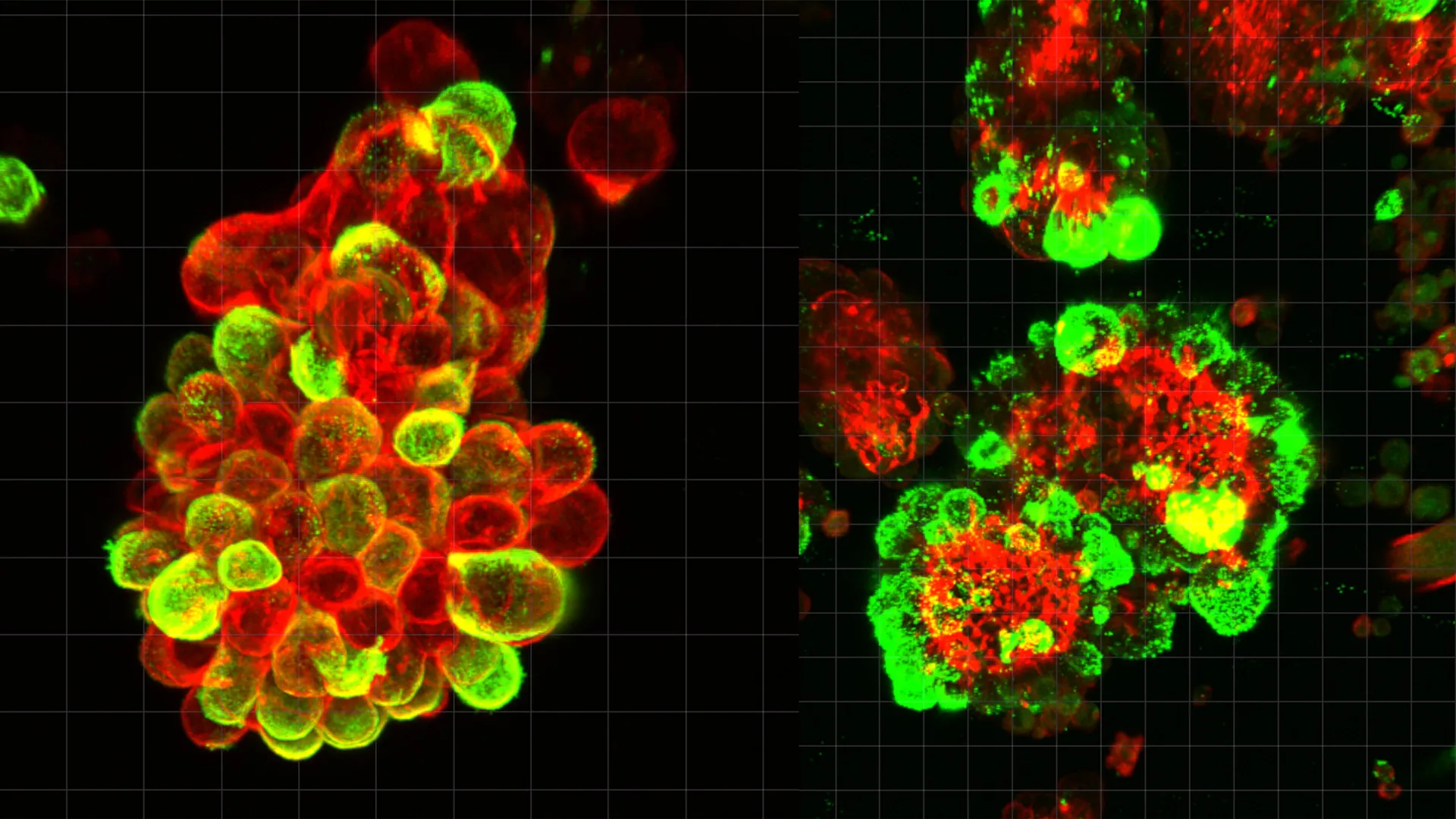
Ovarian cancer has long been characterized by its stealth and its speed, earning a reputation as one of the most formidable challenges in modern oncology. For decades, the medical community has struggled to understand why this specific malignancy spreads with such aggressive efficiency throughout the abdominal cavity, often appearing in advanced stages despite regular medical checkups. A groundbreaking study led by researchers at Nagoya University in Japan has finally provided a definitive biological explanation for this phenomenon. The research, recently published in the prestigious journal Science Advances, reveals that ovarian cancer cells do not operate in isolation; instead, they hijack healthy mesothelial cells to act as "pioneers" that drill through tissue, creating pathways for the cancer to follow and forming hybrid clusters that are significantly more resistant to traditional chemotherapy.
The Silent Progression of Ovarian Malignancy
Ovarian cancer remains the leading cause of death among gynecological cancers globally. According to the World Health Organization and various oncological databases, the five-year survival rate for ovarian cancer drops precipitously when the disease is diagnosed at Stage III or IV, which is the case for the vast majority of patients. The primary reason for this high mortality rate is the "silent" nature of the disease’s progression. Unlike other cancers that may produce localized pain or detectable lumps early on, ovarian cancer often presents with vague symptoms such as bloating or abdominal discomfort, which are frequently dismissed by both patients and clinicians.
The Nagoya University study addresses the central mystery of how these cancer cells transition from a localized tumor to a widespread abdominal infection so rapidly. While most cancers, such as breast or lung cancer, typically utilize the circulatory or lymphatic systems to metastasize to distant organs, ovarian cancer employs a unique method of dissemination. It utilizes the peritoneal fluid—the lubricating liquid found within the abdominal cavity—to drift toward new sites.
The Role of Mesothelial Cells in Cancer Dissemination
The research team, spearheaded by Dr. Kaname Uno, discovered that the key to this rapid spread lies in the interaction between cancer cells and mesothelial cells. Mesothelial cells are a specialized type of epithelial cell that forms the mesothelium, a protective membrane lining the lungs, heart, and abdominal cavity. Under normal circumstances, these cells provide a smooth, slippery surface that allows organs to move against one another without friction.
However, the study found that when ovarian cancer cells break away from the primary tumor and enter the abdominal fluid, they actively seek out and "recruit" these mesothelial cells. By analyzing abdominal fluid (ascites) from human patients, the researchers observed that cancer cells were rarely floating as individual units. Instead, they were found in compact, spherical clusters. Approximately 60% of these spheres were "hybrid clusters," composed of both malignant cancer cells and recruited healthy mesothelial cells.
The Molecular Mechanism: TGF-β1 and the Creation of Invadopodia
The mechanism behind this recruitment is a sophisticated form of molecular manipulation. The study identified that ovarian cancer cells secrete a specific signaling protein known as Transforming Growth Factor-beta 1 (TGF-β1). When this protein reaches the mesothelial cells, it triggers a transformation in the healthy cells’ behavior and morphology.
Under the influence of TGF-β1, the mesothelial cells develop "invadopodia." These are actin-rich, spike-like protrusions on the cell surface that function essentially as biological drills. The research demonstrated that these hybrid clusters use the mesothelial cells as a vanguard. The mesothelial cells, equipped with invadopodia, lead the way by physically penetrating the protective lining of abdominal organs. The cancer cells then follow through the breaches created by their healthy "accomplices," allowing the malignancy to take root in new locations with minimal effort.
This discovery challenges the traditional view of cancer metastasis, which assumes that cancer cells must undergo extensive genetic mutations to become invasive themselves. Dr. Uno’s research suggests that ovarian cancer cells effectively "outsource" the hard work of tissue invasion to the mesothelial cells, allowing the cancer cells to remain relatively unchanged while still spreading aggressively.
A Comparative Analysis of Metastatic Pathways
The study highlights a critical distinction between ovarian cancer and other common malignancies. In cancers that spread through the bloodstream (hematogenous metastasis), the path is somewhat predictable based on blood flow patterns. This allows for the development of liquid biopsies that can detect circulating tumor cells (CTCs) in a patient’s blood.
Ovarian cancer’s reliance on the peritoneal fluid (peritoneal dissemination) makes it far more difficult to track. The fluid within the abdomen is in constant, unpredictable motion, driven by the diaphragm’s movement during breathing and the peristaltic motion of the intestines. This "floating phase" allows cancer clusters to reach any organ within the cavity—including the liver, stomach, and intestines—without ever entering the bloodstream. The Nagoya University study provides the first clear look at what happens during this floating phase, showing that the formation of hybrid spheres is a prerequisite for successful colonization of new tissue.
Resistance to Chemotherapy and Clinical Implications
One of the most concerning findings of the study is the impact these hybrid clusters have on treatment efficacy. The researchers conducted comparative tests between pure cancer cell clusters and the hybrid cancer-mesothelial clusters. The results showed that the hybrid spheres were significantly more resilient when exposed to standard chemotherapy drugs.
This resistance is believed to be a result of the structural integrity and the biological signaling within the hybrid cluster. The mesothelial cells appear to provide a protective environment for the cancer cells, shielding them from the cytotoxic effects of medication. This explains why many ovarian cancer patients initially respond well to chemotherapy—which kills off the less-protected, individual cancer cells—only to suffer a rapid relapse as the resistant hybrid clusters continue to grow and invade.
The Human Element: From the Clinic to the Laboratory
The impetus for this research was deeply personal for the lead author. Before becoming a researcher at Nagoya University’s Graduate School of Medicine, Dr. Kaname Uno spent eight years as a practicing gynecologist. His transition to full-time research was sparked by a specific clinical case that highlighted the limitations of current diagnostic technology.
Dr. Uno recounted the story of a patient who had undergone a routine gynecological screening and received a clean bill of health. Only three months later, she returned with advanced, Stage IV ovarian cancer. Despite the best available medical intervention, the speed at which the cancer had colonized her abdomen was faster than the diagnostic and treatment tools available. This experience drove Dr. Uno to investigate the "why" behind the speed of ovarian cancer spread, eventually leading to the discovery of the mesothelial recruitment mechanism.
Methodology: Real-Time Observation and Genetic Analysis
To validate their findings, the Nagoya University team employed a multi-disciplinary approach. They utilized advanced live-cell imaging and microscopy to watch the formation of hybrid clusters in real-time using samples taken directly from patients. This was complemented by experiments using mouse models, which confirmed that the presence of mesothelial cells significantly increased the rate of tumor implantation and growth.
Furthermore, the team performed single-cell RNA sequencing to analyze gene activity within the clusters. This allowed them to pinpoint the exact molecular pathways being activated by TGF-β1 and confirm that the mesothelial cells were indeed the ones expressing the genes necessary for tissue degradation and invasion.
Future Outlook: New Frontiers in Treatment and Diagnostics
The implications of this study for the future of oncology are profound. By identifying the mesothelial cell as a critical "enabler" of ovarian cancer spread, the research opens up several new avenues for therapeutic intervention:
- Targeting Signaling Pathways: Future drugs could be developed to specifically block the TGF-β1 signal, preventing cancer cells from being able to "recruit" or transform healthy mesothelial cells.
- Disrupting Cluster Formation: If therapies can be designed to prevent the adhesion of cancer cells to mesothelial cells, the cancer might remain in a "floating" state where it is more vulnerable to the body’s immune system or standard chemotherapy.
- Enhanced Monitoring: The study suggests that analyzing the composition of abdominal fluid could serve as a new diagnostic tool. By monitoring the ratio of hybrid clusters in the ascites, doctors could better predict a patient’s prognosis and tailor treatment plans more effectively.
- Redefining Chemotherapy: Current treatments focus almost exclusively on the malignant cells. This research suggests that a dual-target approach—treating both the cancer and the supporting stromal cells (like the mesothelial cells)—may be necessary to overcome chemoresistance.
Conclusion
The work of Dr. Uno and his colleagues at Nagoya University marks a significant turning point in our understanding of gynecological oncology. By revealing that ovarian cancer’s lethality is rooted in its ability to manipulate the body’s own protective cells, the study moves the medical community closer to developing targeted therapies that can finally outpace this "silent killer." As researchers continue to explore the complex relationship between tumors and their microenvironment, the discovery of the mesothelial-cancer hybrid cluster stands as a testament to the importance of looking beyond the cancer cell itself to understand the full scope of the disease.
