
 By George Ongkojoyo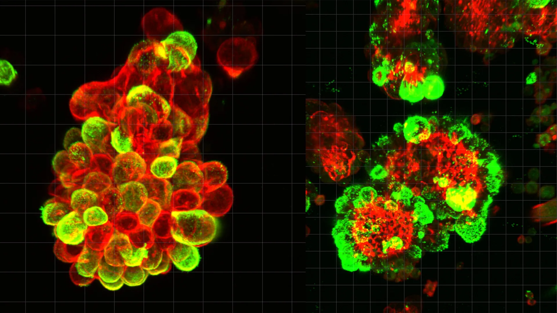
Ovarian cancer has long been classified by oncologists as one of the most formidable challenges in women’s healthcare, primarily due to its reputation as a "silent killer" that evades detection until it has reached an advanced stage. For decades, the medical community has struggled to understand why this specific malignancy spreads with such devastating speed once it enters the abdominal cavity. A groundbreaking study led by a research team at Nagoya University in Japan has finally provided a molecular explanation for this phenomenon. Published in the prestigious journal Science Advances, the research reveals that ovarian cancer cells do not operate in isolation; rather, they hijack healthy mesothelial cells to serve as "pathfinders" and "bodyguards," creating hybrid clusters that are significantly more aggressive and resistant to traditional treatments than independent cancer cells.
The Biological Mystery of Rapid Ovarian Metastasis
Ovarian cancer is the leading cause of death from gynecological malignancies globally. Unlike breast or lung cancers, which typically spread through the lymphatic system or the bloodstream—a process known as hematogenous metastasis—ovarian cancer utilizes a unique mechanism called transcoelomic spread. In this process, cancer cells detach from the primary tumor on the ovary or fallopian tube and enter the peritoneal fluid, the lubricating liquid found within the abdominal cavity.
Until now, the scientific consensus suggested that these floating cancer cells eventually adhered to distant organs by chance or through individual cellular mutations. However, this did not account for the sheer velocity of the disease’s progression. The Nagoya University study, led by Dr. Kaname Uno, demonstrates that the process is far more coordinated. The research indicates that cancer cells actively recruit mesothelial cells—the cells that form the protective lining of the abdomen—and transform them into accomplices that facilitate the invasion of healthy tissue.
The Mechanism of Recruitment: TGF-β1 and the Formation of Hybrid Spheres
The core of the discovery lies in how cancer cells manipulate their environment while suspended in abdominal fluid. By analyzing samples from human patients, the Nagoya team observed that cancer cells were rarely found drifting alone. Instead, they were encased in compact, spherical clusters alongside mesothelial cells.
The researchers discovered that ovarian cancer cells secrete a specific signaling protein known as Transforming Growth Factor-beta 1 (TGF-β1). When mesothelial cells, which naturally shed into the abdominal fluid due to inflammation or normal physiological turnover, encounter this protein, they undergo a radical transformation. Under the influence of TGF-β1, these once-protective cells develop "invadopodia"—sharp, spike-like protrusions composed of actin filaments.
These invadopodia act as biological drill bits. When a hybrid sphere lands on the surface of an organ, such as the liver, intestine, or omentum, the recruited mesothelial cells use these spikes to puncture and degrade the extracellular matrix of the host tissue. This creates a pathway through which the cancer cells can easily flow and establish new colonies. Data from the study suggests that approximately 60% of all observed cancer cell clusters in the peritoneal fluid of patients contained these hijacked mesothelial cells, highlighting the ubiquity of this survival strategy.
A Shift in the Metastatic Paradigm: From Bloodstreams to Abdominal Fluid
One of the most significant implications of this research is the distinction it draws between ovarian cancer and other major cancers. In many malignancies, doctors can utilize "liquid biopsies" to track circulating tumor cells (CTCs) in the blood. Because the circulatory system follows a fixed and predictable map of vessels, the spread of these cancers can sometimes be monitored or intercepted.
Ovarian cancer, however, bypasses the blood-vessel highway. The abdominal fluid in which it travels moves unpredictably, influenced by the mechanical motions of the diaphragm during breathing and the peristaltic movements of the digestive tract. This creates a "chaotic" environment where cancer cells can be deposited anywhere in the abdomen within a very short timeframe.
The Nagoya University study clarifies what occurs during this "floating phase." By forming hybrid spheres, the cancer cells are not merely waiting to land; they are actively preparing for invasion while in transit. Furthermore, these clusters provide a collective defense mechanism. The study found that the physical structure of the hybrid sphere, combined with the altered gene expression of the mesothelial cells, makes the cluster more resistant to chemotherapy. This explains why many patients experience a recurrence of the disease even after undergoing aggressive cycles of platinum-based chemotherapy.
The Human Element: Dr. Kaname Uno’s Path from Clinic to Laboratory
The impetus for this research was rooted in clinical tragedy. Dr. Kaname Uno, the lead author of the study, served as a practicing gynecologist for eight years before transitioning into full-time molecular research. During his time in the clinic, he treated a patient whose case became a catalyst for his scientific inquiry.
The patient had undergone a routine gynecological screening and received a clean bill of health. However, only three months later, she returned with severe symptoms and was diagnosed with Stage IV ovarian cancer that had already disseminated throughout her abdomen. The speed of the progression was so great that existing medical interventions were powerless.
"She had been told she was healthy just ninety days prior," Dr. Uno recalled. "That experience stayed with me. It was clear that our current understanding of how this cancer moves was incomplete, and our diagnostic tools were looking for the wrong things."
Driven by this experience, Dr. Uno joined the Graduate School of Medicine at Nagoya University to investigate the "floating stage" of ovarian cancer. His team utilized advanced live-cell imaging and single-cell RNA sequencing to watch the interaction between cancer and mesothelial cells in real-time, confirming that the cancer cells themselves remain relatively "passive" while the hijacked mesothelial cells perform the labor of tissue penetration.
Supporting Data and Methodology
The Nagoya University study employed a multi-faceted approach to validate its findings. The researchers utilized:
- Patient-Derived Samples: Analysis of ascites (abdominal fluid buildup) from patients with advanced ovarian cancer confirmed the presence of TGF-β1 and the prevalence of hybrid cell spheres.
- Mouse Models: Researchers injected hybrid spheres into mice and compared the rate of metastasis against a control group injected with pure cancer cell clusters. The hybrid spheres demonstrated a significantly higher rate of successful organ colonization and a faster rate of growth.
- Single-Cell Level Analysis: By examining the genetic activity of individual cells within the clusters, the team identified the specific pathways activated by TGF-β1, proving that the mesothelial cells were being genetically "reprogrammed" by the cancer.
- Advanced Microscopy: Using high-resolution time-lapse microscopy, the team captured the exact moment invadopodia from mesothelial cells began to penetrate a simulated peritoneal lining.
The data indicated that when mesothelial cells were present, the "invasion index"—a measure of how effectively cells move into a substrate—increased by more than twofold compared to cancer cells acting alone.
Implications for Future Oncology and Treatment
The discovery of the "mesothelial accomplice" opens several new doors for the treatment of ovarian cancer, which has seen relatively few breakthroughs in survival rates over the last two decades.
1. Targeting the Signal, Not Just the Cell:
Current chemotherapy regimens are designed to kill rapidly dividing cells. However, if the mesothelial cells are the ones driving the invasion, killing the cancer cells alone may not be enough to stop the spread. Future therapies could focus on inhibiting the TGF-β1 signaling pathway, essentially "deafening" the mesothelial cells so they do not respond to the cancer’s recruitment signals.
2. Disrupting the Hybrid Spheres:
If drugs can be developed to prevent the adhesion of cancer cells to mesothelial cells, the cancer cells would remain in a less aggressive, solitary state, making them more vulnerable to the body’s immune system and standard chemotherapy.
3. Enhanced Diagnostic Monitoring:
The study suggests that monitoring the ratio of hybrid spheres in a patient’s abdominal fluid could serve as a predictive marker for how aggressive the cancer is likely to be. This could allow for more personalized treatment plans, where patients with high levels of hybrid clusters receive more intensive or specialized interventions.
Conclusion: A New Frontier in Gynecological Research
Ovarian cancer remains a global health crisis, with the World Health Organization estimating nearly 300,000 new cases annually and a five-year survival rate that hovers below 50% for late-stage diagnoses. The findings from Nagoya University represent a paradigm shift in how scientists view the "peritoneal microenvironment."
By shifting the focus from the cancer cell as a lone actor to the cancer cell as a master manipulator of its environment, Dr. Uno and his colleagues have provided a new roadmap for drug development. The study underscores the importance of the "floating stage" of cancer, suggesting that the battle against metastasis is won or lost not just at the site of the tumor, but in the fluid spaces between organs.
As the medical community moves toward "precision medicine," the ability to intercept the formation of these hybrid cell clusters could eventually turn ovarian cancer from a rapid and unpredictable killer into a manageable condition. For now, the research serves as a poignant reminder of the power of clinical observation—where a single patient’s story can lead to a discovery that may eventually save thousands of lives.
