
 By Gabriel Paijo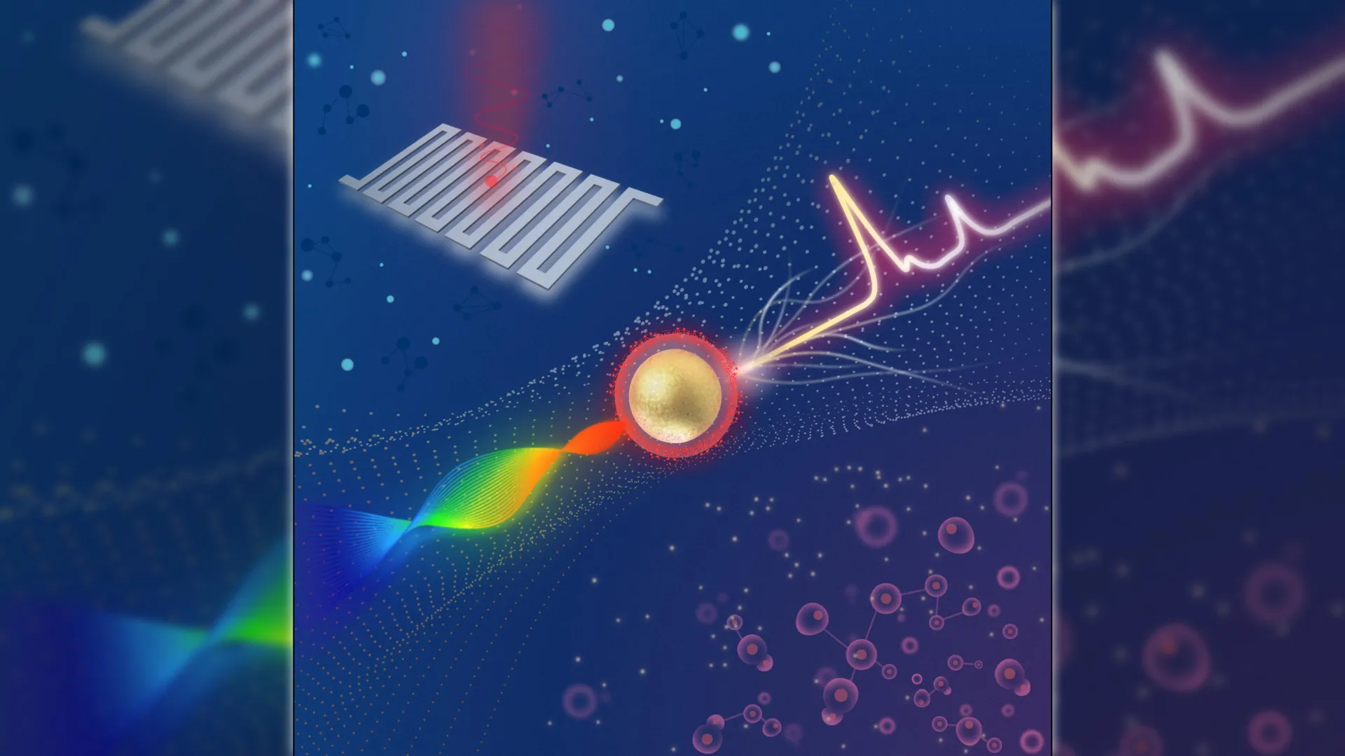
Researchers at Michigan State University have unveiled a significant breakthrough in medical imaging technology by developing a compact Raman imaging system that utilizes advanced superconducting nanowire single-photon detectors (SNSPDs) to identify cancerous tissues with unprecedented precision. This innovation, led by the Institute for Quantitative Health Science and Engineering (IQ), represents a major step forward in the quest for real-time, non-invasive diagnostic tools that can differentiate between malignant and healthy cells at the molecular level. By combining high-sensitivity photon counting with surface-enhanced Raman scattering (SERS) nanoparticles, the system provides a detection threshold significantly lower than current commercial standards, potentially transforming the landscape of early cancer detection and intraoperative surgical guidance.
The core of this technological leap lies in the system’s ability to capture and process extremely weak optical signals that traditional imaging methods often miss. Conventional Raman spectroscopy, while highly specific in identifying chemical compositions, typically suffers from low signal intensity, making it difficult to use in fast-paced clinical environments. The new system overcomes this hurdle by integrating a swept-source laser architecture with ultra-sensitive SNSPDs, allowing for the detection of Raman signals that are approximately four times weaker than those measurable by the best comparable commercial systems currently on the market.
The Evolution of Molecular Imaging and the Raman Effect
To understand the significance of this development, it is necessary to examine the history and limitations of Raman spectroscopy in medicine. Discovered by C.V. Raman in 1928, the Raman effect occurs when light interacts with the chemical bonds of a molecule, resulting in a shift in the energy of the photons. This "fingerprint" allows scientists to identify substances with high specificity. However, because only about one in every ten million photons undergoes Raman scattering, the signal is inherently faint.
In recent decades, researchers have utilized Surface-Enhanced Raman Scattering (SERS) to amplify these signals. By using metallic nanoparticles—often gold or silver—the local electromagnetic field is intensified, boosting the Raman signal by several orders of magnitude. While SERS improved the feasibility of medical Raman imaging, the detection hardware remained a bottleneck. Traditional systems relied on bulky spectrometers and charge-coupled device (CCD) cameras, which required long exposure times and lacked the sensitivity needed for rapid, real-time clinical applications.
The Michigan State University team, led by Zhen Qiu, addressed these hardware limitations by reimagining the detection pipeline. By replacing traditional cameras with SNSPDs—devices typically used in quantum communication and deep-space optical tracking—the researchers have introduced a level of sensitivity that allows for the detection of individual photons with minimal background noise.
Technical Architecture: Swept-Source Lasers and Superconducting Detectors
The innovative system design departs from the static laser approach used in most Raman devices. Instead, it employs a swept-source laser that rapidly changes its wavelength during the analysis process. This movement, when synchronized with the SNSPD, allows the system to map the Raman spectrum without the need for a traditional, bulky spectrometer.
The SNSPD itself is a marvel of modern physics. It consists of a thin film of superconducting material, such as niobium nitride, cooled to cryogenic temperatures. When a single photon strikes the nanowire, it disrupts the superconducting state, creating a brief "hotspot" of electrical resistance. This change is recorded as a digital pulse. Because SNSPDs have near-zero dark count rates—meaning they rarely register "false" photons from heat or electronic noise—the signal-to-noise ratio is vastly superior to that of silicon-based detectors.
"Combining this advanced detector with a swept-source Raman architecture that replaces a bulky camera and collects light more efficiently resulted in a system with a detection limit well beyond that of comparable commercial systems," explained Zhen Qiu. The fiber-coupled configuration of the device also facilitates miniaturization, a critical factor for moving the technology from a laboratory benchtop to a clinical setting where space is at a premium.
Chronology of Development and Experimental Validation
The development of this imaging platform followed a rigorous multi-stage validation process. The research team first focused on engineering the SERS nanoparticles to ensure they could effectively target cancer biomarkers. They selected CD44, a cell-surface glycoprotein that is overexpressed in a wide variety of cancers, including breast, colon, and prostate tumors. The nanoparticles were coated with hyaluronan acid, which possesses a high affinity for the CD44 receptor.
The timeline of the project’s experimental phase progressed through three distinct tiers:
- Sensitivity Benchmarking: Initial testing involved simple nanoparticle solutions to determine the absolute detection limit. The system successfully achieved femtomolar (10^-15 moles per liter) sensitivity, confirming that it could detect incredibly sparse concentrations of markers.
- In Vitro Cell Studies: The system was then used to image cultured breast cancer cells. The high-sensitivity detectors were able to map the distribution of the nanoparticles across the cell surfaces, providing a clear visual contrast between cells expressing CD44 and those that were not.
- Ex Vivo and Animal Models: Finally, the researchers applied the platform to mouse tumor models and healthy tissue samples. The results showed that SERS signals were heavily concentrated within the tumor regions, while healthy tissues showed negligible background interference.
This progression demonstrated not only the sensitivity of the hardware but also the reliability of the biological targeting mechanism. The ability to distinguish between tumor and healthy tissue in complex biological environments is the primary requirement for any diagnostic tool intended for human use.
Supporting Data and Comparative Performance
The data published in the journal Optica highlights the system’s competitive edge. In head-to-head comparisons with high-end commercial Raman spectrometers, the MSU system demonstrated a fourfold improvement in signal detection capability. This means that in a clinical scenario where a tumor might be shedding very few markers, or where a surgeon is looking for microscopic "margins" of cancer at the edge of a resection, this system could identify threats that other machines would miss.
Furthermore, the integration of SNSPDs allows for high-speed data acquisition. Traditional Raman mapping can take minutes or even hours to scan a single square centimeter of tissue. The MSU team’s architecture aims to reduce this to a timeframe compatible with active surgery, where every second counts. The use of a swept-source laser also eliminates the need for post-processing steps like baseline subtraction and fluorescence quenching, which are often required in standard Raman spectroscopy to clean up the "noisy" data.
Clinical Context and the Path to Implementation
The current gold standard for cancer diagnosis remains histopathology—the process of taking a tissue biopsy, staining it with dyes (such as Hematoxylin and Eosin), and having a pathologist examine it under a microscope. While highly accurate, this process is inherently slow. It often takes days to receive results, and during surgery, "frozen section" analysis can still take 20 to 30 minutes, keeping the patient under anesthesia longer than necessary.
The Michigan State University system is not intended to replace the pathologist but to act as a "rapid screening tool." In a surgical context, it could provide a "heat map" of a surgical cavity, showing the surgeon exactly where the cancerous tissue ends and healthy tissue begins. This could significantly reduce the rate of "positive margins," where cancer cells are accidentally left behind, necessitating a second surgery.
Industry experts have reacted positively to the findings. Collaborators at Quantum Opus, the firm that provided the SNSPD devices, noted that the application of quantum-grade sensors to medical imaging is a burgeoning field. By proving that these sensors can operate effectively in a biological context, the MSU team has opened the door for a new generation of "quantum medicine."
Future Directions and Broader Impact
Despite the promising results, the transition to clinical use requires several more steps. The research team has outlined a roadmap for the next three to five years, focusing on:
- Miniaturization of Cooling Systems: Currently, SNSPDs require cryogenic cooling to reach their superconducting state. While compact cryocoolers exist, the team is working on further integrating these into a portable unit that can fit on a standard hospital equipment cart.
- Multiplexing Capabilities: The researchers plan to use different types of nanoparticles, each "tuned" to a different Raman frequency and targeting a different biomarker. This would allow the system to look for multiple types of cancer or different characteristics of a single tumor (such as its aggressiveness or its likely response to specific drugs) simultaneously.
- Speed Optimization: By exploring faster laser sources, such as Vertical-Cavity Surface-Emitting Lasers (VCSELs), the team hopes to achieve real-time video-rate Raman imaging.
- Human Clinical Trials: The team is preparing for larger-scale validation studies using human tissue samples obtained from surgical resections to ensure the system performs across diverse patient populations and cancer subtypes.
The implications of this technology extend beyond oncology. The ability to detect molecular markers with femtomolar sensitivity could be applied to infectious disease testing, neurodegenerative disease monitoring, and even environmental sensing.
Ultimately, the goal is to reduce the "diagnostic lag" that often dictates a patient’s prognosis. As Zhen Qiu noted, "Such advances could enhance patient outcomes and reduce diagnostic delays, accelerating the path from detection to treatment." By bridging the gap between high-end physics and practical medicine, the MSU team is paving the way for a future where cancer detection is faster, more accurate, and more accessible than ever before. This research marks a pivotal moment where quantum-level sensitivity meets the urgent needs of global healthcare, promising a more precise era of molecular medicine.
