
 By Lina carolina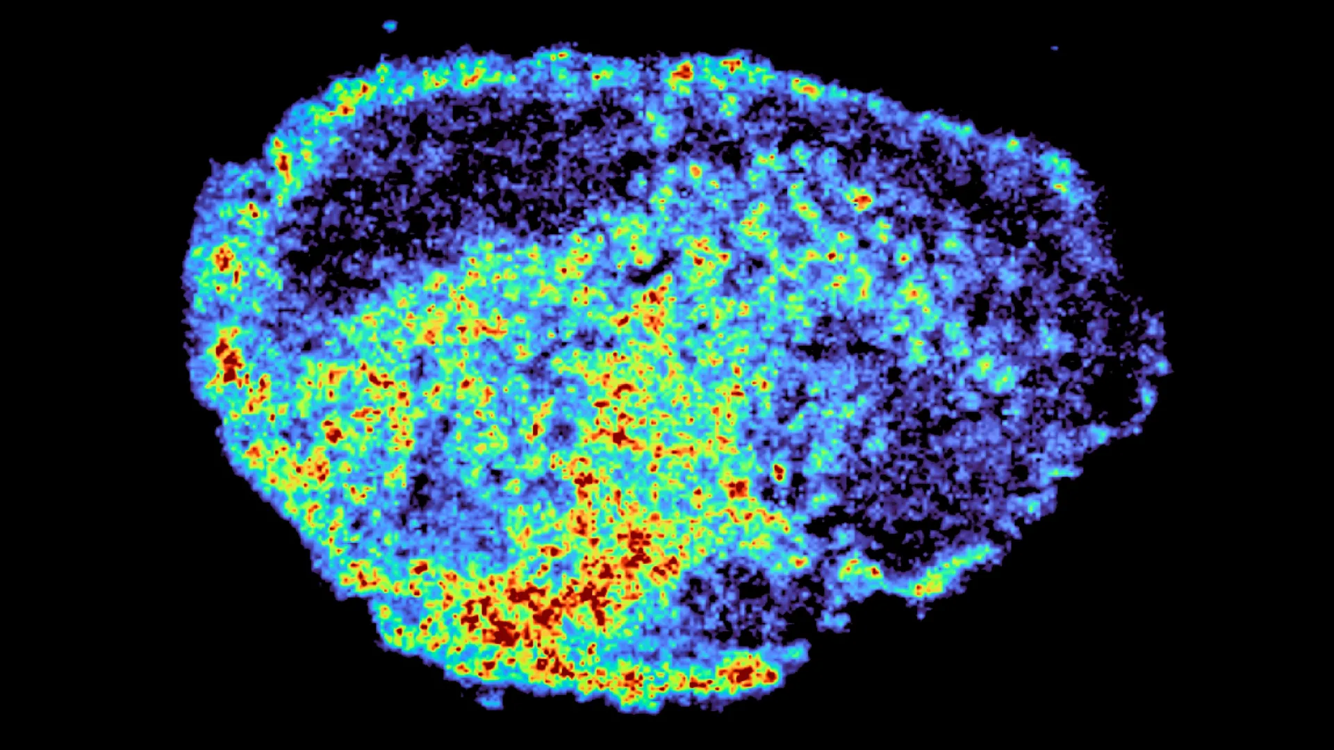
The persistent enigma of cancer therapy—why a single drug can be a life-saving breakthrough for one patient and a disappointing failure for another—is beginning to yield its secrets, thanks to groundbreaking research from the MRC Laboratory of Medical Sciences (LMS). A pivotal study, published in the prestigious journal Nature Communications, led by Dr. Louise Fets, has illuminated a critical mechanism influencing the effectiveness of PARP inhibitors, a vital class of targeted cancer drugs. By employing sophisticated imaging techniques to meticulously track the journey of these drugs within ovarian tumor samples, scientists have uncovered a surprising role for cellular "recycling centers" in dictating treatment outcomes.
The Uneven Landscape of Cancer Drug Distribution
The landscape of cancer treatment has been dramatically reshaped in recent years, offering unprecedented hope and improved prognoses for a multitude of patients. Among the most significant advancements has been the development of PARP inhibitors. These drugs have revolutionized the management of ovarian cancer, offering a potent weapon against a disease that has historically presented formidable challenges. However, the clinical reality is that not all patients respond uniformly. Some experience remarkable benefits, while others see their disease progress or develop resistance to the therapy over time.
For PARP inhibitors to exert their therapeutic effect, they must achieve and maintain sufficiently high concentrations within cancer cells to trigger programmed cell death, a process known as apoptosis. Despite this fundamental requirement, a comprehensive understanding of how these drugs are distributed throughout the complex architecture of a tumor, and the factors that govern this distribution at a cellular level, has remained elusive. This knowledge gap has been a significant hurdle in optimizing treatment strategies and overcoming acquired resistance.
The research spearheaded by Dr. Fets and her team directly addresses this deficit. Their findings underscore a critical paradigm shift: a drug’s efficacy is not solely determined by its ability to reach the tumor, but crucially, by its intricate distribution within the tumor mass and, even more granularly, inside individual cancer cells. To investigate this complex interplay, the researchers ingeniously utilized patient-derived ovarian tumor samples, meticulously maintained in a viable state ex vivo. These "explants" provided an authentic human tumor microenvironment, allowing scientists to directly observe and quantify the behavior of PARP inhibitors in real-time, as they navigated through native tumor tissue.
Mapping the Invisible: Advanced Imaging Reveals Drug Depots
The study’s methodology represented a significant leap forward in visualizing drug pharmacokinetics within solid tumors. By employing cutting-edge mass spectrometry imaging (MSI), the team generated highly detailed, spatially resolved maps. These maps precisely delineated regions of drug accumulation within the tumor explants, providing an unprecedented level of detail about drug localization. MSI works by ionizing molecules within a tissue sample and then analyzing the mass-to-charge ratio of the resulting ions, allowing for the identification and quantification of specific compounds at specific locations.
Complementing this powerful imaging technique, the researchers integrated spatial transcriptomics. This innovative approach allowed them to simultaneously examine gene expression patterns in areas identified as having high versus low drug concentrations within the same tissue slice. Spatial transcriptomics provides a snapshot of cellular activity, revealing which genes are active or inactive in distinct spatial locations. The synergistic application of MSI and spatial transcriptomics yielded striking insights, revealing profound heterogeneity in drug distribution. This variability was not only observed between different tumor samples from distinct patients but also within individual tumors, even when exposed to identical drug dosages.
"A novel aspect of this study was the use of mass spectrometry imaging to directly measure and visualize drug uptake in patient tumour tissue," stated Dr. Zoe Hall, a senior author on the study and Associate Professor at Imperial’s Department of Metabolism, Digestion and Reproduction. "Through the spatial mapping of drug molecules, we could pinpoint regions of high and low drug and compare gene expression, from the same tissue slice, using spatial transcriptomics." This quote highlights the pioneering nature of the imaging approach and its ability to correlate drug presence with cellular responses at a micro-spatial level.
Lysosomes: Unveiling Hidden Drug Reservoirs
The investigation’s most compelling revelation centered on the unexpected role of lysosomes, small, membrane-bound organelles within cells that are primarily known for their function as cellular "recycling centers." The researchers discovered that certain PARP inhibitors exhibit a strong affinity for these lysosomal compartments. Instead of diffusing evenly throughout the cytoplasm, these drugs were actively sequestered within lysosomes, effectively becoming trapped. This lysosomal accumulation created localized "hotspots" of drug concentration within the cells.
These findings suggest that lysosomes are not merely passive storage sites but rather function as dynamic, slow-release reservoirs. By holding onto the drug and gradually releasing it, lysosomes can lead to prolonged and fluctuating exposure levels in different cells. This dynamic release mechanism results in a scenario where some cells experience significantly higher drug exposure, potentially leading to enhanced therapeutic efficacy, while others in close proximity may have substantially lower drug levels, rendering them less susceptible to treatment.
Crucially, the study identified that this lysosomal sequestration is not a universal phenomenon across all PARP inhibitors. While drugs such as rucaparib and niraparib were demonstrably affected by this lysosomal trapping mechanism, olaparib, another widely used PARP inhibitor, appeared to be largely exempt from this particular mode of intracellular distribution. This differential behavior underscores the complexity of drug-target interactions and highlights the need for a nuanced understanding of each drug’s unique pharmacokinetic profile.
"We were surprised to see large variability in drug accumulation at the single-cell level," commented Dr. Carmen Ramirez Moncayo, the study’s first author and a Postdoctoral Researcher at the LMS. "This variability was driven by the build-up of a drug in lysosomes, which are acting as reservoirs, increasing the exposure of cancer cells to drugs, by storing and releasing the drug when needed." Dr. Ramirez Moncayo’s statement emphasizes the unexpected discovery of lysosomes as active modulators of drug availability and the potential implications for therapeutic response.
Implications for Personalized Cancer Therapy and Future Directions
The therapeutic implications of these findings are profound and far-reaching. PARP inhibitors are not only a cornerstone of ovarian cancer treatment but are also gaining traction in the management of breast, prostate, and a growing array of other cancer types. A deeper understanding of the intracellular mechanisms governing their distribution and retention, such as lysosomal sequestration, opens exciting avenues for developing more precise and personalized therapeutic strategies.
By unraveling the complexities of drug uptake and intracellular trafficking, researchers can begin to correlate these mechanisms with observed patient responses. This knowledge could pave the way for predicting which patients are most likely to benefit from specific PARP inhibitors, thereby maximizing treatment effectiveness while simultaneously mitigating the development of resistance and reducing the risk of disease relapse.
"By understanding how drugs are taken up into cells, we can understand whether this influences why cancer drugs work for some people and not for others," stated Dr. Louise Fets, a senior author and Head of the LMS’ Drug Transport and Tumour Metabolism Group. "Eventually, we hope to be able study the molecular signature of a patient’s tumor to help to tailor therapeutic approaches in a more personalized way." Dr. Fets articulates the long-term vision of translating these fundamental discoveries into actionable clinical tools for personalized medicine.
It is important to acknowledge that the current study was conducted using tumor tissue maintained outside the body. In living patients, drug delivery is mediated by the bloodstream, and the often disorganized and leaky vasculature within tumors can introduce further complexities to drug distribution. These in vivo factors, such as tumor vascularization patterns and host immune responses, could further exacerbate or modify the observed drug distribution patterns.
To bridge this gap and translate these ex vivo findings into clinical relevance, future research will undoubtedly involve sophisticated animal models that more closely mimic the human tumor microenvironment and drug delivery dynamics. Expanding these investigations to larger patient cohorts will be essential to validate these observations and explore how drug delivery, tumor architecture, and lysosomal storage interact in real-world clinical settings, particularly in the challenging context of relapsed and refractory cancers.
The research was made possible through significant financial support from a consortium of leading research organizations. Funding was provided by the Medical Research Council (MRC), Cancer Research UK, a PhD studentship from the Integrative Toxicology Training Partnership administered by the MRC Toxicology Unit, and a Victoria’s Secret Global Fund for Women’s Cancers Career Development Award, in partnership with Pelotonia and the American Association for Cancer Research (AACR). This collaborative effort underscores the multi-faceted commitment required to tackle complex scientific challenges in cancer research.
This study represents a significant stride in demystifying the variability in cancer drug response. By illuminating the intricate dance of drugs within cellular compartments, particularly the unexpected role of lysosomes as drug reservoirs, scientists are charting a course towards more intelligent, tailored, and ultimately, more effective cancer therapies for patients worldwide. The journey from fundamental discovery to clinical application is often long and arduous, but insights like these provide crucial beacons of hope, illuminating the path towards a future where cancer treatment is as unique as the patient it aims to heal.
