
 By George Ongkojoyo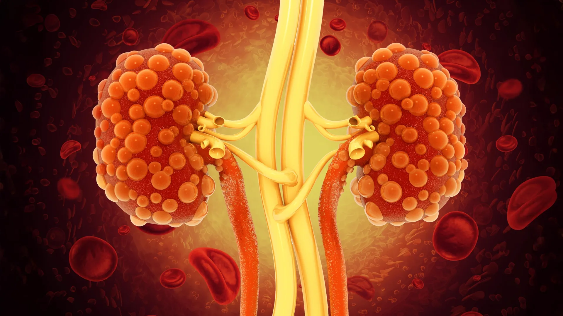
Polycystic kidney disease (PKD) represents one of the most significant challenges in modern nephrology, affecting millions of individuals worldwide with a progressive and often relentless decline in renal function. Characterized by the development of numerous fluid-filled cysts that distort the architecture of the kidneys, the condition frequently culminates in end-stage renal disease (ESRD), necessitating life-sustaining interventions such as dialysis or organ transplantation. However, a pioneering study led by researchers at the University of California, Santa Barbara (UCSB), has unveiled a potential therapeutic breakthrough that utilizes specially engineered monoclonal antibodies to penetrate these cysts and disrupt the biological signaling pathways that drive their expansion.
Published in the journal Cell Reports Medicine, the research introduces a novel method for delivering medication directly into the interior of kidney cysts—a feat that has historically eluded traditional antibody therapies. By redesigning the structural "backbone" of laboratory-grown proteins, the team, headed by UCSB biologist Thomas Weimbs, has demonstrated a way to bypass the biological barriers that protect cyst growth, offering a new glimmer of hope for a condition that currently has no known cure.
Understanding the Pathophysiology of Polycystic Kidney Disease
To appreciate the significance of the UCSB discovery, one must first understand the biological mechanism of PKD. The disease is primarily an inherited disorder, occurring in two main forms: Autosomal Dominant PKD (ADPKD), which is more common and often manifests in adulthood, and Autosomal Recessive PKD (ARPKD), a rarer and more severe form that typically appears in infancy or childhood.
In both forms, mutations in specific genes—primarily PKD1 and PKD2—cause the epithelial cells lining the kidney tubules to proliferate abnormally. Instead of maintaining the structure of healthy tubules, these cells form "pockets" that fill with fluid, eventually detaching from the tubule to become independent, sealed cysts. As these cysts grow, they exert pressure on the surrounding healthy kidney tissue, leading to inflammation, fibrosis, and the eventual loss of the organ’s ability to filter waste from the blood.
According to global health data, PKD affects approximately 12.5 million people worldwide. In the United States alone, it is the fourth leading cause of kidney failure. Despite the high prevalence, therapeutic options have remained tragically limited.
The Limitations of Current Pharmacological Interventions
Until recently, the primary approach to managing PKD involved controlling blood pressure and maintaining hydration to slow the progression of the disease. In 2018, the U.S. Food and Drug Administration (FDA) approved Tolvaptan (marketed as Jynarque), the first drug specifically designed to slow kidney function decline in adults at risk of rapidly progressing ADPKD.
While Tolvaptan has been a milestone in PKD treatment, it is far from a perfect solution. The drug works by blocking the activity of vasopressin, a hormone that promotes cyst growth. However, this mechanism also leads to significant side effects, including extreme thirst and frequent urination, which can severely impact a patient’s quality of life. More critically, Tolvaptan carries a risk of serious liver toxicity, requiring patients to undergo frequent and lifelong liver function monitoring.
"There are several small-molecule drugs that show potential for slowing cyst expansion," noted Thomas Weimbs, the senior author of the UCSB study. "However, the only approved drug that offers some benefit also brings significant side effects and toxicity to nearby kidney tissue."
This gap in treatment efficacy led the UCSB team to look toward immunotherapy and monoclonal antibodies, which have revolutionized the treatment of various cancers and autoimmune diseases due to their high specificity and low off-target toxicity.
The Barrier Problem: Why Traditional Antibodies Fail
Monoclonal antibodies are typically designed using Immunoglobulin G (IgG), the most common type of antibody found in human circulation. While IgG antibodies are highly effective at identifying and neutralizing targets in the bloodstream or on the surface of cells, they face a physical limitation when it comes to PKD.
Kidney cysts are essentially sealed chambers lined by a layer of epithelial cells. These cells form "tight junctions," creating a nearly impermeable barrier. IgG antibodies are simply too large and lack the necessary transport mechanisms to cross this epithelial layer. Consequently, they cannot reach the fluid inside the cyst where the disease-driving activity is most concentrated.
Within these cysts, a "vicious cycle" occurs. The cyst-lining cells secrete various growth factors into the internal fluid. These factors then bind back to receptors on the same cells, creating a self-sustaining loop of activation and expansion. "It’s like a never-ending scheme in which the cells just keep activating themselves and other cells in there," Weimbs explained. "Our premise was that if you block either the growth factor or the receptor for the growth factor, you should be able to stop this constant activation."
A Novel Solution: Engineering dIgA for Targeted Delivery
To overcome the barrier posed by the cyst wall, the UCSB researchers turned to a different class of antibody: Dimeric Immunoglobulin A (dIgA). In the human body, dIgA plays a vital role in mucosal immunity. It is the primary antibody found in secretions such as tears, saliva, and mucus, and it possesses a unique ability that IgG lacks: it can be actively transported across epithelial cell layers.
This transport is facilitated by the polymeric immunoglobulin receptor (pIgR), which sits on the "basolateral" side of epithelial cells. When dIgA binds to this receptor, the cell engulfs the antibody and moves it through its interior to the other side (the "apical" side) in a process known as transcytosis.
In a theoretical paper published in 2015, Weimbs and his colleagues hypothesized that because kidney cysts express pIgR, dIgA could be used as a "Trojan horse" to enter the cyst interior. The current study represents the successful validation of that hypothesis.
Chronology of the Discovery and Experimental Results
The journey from hypothesis to experimental proof involved several years of rigorous laboratory work and genetic engineering. The research team, which included lead author Margaret F. Schimmel and contributors Bryan C. Bourgeois, Alison K. Spindt, and others, began by altering the DNA sequence of a standard IgG antibody. They replaced its structural "backbone" with that of a dIgA antibody while maintaining its ability to target a specific receptor.
The target chosen for this study was the cell mesenchymal-epithelial transition (cMET) receptor. cMET is a well-known driver of cell proliferation and is found in high concentrations on the cells lining PKD cysts.
The experimental timeline and findings can be summarized as follows:
- Antibody Redesign: The team successfully engineered the dIgA antibody to recognize and bind to the cMET receptor.
- In Vitro Validation: Initial tests confirmed that the redesigned protein maintained its affinity for the target and could be recognized by the transport receptors.
- Mouse Model Testing: The antibody was administered to mouse models of PKD. Imaging and tissue analysis confirmed that the dIgA antibody successfully crossed into the cyst interior and remained there, whereas traditional IgG antibodies were excluded.
- Efficacy and Safety: Once inside, the antibody successfully inhibited the cMET receptor. This led to a significant reduction in the signals that promote cell growth.
- Selective Apoptosis: Perhaps most significantly, the treatment triggered a "dramatic onset of apoptosis" (programmed cell death) specifically in the cyst-lining cells. Crucially, this effect was not observed in healthy kidney tissue, suggesting a high level of safety and selectivity.
Supporting Data and Institutional Support
The success of the preclinical trials is backed by data showing that the reduction in cMET activity directly correlates with a slowing of cyst enlargement. The researchers observed that by disrupting the internal growth-factor loop, the "pressure" for the cysts to expand was mitigated.
The study received significant backing from major federal institutions, reflecting the high priority placed on finding a solution for PKD. Partial support was provided by the National Institutes of Health (NIH) and the U.S. Department of Defense (DoD). This funding underscores the recognition of PKD not only as a public health crisis but also as a condition that affects the long-term health of veterans and service members.
Official Responses and Expert Analysis
The scientific community has reacted with cautious optimism to the UCSB findings. While the work remains in the preclinical phase, the ability to target the interior of a "sealed" organ structure represents a paradigm shift in drug delivery.
"The results are compelling because they address the fundamental delivery problem that has hamstrung antibody therapy for kidney diseases for decades," said one independent analyst in the field of nephrology. "By leveraging a natural transport mechanism (dIgA), the researchers have turned a biological barrier into a gateway."
Thomas Weimbs remains focused on the next steps, noting that the diversity of growth factors in cyst fluid presents further opportunities. "In the literature, there are dozens of growth factors that have been shown to be active in these cyst fluids," Weimbs said. "So it would be a good idea to compare blocking of several different growth factors and several receptors… We can also combine different antibodies against different receptors at the same time."
Broader Impact and Future Implications
The implications of this research extend beyond Polycystic Kidney Disease. The successful use of dIgA to penetrate epithelial barriers could potentially be applied to other diseases involving "shielded" environments, such as certain types of glandular cancers or inflammatory conditions of the mucosal linings.
For PKD patients, however, the road to a human treatment remains long. Transitioning from mouse models to human clinical trials requires extensive safety testing, the scaling of manufacturing processes for dIgA antibodies (which are more complex to produce than IgG), and the identification of the most effective combinations of targets.
The researchers are now actively seeking partners in the biotechnology and pharmaceutical industries to help navigate the complex regulatory and production landscape. The goal is to develop a suite of dIgA-based therapies that could be tailored to the specific genetic profile or disease stage of the patient.
If successful, this approach could move the medical community away from "managing" PKD toward "reversing" it. By selectively inducing cell death in the cysts while leaving healthy tissue intact, scientists may finally have a way to restore kidney function and eliminate the need for dialysis for thousands of patients.
In the words of the research team, this study is a "first step" toward a future where the relentless growth of kidney cysts is no longer an inevitable path toward organ failure, but a manageable—and perhaps even curable—biological process.
