
 By Budi Oetomo Narendra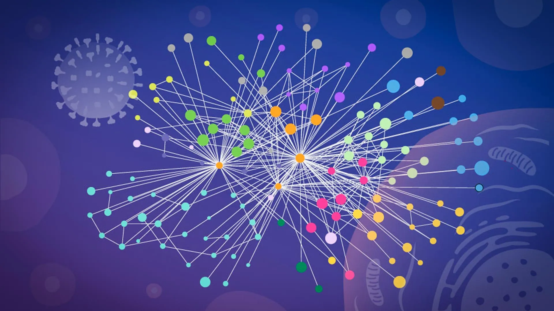
This groundbreaking research marks a significant leap in understanding the intricate mechanisms by which influenza A, a formidable viral pathogen, commandeers human cellular machinery. The ability to peer into the living, infected cell, observing protein-protein interactions in their native context, provides an unprecedented level of detail crucial for developing more effective countermeasures against a virus responsible for widespread illness and death globally.
The Enduring Threat of Influenza A: A Global Health Challenge
Influenza A stands as a perennial global health threat, responsible for millions of severe illnesses and hundreds of thousands of fatalities annually. Seasonal influenza epidemics cause an estimated 3 to 5 million cases of severe illness worldwide each year, leading to a staggering 290,000 to 650,000 respiratory deaths. Beyond its seasonal toll, influenza A is notoriously recognized for its capacity to drive devastating pandemics, reshaping public health landscapes and economies.
The most infamous example remains the 1918 "Spanish Flu" pandemic, caused by an H1N1 strain of influenza A, which infected an estimated one-third of the world’s population and claimed between 50 and 100 million lives. More recently, the 2009 H1N1 pandemic underscored the continuous threat, demonstrating influenza A’s ability to evolve and jump between species, causing widespread disruption. Currently, strains like H5N1, primarily circulating in avian populations, are closely monitored by global health organizations due to their high pathogenicity and pandemic potential should they acquire efficient human-to-human transmission capabilities.
Upon entering a host cell, the influenza A virus initiates a rapid and ruthless takeover. It releases its RNA genome, containing the genetic blueprint for a small but potent set of viral proteins. These viral proteins then disseminate throughout the host cell, systematically redirecting its complex molecular systems. The cell, once a functional unit of the human body, is transformed into an efficient factory for producing new virus particles, perpetuating the infection cycle. Understanding the precise choreography of this cellular hijack is paramount for scientific and medical advancement.
Pioneering a Clearer View: Mapping the Flu Virus Inside Intact Cells
For decades, scientists have grappled with a fundamental challenge in virology: how to observe the dynamic, fleeting interactions between viral and human proteins within a living, infected cell without disturbing its delicate internal architecture. A clearer, undistorted view of this viral takeover is not merely an academic pursuit; it is a critical step towards developing more effective flu vaccines that can elicit broader, more durable immunity, and novel antiviral drugs that can precisely target essential viral-host protein interfaces. To achieve this, researchers need comprehensive answers: which specific viral proteins engage with which human proteins, exactly where within the cell these contacts occur, and precisely how the virus exploits these interactions to support its own replication.
The new study, published in the prestigious journal Nature Microbiology, represents a landmark achievement in this quest. It is the first large-scale mapping of direct contacts between influenza proteins and human proteins conducted inside intact infected cells. Crucially, the structural detail obtained was so precise that researchers were able to computationally model how these interacting proteins likely fit together, offering a three-dimensional perspective on the molecular battleground.
"Our work provides a new way to study flu-host interactions in their native context and with structural insight," stated Jan Kosinski, Group Leader at EMBL Hamburg and the Centre for Structural Systems Biology (CSSB). He emphasized the forward-looking nature of the findings: "The current results are a snapshot of a moment during infection, and it opens the door to studying flu-host interactions across the entire infection cycle." This holistic understanding, moving beyond static observations, promises to unlock deeper insights into the virus’s lifecycle and vulnerabilities.
Overcoming a Major Experimental Hurdle: The Power of Specialized XL-MS
Tracking the intricate web of protein-protein interactions during an active viral infection has historically been an extremely difficult endeavor. Many earlier studies, while valuable, were constrained by the limitations of conventional biochemical techniques. These methods typically required scientists to physically break cells open—a process known as lysis—before they could isolate and measure protein contacts.
This destructive process often introduces significant artifacts and can distort the true picture of what was happening inside the living cell. Once internal compartments are ruptured, proteins that were naturally separated by membranes or cellular architecture may artificially come into contact in the laboratory environment. Conversely, weak, transient, or highly location-specific interactions, which are often critical for viral function, can easily disappear or be missed entirely during the harsh extraction process. As a result, researchers frequently struggled to definitively determine which connections genuinely existed and were functionally relevant during the actual infection. The challenge was akin to trying to understand a complex machine by only examining its disassembled parts.
The breakthrough came through a specialized adaptation of an established technique. "This is when we learned that our collaborators, Boris Bogdanow and Fan Liu, at FMP Berlin had developed a specialized version of cross-linking mass spectrometry (XL-MS), a long-established technique for mapping protein contacts, tailored specifically to virus-infected cells," Kosinski explained.
Cross-linking Mass Spectrometry (XL-MS) is a powerful biochemical method that uses chemical reagents to create covalent bonds (cross-links) between proteins that are in close proximity within a sample. These cross-links essentially "freeze" the interactions in place. The proteins are then digested into smaller peptides, and mass spectrometry is used to identify the cross-linked peptides, thereby pinpointing which proteins were interacting and, in some cases, which specific regions were in contact.
The innovation by Bogdanow and Liu was to meticulously optimize this technique for the unique environment of virus-infected cells, enabling the capture of interactions in situ. This specialized method provided precisely the breakthrough the team needed, making it possible to capture interactions that occur only briefly or within particular, often transient, regions of an infected cell.
Bogdanow, now a Junior Research Group Leader at the Institute of Virology, Charité – Universitätsmedizin Berlin, elaborated on its advantages: "XL-MS allows us to capture protein-protein interactions directly in infected intact cells, while also providing structural information about how these interactions are happening." He underscored the potential impact: "This gives us insight into the interface between the virus and the human cell and may, through structural modeling, help identify actionable targets for future pharmaceutical interventions."
Synergistic Science: Combining Experimental Data with AlphaFold
The power of this study was further amplified by the innovative pairing of the experimental XL-MS results with advanced computational structural modeling. This integrated approach allowed the researchers to not only identify which viral and human proteins interact but also to estimate with high precision how those proteins are positioned relative to each other when they connect, providing crucial three-dimensional context.
To construct these detailed structural models, the team leveraged a modified version of AlphaFold, the revolutionary artificial intelligence system developed by DeepMind. AlphaFold, awarded the Nobel Prize in Chemistry for its unparalleled accuracy in predicting protein structures from amino acid sequences, has transformed structural biology. Its original design, however, focuses on predicting the structure of individual proteins or stable protein complexes based purely on sequence data.
"The key advantage of the modified AlphaFold approach is that it allowed us to feed our experimental cross-linking data directly into the structural modeling," explained Kosinski. This critical modification meant that the computational model was not relying solely on theoretical predictions but was guided by empirical evidence of actual physical proximity within the infected cell. "This tells the model which parts of the viral and host proteins are close to each other inside infected cells. This was especially useful for virus-host complexes, which are often difficult to predict reliably," he added. By incorporating the XL-MS data as constraints, the researchers could refine AlphaFold’s predictions, generating highly accurate models of these complex, often transient, virus-host interfaces.
Unveiling Influenza’s Two-Pronged Cellular Hijack Strategy
The meticulous findings, meticulously detailed in Nature Microbiology, brought to light two particularly striking strategies that influenza A appears to employ in its sophisticated campaign to seize control of human cells. These discoveries offer new avenues for therapeutic intervention.
Strategy 1: Hijacking the Cell’s Protein Processing Network for Hemagglutinin Maturation
The first strategy revolves around hemagglutinin (HA), a crucial glycoprotein found on the surface of the influenza virus. Hemagglutinin is not merely a structural component; it is the molecular key that allows the virus to attach to and subsequently enter host cells by binding to sialic acid receptors on the cell surface. After entry, HA undergoes a conformational change, facilitating the fusion of the viral and endosomal membranes, thereby releasing the viral genome into the host cytoplasm.
The researchers meticulously tracked hemagglutinin as it journeyed through the cell’s intricate internal transport and processing network. This network, a highly organized system of membrane-bound compartments including the endoplasmic reticulum (ER) and Golgi apparatus, is responsible for the proper folding, modification (such as glycosylation), and sorting of proteins destined for the cell surface or secretion. It is a vital quality control system.
The detailed analysis revealed that several human host proteins actively participated in helping hemagglutinin fold correctly and undergo necessary modifications during the infection process. These host proteins, essentially co-opted by the virus, ensure that HA is properly presented on the surface of newly formed virions, making them infectious. Intriguingly, some of these identified host proteins previously had poorly understood functions, highlighting how viral investigations can shed light on fundamental cellular biology. Disrupting these host-assisted folding pathways could potentially cripple the virus’s ability to produce infectious progeny.
Strategy 2: Dissolving Nuclear Paraspeckles to Release RNA-Binding Proteins
The second profound discovery involved paraspeckles, small, dynamic, droplet-like compartments located within the nucleus of human cells. These organelles are known to play roles in RNA metabolism, gene expression regulation, and stress responses, acting as reservoirs for specific RNA-binding proteins.
The team’s observations showed that influenza A infection consistently caused these paraspeckle structures to dissolve. When the paraspeckles broke apart, they released a specific subset of RNA-binding proteins that had been sequestered within them. The researchers hypothesize that the influenza virus may then exploit these newly freed host proteins, repurposing them to support its own replication and propagation. This represents a clever, targeted manipulation of host cellular resources.
Iuliia Kotova, a former predoctoral fellow at the Kosinski Group at EMBL Hamburg, now at ETH Zurich and first author of the publication, expressed her surprise and conviction: "What surprised us most was the paraspeckles. Watching these tiny organelles in the nucleus dissolve, consistently across every cell line and every flu strain we tested, told us this isn’t a side effect of infection — it might be a strategy." The consistency of the observation across different experimental conditions strongly suggests an active, deliberate viral mechanism rather than an accidental byproduct of cellular stress.
Furthermore, the disruption of paraspeckles may provide influenza with more than one advantage. Kosinski elaborated on a potential dual benefit for the virus: "There may also be a second benefit for the virus: some evidence suggests paraspeckles contribute to cellular stress responses and antiviral gene regulation, so disrupting them could also weaken parts of the cell’s defense response." By dismantling these nuclear defense outposts, the virus could effectively disarm the cell’s ability to mount an effective antiviral response, creating a more permissive environment for its own multiplication. This dual attack — acquiring essential host proteins and simultaneously suppressing host defenses — exemplifies the sophisticated evolutionary strategies employed by successful pathogens.
A Collaborative Triumph: Expertise Across Three Institutions
The success of this ambitious project was a testament to the power of interdisciplinary collaboration and shared technological expertise across multiple leading institutions. The complex and specialized cross-linking mass spectrometry work, central to capturing the protein interactions, was meticulously carried out at Charité – Universitätsmedizin Berlin, leveraging their cutting-edge facilities and Bogdanow’s specialized methodology. Crucial glycoproteomics analyses, essential for understanding modifications like glycosylation on proteins such as hemagglutinin, were expertly completed at the EMBL Proteomics Core Facility, a renowned hub for advanced proteomics research.
The intensive computational demands of the modified AlphaFold modeling, requiring significant processing power, were met by the robust infrastructure of the EMBL Compute Cluster. Finally, the critical microscopy imaging, providing visual confirmation and contextualizing the molecular findings, took place at CSSB’s Advanced Light and Fluorescence Microscopy (ALFM) Facility, ensuring high-resolution visualization of cellular events. This seamless integration of diverse scientific disciplines and state-of-the-art technologies was indispensable for achieving the depth and breadth of this study’s findings.
A New Paradigm for Studying Potential Pandemic Viruses
The profound findings from this research unequivocally demonstrate how the study of molecular contacts within intact infected cells can reveal not only where but also how a virus systematically takes control of human cellular machinery. This innovative approach, termed ‘mapping in context,’ promises to revolutionize our understanding of viral pathogenesis and may well explain the operational strategies of numerous other viruses.
"While the exact host factors and mechanisms often differ from virus to virus, we think our overall approach — combining in-cell cross-linking, structural modeling, and targeted cell-biology follow-up to map native virus-host interactions at specific stages of infection — remains broadly applicable," Kosinski affirmed. This suggests a powerful new template for virological research, capable of being deployed against a wide array of pathogens.
Although the current study meticulously examined a laboratory-adapted strain of influenza A, the researchers are confident that the same robust methodology can eventually be applied to investigate viruses with far greater pandemic potential. This is a crucial step towards proactive pandemic preparedness.
Bogdanow strongly concurred, emphasizing the forward trajectory: "Although this study has focused on a lab-adapted strain, this study lays the groundwork to apply the methodology to viruses of potential pandemic relevance, such as H5N1, and for uncovering the interaction networks that support their multiplication in human cells." The immediate implication is the potential to identify critical vulnerabilities in highly pathogenic avian influenza strains like H5N1, which pose a significant zoonotic threat. By mapping their cellular hijacking strategies, scientists could pinpoint novel drug targets or design more effective vaccines, offering a preemptive defense against future outbreaks.
In essence, this research not only deepens our understanding of influenza A but also provides a powerful methodological framework. This framework is poised to accelerate the discovery of actionable targets for pharmaceutical interventions, paving the way for the development of next-generation antiviral drugs and more broadly protective vaccines, ultimately strengthening humanity’s defenses against the persistent and evolving threat of viral pandemics.
