
 By Nana O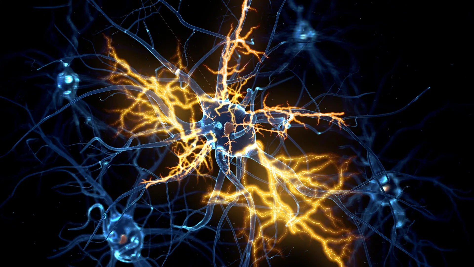
Scientists at the University of Cambridge have engineered sophisticated, lab-grown brain and spinal cord systems that meticulously replicate the intricate pathways of movement signals within the human nervous system. This pioneering research, utilizing miniature models that mimic human development, has yielded a potentially revolutionary discovery: nerve damage, long considered irreversible, may indeed be amenable to repair under specific conditions. The findings offer a beacon of hope for individuals living with paralysis and a range of debilitating neurological conditions.
The Intricate Dance of Neural Communication and the Challenge of Regeneration
From the earliest stages of embryonic development, the human nervous system embarks on an extraordinary journey of growth and connection. Neurons, the fundamental building blocks of this system, forge complex communication networks that span the brain and spinal cord. These vital messages are transmitted along axons, the elongated nerve fibers that act as conduits, enabling neurons to send signals and ultimately orchestrate voluntary muscle movements.
However, as the central nervous system matures, it largely relinquishes its remarkable capacity for axonal regrowth. This inherent limitation means that injuries to the brain or spinal cord, which disrupt these crucial connections, often result in permanent damage. The consequences can be devastating, leading to profound disabilities such as paralysis, loss of motor control, and sensory deficits. Furthermore, this diminished regenerative potential is also implicated in the progression of neurodegenerative diseases, including motor neurone disease (also known as amyotrophic lateral sclerosis or ALS) and multiple sclerosis (MS), conditions that relentlessly erode nerve function.
Genesis of the Miniature Neural Worlds: A Leap in Organoid Technology
The foundation for this latest breakthrough was laid in 2021 when Dr. András Lakatos and his team at the University of Cambridge first unveiled miniature human brain models. These intricate structures, crafted from patient-derived stem cells, were no larger than a pea but bore striking resemblances to specific regions of the cerebral cortex. Dubbed "brain organoids," these models provided researchers with an unprecedented platform to investigate the molecular underpinnings of conditions like motor neurone disease and to explore preventative strategies.
Building upon this significant achievement, the current study, published in the prestigious journal Cell Reports, represents a natural and ambitious extension of their earlier work. The Cambridge team has now successfully constructed a miniature, interconnected system that mimics the functional relationship between the human brain and spinal cord.
Recognizing the anatomical separation yet functional linkage of these two critical components in vivo, the researchers ingeniously maintained the brain and spinal cord organoids as physically distinct entities within their laboratory environment. The remarkable observation that followed was the directed growth of axons from the brain tissue, spanning the meticulously created gap to establish direct connections with the spinal cord tissue. The resulting neural circuit was not merely structural; it demonstrated functional capability, eliciting measurable contractions in minuscule clusters of muscle cells – a compelling testament to its nascent ability to transmit motor commands.
A Developmental Timeline of Regenerative Capacity: Unveiling the Critical Window
The researchers meticulously monitored these miniature neural systems for an extended period, exceeding one year in laboratory conditions. Their extended observation period revealed a critical developmental window for axonal regeneration. They discovered that up to approximately day 150 of development – a timeframe that broadly corresponds to the mid-pregnancy stage in human gestation – damaged axons retained a significant capacity for regrowth. Beyond this developmental milestone, however, the neurons exhibited a dramatic and precipitous decline in their regenerative abilities.
George Gibbons, a researcher from the Department of Clinical Neurosciences at the University of Cambridge and the first author of the study, articulated the significance of this finding: "Neurons taken from less mature organoids regrew long fibers after injury, but those from more mature organoids showed a sharp drop in their ability to regrow. In other words, poor regeneration is built into human neurons as they mature in the central nervous system." This statement underscores a fundamental, developmentally programmed limitation that appears to be intrinsically embedded within human neurons as they progress towards maturity within the central nervous system.
Decoding the Genetic Switch: Identifying the Molecular Regulators of Axon Growth
To unravel the mechanisms behind this developmental decline in regenerative capacity, the team undertook a detailed analysis of gene activity within the neurons responsible for connecting the brain and spinal cord. Their investigations pinpointed a complex network of genes that functions akin to a biological switch. This genetic circuitry appears to be intricately involved in regulating and ultimately limiting axon growth as neurons mature and establish synaptic connections.
In a truly remarkable and potentially transformative development, the researchers demonstrated that by selectively blocking key regulatory elements within this identified gene network, they could effectively restore the neurons’ ability to grow axons. This experimental intervention effectively reversed the age-dependent decline in regeneration, suggesting a direct and targetable mechanism for promoting nerve regrowth.
A Promising Pharmaceutical Avenue: Lynestrenol’s Regenerative Potential
In parallel with their genetic investigations, the scientists scoured a comprehensive database of drug compounds, searching for existing medicines that might influence this newly characterized gene network. Their search yielded a particularly promising candidate: lynestrenol. Lynestrenol is a progestogen hormone drug that is currently approved and utilized for specific menstrual disorders and as a form of contraception.
When lynestrenol was administered to damaged neurons in their organoid model, the results were striking. The drug significantly enhanced axon regrowth, demonstrating a clear therapeutic effect on the regenerative process. This finding opens up a tangible pharmaceutical pathway for potential future interventions aimed at restoring nerve function.
The researchers acknowledged that factors beyond intrinsic neuronal capacity, such as scar tissue formation and inflammation, also pose significant barriers to nerve repair following injury. However, they emphasized the paramount importance of understanding the neuron-specific biological mechanisms that inherently limit regeneration. Existing research has already provided evidence that younger neurons possess the capability to navigate and grow through environments that typically impede repair at injury sites. This suggests that overcoming the intrinsic developmental limitations of neurons may be a more fundamental prerequisite for successful regeneration.
Expert Commentary: A Paradigm Shift in Neurological Repair
Dr. András Lakatos, the senior author who spearheaded this groundbreaking study at the Department of Clinical Neurosciences, provided crucial context and insight into the implications of their work: "When the brain and spinal cord are damaged, the nerve fibers that carry movement signals from the brain to the spinal cord rarely grow back. That’s why paralysis is usually permanent. But we didn’t know exactly when the ability of axons to regenerate becomes limited. Our model provides a good indication that this block happens during development, and it can still be reversed after this point."
He further elaborated on the potential of their findings: "Lynestrenol itself may not be the answer to spinal cord repair, but it shows us that, in principle, it should be possible to directly target human neurons and regenerate their axons. Although we still need to show that this strategy will also help to re-establish appropriate connections between the brain and spinal cord cells, this gives us hope that one day we may be able to treat conditions previously thought untreatable." This measured optimism highlights both the significant progress made and the necessary future research to translate these findings into clinical reality.
The Indispensable Role of Human Organoids in Medical Research
The development and application of organoid technology are proving increasingly invaluable in advancing our understanding of human biology and disease. While animal models, such as mice and rats, have historically played a crucial role in scientific inquiry, inherent biological differences between species can limit their predictive accuracy for human nervous system function.
Human stem cell-derived organoids, by contrast, offer a more faithful recapitulation of human biological processes. This enhanced fidelity helps to bridge the critical gap between findings in animal studies and the eventual outcomes observed in human patients.
Dr. Lakatos underscored the importance of this translational aspect: "Much of what we know about nerve regeneration comes from rodents, whose neurons behave differently from human neurons. Our sophisticated organoid models help bridge the knowledge gap from animal models to what we see in patients. They are also an important contribution to efforts to reduce the use of animals in research." This statement not only highlights the scientific advantages but also touches upon the ethical considerations driving the shift towards more human-relevant research models.
The University of Cambridge is at the forefront of organoid research, employing these advanced models across a diverse spectrum of medical investigations. Their applications range from pioneering efforts to repair damaged livers and unraveling the complexities of Crohn’s disease in children to studying the nascent stages of human pregnancy.
This transformative research was generously supported by funding from the UK Research and Innovation Medical Research Council and Spinal Research, underscoring the collaborative and well-supported nature of such significant scientific endeavors.
Broader Implications: A Future of Restorative Therapies
The implications of this Cambridge study extend far beyond the immediate discovery of lynestrenol’s potential. It fundamentally shifts our understanding of nerve damage, moving it from a category of irreversible injury to one that may, in certain contexts, be amenable to targeted therapeutic intervention.
Key implications include:
- Revisiting Spinal Cord Injury Treatment: For decades, treatments for spinal cord injuries have focused on managing symptoms and preventing secondary damage, with limited options for restoring lost function. This research opens a new frontier, suggesting that regaining lost motor control might become a tangible goal.
- Potential for Neurodegenerative Disease Therapies: Conditions like motor neurone disease and multiple sclerosis, characterized by progressive neuronal degeneration, could potentially benefit from therapies that enhance intrinsic regenerative capabilities.
- Accelerated Drug Discovery: The identification of a specific gene network and a potential drug target provides a clear roadmap for the development of novel regenerative therapies. Future research can now focus on optimizing drugs that modulate this network.
- Enhanced Understanding of Neural Development: The study provides invaluable insights into the critical developmental processes that govern neuronal growth and plasticity, offering a deeper understanding of how the nervous system forms and matures.
- Ethical Advancements in Research: The reliance on human organoids, as highlighted by Dr. Lakatos, contributes to the ongoing effort to reduce and refine the use of animals in research, aligning with global trends towards more ethically sound scientific practices.
While significant hurdles remain before these findings can be translated into clinical treatments for patients, the Cambridge team’s work represents a monumental leap forward. The creation of functional brain-spinal cord organoid systems and the discovery of a mechanism to potentially reverse developmental limitations in nerve regeneration offer profound hope and a renewed sense of possibility in the quest to combat debilitating neurological conditions. The scientific community will undoubtedly be closely watching as this promising research progresses towards clinical application.
