
 By Budi Oetomo Narendra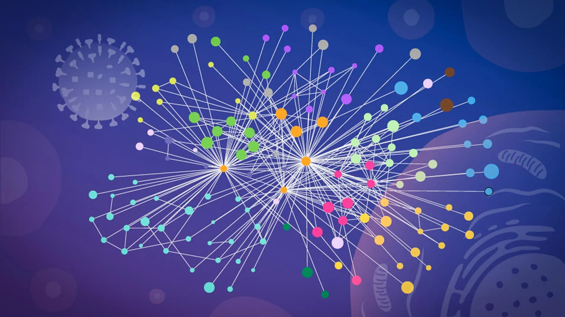
Researchers at EMBL Hamburg, in a pivotal collaboration with scientists at the Leibniz Research Institute for Molecular Pharmacology (FMP), have unveiled an unprecedentedly detailed map illustrating precisely how influenza A meticulously reshapes and commandeers infected human cells. This groundbreaking study leverages a sophisticated, customized workflow that allowed scientists to directly observe intricate protein interactions within the natural environment of intact cells, sidestepping the limitations of traditional methods that often required cellular disruption. This "in-context" mapping represents a significant leap forward in understanding viral pathogenesis and holds immense promise for the development of more effective antiviral treatments and vaccines against one of humanity’s most persistent infectious threats.
The Pervasive Threat of Influenza A
Influenza, commonly known as the flu, remains a formidable global health challenge. Seasonal influenza alone accounts for an estimated 3 to 5 million cases of severe illness and contributes to as many as 290,000 to 650,000 respiratory deaths worldwide each year, according to figures from the World Health Organization (WHO). Beyond its annual toll, influenza A viruses are particularly notorious for their pandemic potential, having been the causative agent behind some of history’s most devastating global outbreaks. The infamous 1918 Spanish Flu pandemic, caused by an H1N1 strain of influenza A, claimed an estimated 50 million to 100 million lives globally, fundamentally reshaping public health approaches and underscoring the virus’s capacity for widespread devastation. More recently, the 2009 H1N1 pandemic and ongoing concerns about highly pathogenic avian influenza strains like H5N1 highlight the continuous need for deeper scientific understanding to bolster preparedness and response strategies.
Upon gaining entry into a human cell, the influenza A virus initiates a rapid and systematic takeover. It releases its RNA, which acts as a blueprint containing the genetic instructions for synthesizing a small but potent arsenal of viral proteins. These proteins then disperse throughout the host cell, swiftly redirecting its intricate molecular systems. The cell’s natural functions are subverted, transforming it from a healthy, functioning unit into a dedicated factory for producing new virus particles, perpetuating the infection cycle. Understanding the precise mechanisms of this cellular reprogramming is paramount for devising interventions that can effectively disrupt the virus’s lifecycle.
Overcoming a Major Scientific Hurdle: Mapping Interactions in Vivo
For decades, a significant hurdle in virology research has been the challenge of accurately mapping the dynamic interactions between viral and human proteins within the living, infected cell. Traditional biochemical techniques, while valuable, often necessitate the physical disruption of cells – breaking them open – before protein contacts can be measured. This destructive process introduces a critical problem: it can inadvertently distort the true biological reality within the living cell. Internal cellular compartments, which naturally segregate proteins, are destroyed, leading to artificial interactions between proteins that would never normally meet. Conversely, weak, transient, or highly localized interactions, which are often crucial for viral manipulation, can be lost entirely in the chaotic aftermath of cell lysis. Consequently, researchers have often struggled to definitively determine which protein connections genuinely existed and were functionally relevant during an active infection.
To address this profound limitation, scientists recognized the urgent need for a methodology that could capture these interactions in their native context, providing a clearer, more accurate "snapshot" of the viral takeover. This pursuit led to the development of a specialized approach, marking a turning point in the study of virus-host dynamics.
The Breakthrough Methodology: Specialized XL-MS and AI-Powered Modeling
The pivotal breakthrough came through a customized version of cross-linking mass spectrometry (XL-MS), a well-established technique for mapping protein contacts, which was specifically tailored for virus-infected cells by collaborators Boris Bogdanow and Fan Liu at FMP Berlin. As Jan Kosinski, Group Leader at EMBL Hamburg and Centre for Structural Systems Biology (CSSB), explained, "This is when we learned that our collaborators… had developed a specialized version of cross-linking mass spectrometry (XL-MS), a long-established technique for mapping protein contacts, tailored specifically to virus-infected cells."
This specialized XL-MS method provided the precision and context the research team needed. By chemically "freezing" protein interactions within intact cells, it allowed for the capture of contacts that occur only briefly or within particular, highly localized regions of an infected cell – interactions that would be lost with conventional methods. "XL-MS allows us to capture protein-protein interactions directly in infected intact cells, while also providing structural information about how these interactions are happening," explained Bogdanow, now a Junior Research Group Leader at the Institute of Virology, Charité – Universitätsmedizin Berlin. This capability is critical because the exact location and duration of an interaction can dictate its biological significance.
The experimental power of specialized XL-MS was then synergistically paired with advanced computational structural modeling. This innovative combination allowed the researchers to not only identify which viral and human proteins were interacting but also to estimate with remarkable precision how these proteins were physically positioned when they connected. To achieve this high level of structural detail, the team employed a modified version of AlphaFold, a highly acclaimed AI system recognized for its unprecedented ability to predict protein structures from amino acid sequences. While AlphaFold famously won the Breakthrough Prize in Life Sciences for its profound impact on structural biology, its application here was further refined.
"The key advantage of the modified AlphaFold approach is that it allowed us to feed our experimental cross-linking data directly into the structural modeling," Kosinski elaborated. "This tells the model which parts of the viral and host proteins are close to each other inside infected cells. This was especially useful for virus-host complexes, which are often difficult to predict reliably." By integrating empirical data from XL-MS directly into the AlphaFold algorithm, the team enhanced the accuracy and reliability of their structural predictions, providing unparalleled insight into the molecular interfaces between the virus and the host.
Unveiling Influenza’s Hijack Strategies: Hemagglutinin and Paraspeckle Dissolution
The findings, published in the prestigious journal Nature Microbiology, illuminated two particularly notable strategies employed by influenza A as it systematically takes control of a human cell. These discoveries offer concrete examples of the virus’s cunning molecular tactics.
The first strategy centers on hemagglutinin (HA), a crucial protein prominently displayed on the surface of the influenza virus. Hemagglutinin serves as the viral key, enabling the virus to attach to and subsequently enter host cells. The researchers meticulously tracked this protein’s journey as it navigated the cell’s complex internal transport and processing network. This network comprises a series of specialized compartments, including the endoplasmic reticulum and Golgi apparatus, which are responsible for folding, modifying, and preparing proteins before dispatching them to their final cellular destinations. The detailed analysis revealed that several human proteins actively assisted in the correct folding and modification of hemagglutinin during the infection process. Intriguingly, some of these host proteins previously had poorly understood functions, suggesting that influenza A’s reliance on them could unlock new avenues for research into fundamental cell biology, beyond just viral infection. This discovery highlights how the virus cleverly co-opts existing cellular machinery for its own assembly and maturation.
The second significant discovery involved paraspeckles, small, dynamic droplet-like compartments located within the cell nucleus. These subnuclear bodies are known to play roles in RNA processing and gene regulation. The research team made the striking observation that influenza A infection consistently caused these paraspeckle structures to dissolve. When paraspeckles broke apart, they released a variety of RNA-binding proteins that had been sequestered within them. The compelling hypothesis is that the virus then exploits these newly freed proteins, redirecting them to support its own replication cycle, effectively turning host cellular components against the host.
Iuliia Kotova, a former predoctoral fellow at the Kosinski Group at EMBL Hamburg and now at ETH Zurich, and the first author of the publication, expressed her surprise: "What surprised us most was the paraspeckles. Watching these tiny organelles in the nucleus dissolve, consistently across every cell line and every flu strain we tested, told us this isn’t a side effect of infection – it might be a strategy." This consistent observation across different experimental conditions strongly suggests an active, deliberate viral mechanism. The disruption of paraspeckles may offer influenza more than one advantage. As Kosinski added, "There may also be a second benefit for the virus: some evidence suggests paraspeckles contribute to cellular stress responses and antiviral gene regulation, so disrupting them could also weaken parts of the cell’s defense response." This dual benefit – gaining resources for replication and simultaneously impairing host defenses – underscores the sophistication of viral evolution.
A Collaborative Effort Across Leading Institutions
This ambitious project, yielding such profound insights, was the result of a concerted, collaborative effort, pooling diverse technologies and expertise from three leading institutions. The intricate cross-linking mass spectrometry work, forming the experimental backbone of the study, was meticulously carried out at Charité – Universitätsmedizin Berlin. Further essential analyses, specifically glycoproteomics, were completed at the state-of-the-art EMBL Proteomics Core Facility. The complex AlphaFold modeling, which translated experimental data into structural predictions, was performed on the powerful EMBL Compute Cluster, showcasing the critical role of high-performance computing in modern biology. Finally, the crucial microscopy imaging, which provided visual confirmation and contextual information, took place at CSSB’s Advanced Light and Fluorescence Microscopy (ALFM) Facility. This multi-institutional synergy underscores the increasing complexity and interdisciplinary nature of cutting-edge biological research.
A New Paradigm for Studying Potential Pandemic Viruses
The groundbreaking findings from this study fundamentally demonstrate how the detailed examination of molecular contacts within intact infected cells can comprehensively reveal both where and how a virus effectively takes control of human cellular machinery. This innovative "mapping in context" approach is poised to revolutionize our understanding of viral infections, moving beyond mere identification of interacting proteins to a mechanistic understanding of their spatial and temporal dynamics.
"While the exact host factors and mechanisms often differ from virus to virus, we think our overall approach – combining in-cell cross-linking, structural modeling, and targeted cell-biology follow-up to map native virus-host interactions at specific stages of infection – remains broadly applicable," Kosinski affirmed. This suggests that the methodology developed here could serve as a versatile template for investigating a wide array of other pathogenic viruses, including those responsible for coronaviruses (like SARS-CoV-2), HIV, dengue, and countless others that exploit host cells for replication.
Although the current study focused on a laboratory-adapted strain of influenza A, chosen for its tractability in experimental settings, the researchers are confident that the same robust strategy can eventually be deployed to investigate viruses with far greater pandemic potential. Boris Bogdanow shares this optimistic outlook: "Although this study has focused on a lab-adapted strain, this study lays the groundwork to apply the methodology to viruses of potential pandemic relevance, such as H5N1, and for uncovering the interaction networks that support their multiplication in human cells." Applying this detailed mapping to highly virulent strains could rapidly identify unique vulnerabilities, paving the way for targeted pharmaceutical interventions that disrupt the viral lifecycle at its most critical points.
The implications for public health are profound. A clearer, structurally informed view of virus-host interactions can directly inform the design of more effective antiviral drugs that specifically target these crucial interfaces, minimizing off-target effects. Furthermore, understanding the precise mechanisms of viral takeover can guide the development of next-generation vaccines that elicit more potent and durable immune responses. This research marks a significant stride towards a future where infectious disease outbreaks, particularly those driven by influenza A, can be more effectively predicted, prevented, and treated, ultimately saving countless lives and mitigating global health crises. The journey from this detailed map to new therapies is complex, but this study provides an invaluable compass.
