
 By George Ongkojoyo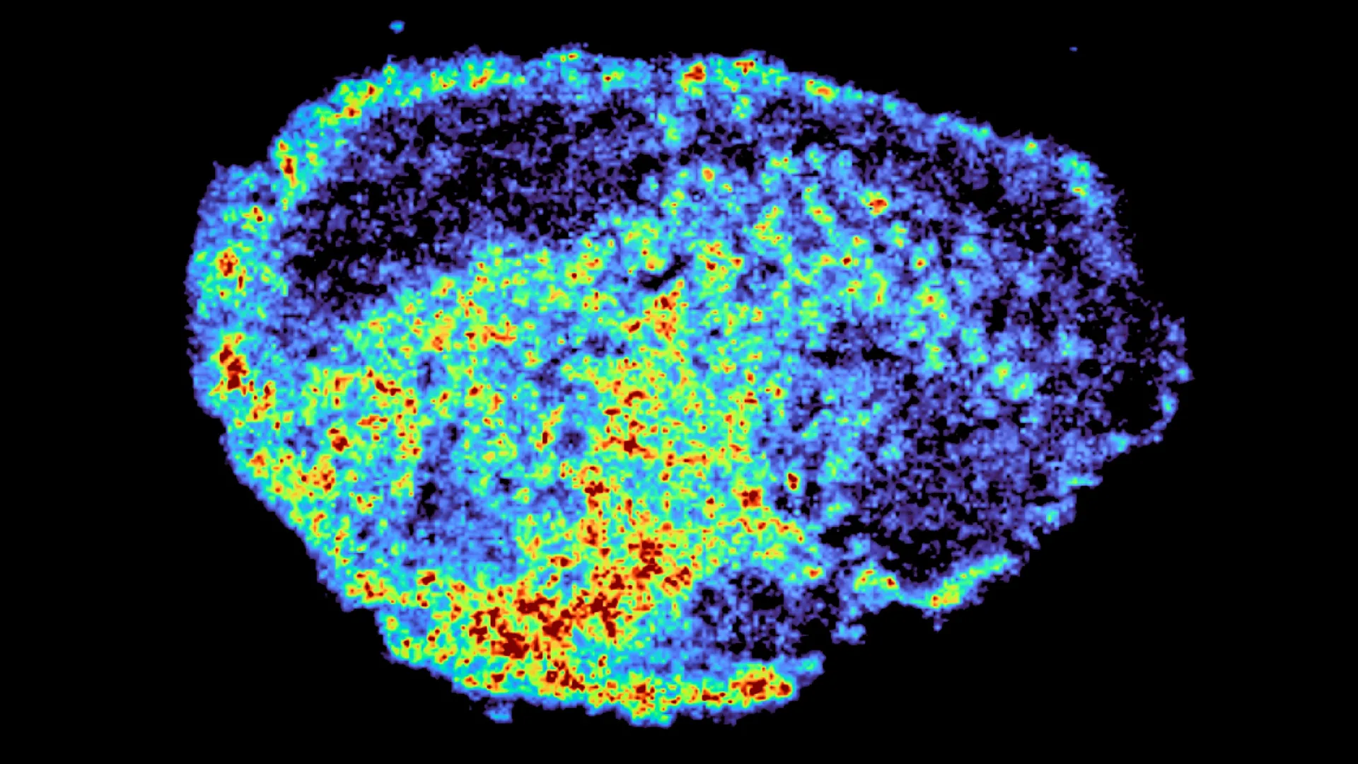
The fundamental challenge in modern oncology remains the unpredictable nature of therapeutic response, where a treatment regimen that proves life-saving for one individual may offer negligible benefits to another with a seemingly identical diagnosis. A landmark study recently published in the journal Nature Communications has provided critical insights into this phenomenon, specifically concerning the use of PARP inhibitors in the treatment of ovarian cancer. Led by Dr. Louise Fets at the MRC Laboratory of Medical Sciences (LMS), the research team utilized pioneering imaging technologies to track the movement of drugs through tumor tissue, uncovering a previously misunderstood mechanism of drug accumulation within cellular "recycling centers" known as lysosomes. This discovery explains how certain drugs can become sequestered within these structures, acting as slow-release reservoirs that fundamentally alter the effectiveness of the treatment and the potential for patient resistance.
The Evolution and Mechanism of PARP Inhibitors in Oncology
To understand the significance of the findings by the MRC Laboratory of Medical Sciences, it is necessary to examine the role of Poly (ADP-ribose) polymerase (PARP) inhibitors in the current landscape of cancer care. PARP inhibitors represent a class of targeted therapies that have revolutionized the management of several malignancies, most notably ovarian, breast, prostate, and pancreatic cancers. These drugs operate on the principle of "synthetic lethality." In healthy cells, multiple pathways exist to repair DNA damage. However, in many cancer cells—particularly those with BRCA1 or BRCA2 mutations—one of these repair pathways is already compromised. PARP inhibitors block a secondary repair pathway, leaving the cancer cell unable to fix its DNA, which eventually triggers programmed cell death (apoptosis).
Since the first PARP inhibitor, olaparib, received regulatory approval in 2014, the clinical use of these agents has expanded. They are now frequently utilized as maintenance therapy to prevent disease recurrence following chemotherapy. Despite their success, oncologists have long grappled with the high degree of variability in patient response. While some patients experience prolonged remission, others see their tumors progress rapidly despite high-dose therapy. The research led by Dr. Fets addresses the "black box" of drug distribution—the question of what happens to the drug once it enters the complex environment of a human tumor.
Innovative Methodology: Mapping the Intratumoral Landscape
The research team at the MRC LMS, in collaboration with Imperial College London, moved beyond traditional cell-line studies, which often fail to replicate the complexity of human biology. Instead, they employed "explants"—thin, live slices of ovarian tumor tissue donated by patients. These samples were maintained in a laboratory environment that preserved the original architecture and cellular diversity of the tumor. By treating these explants with various PARP inhibitors, the scientists were able to observe the real-time movement and localization of drug molecules within genuine human tissue.
The study’s methodology relied on two primary high-tech tools: mass spectrometry imaging (MSI) and spatial transcriptomics. Mass spectrometry imaging allowed the researchers to create high-resolution maps of where the drug molecules were physically located across the tissue sample. This was then integrated with spatial transcriptomics, a cutting-edge technique that measures gene expression in specific locations. By overlaying these two datasets, the team could compare the genetic activity of cells in areas of high drug concentration against those in areas where the drug failed to penetrate.
The results were unexpected. Even when a uniform dose of a drug was applied to a sample, the distribution was remarkably uneven. Some regions of a single tumor were saturated with the medication, while adjacent areas remained virtually untouched. This heterogeneity suggests that the failure of cancer treatment may not always be due to a lack of genetic sensitivity to the drug, but rather a physical failure of the drug to reach its target in sufficient quantities.
The Role of Lysosomes as Hidden Drug Reservoirs
The most significant finding of the study involves the role of lysosomes. Traditionally viewed as the cell’s waste disposal and recycling units, lysosomes are acidic organelles that break down cellular debris. The research revealed that certain PARP inhibitors, specifically rucaparib and niraparib, are "lysosomotropic." This means they are chemically drawn into the acidic environment of the lysosome. Once inside, these drugs become trapped, creating localized "pockets" of high concentration.
Dr. Carmen Ramirez Moncayo, the study’s first author and a Postdoctoral Researcher at the LMS, noted the surprise of the team at the sheer level of variability at the single-cell level. The lysosomes essentially act as reservoirs. Rather than the drug being distributed evenly throughout the cytoplasm where it can reach the nucleus to inhibit the PARP enzyme, it becomes concentrated in these small organelles.
Crucially, the study found that these reservoirs function as a double-edged sword. On one hand, they store the drug and release it slowly over time, which can actually increase the duration of exposure for certain cancer cells. On the other hand, this sequestration can prevent the drug from reaching the necessary threshold in other cells, potentially allowing those cells to survive and develop resistance. Furthermore, the study highlighted a distinction between different PARP inhibitors: while rucaparib and niraparib showed significant lysosomal accumulation, olaparib did not exhibit this behavior, suggesting that the chemical structure of the specific drug determines how it is handled by the cell’s internal machinery.
Data and Observations on Treatment Resistance
The implications of this uneven distribution are profound for understanding cancer relapse. In clinical settings, the primary cause of mortality in ovarian cancer is the development of chemoresistance. Ovarian cancer is often referred to as a "silent killer" because it is frequently diagnosed at an advanced stage (Stage III or IV), where the five-year survival rate remains significantly lower than for cancers detected early.
Data from the study suggests that the "pockets" of low drug concentration created by lysosomal trapping could provide a sanctuary for cancer cells. In these regions, the cells are exposed to sub-lethal doses of the medication—a condition that is known to accelerate the evolution of drug resistance. If a cell is not killed by the initial treatment but is instead "stressed" by a low dose, it may undergo genetic or epigenetic changes that allow it to survive subsequent, more potent rounds of therapy.
The research team’s use of spatial transcriptomics provided further evidence of this. In areas with low drug uptake, cells exhibited different gene expression patterns compared to cells in high-uptake areas. This indicates that the tumor is not a monolithic entity but a collection of diverse microenvironments, each responding differently to the same clinical intervention.
Expert Reactions and the Path to Personalized Medicine
The scientific community has responded to these findings with cautious optimism, viewing them as a vital step toward more personalized oncology. Dr. Zoe Hall, a senior author of the study and Associate Professor at Imperial College London, emphasized that the ability to visualize drug uptake directly in patient tissue represents a major technological leap. According to Dr. Hall, the spatial mapping of drug molecules allows researchers to pinpoint exactly why certain regions of a tumor may be resisting treatment.
Dr. Louise Fets, who heads the Drug Transport and Tumour Metabolism Group at the LMS, highlighted the long-term goal of the research: using the molecular signature of a patient’s tumor to tailor therapy. If clinicians can determine beforehand how a specific patient’s tumor cells utilize lysosomes or how their unique tumor architecture might hinder drug distribution, they could choose the specific PARP inhibitor—or a combination of therapies—most likely to succeed. For example, if a patient’s tumor is found to have high lysosomal activity, a non-lysosomotropic drug like olaparib might be preferred over rucaparib.
Broader Implications for Clinical Practice and Future Research
While the study focused on ovarian cancer, the findings have broader implications for the use of PARP inhibitors in breast and prostate cancers. The mechanism of lysosomal trapping is likely not unique to ovarian tissue and could be a factor in treatment failure across various cancer types.
However, the researchers acknowledge that there are limitations to studying tissue outside the human body. In a living patient, the delivery of drugs is further complicated by the circulatory system. Tumor blood vessels are notoriously "leaky" and disorganized, which creates physical barriers to drug delivery even before the medication reaches the individual cell membranes.
The next phase of this research will involve moving into animal models to observe how these cellular reservoirs interact with the complex blood flow of a living organism. Additionally, larger-scale patient studies are planned to correlate lysosomal drug accumulation with actual clinical outcomes and survival rates. The research team also intends to investigate whether "lysosome-targeted" therapies could be used in conjunction with PARP inhibitors to prevent drug trapping and ensure a more uniform distribution of the medication.
Funding and Institutional Support
This research was made possible through a multi-institutional effort and significant financial backing from major health organizations. Supporters include the Medical Research Council (MRC), Cancer Research UK, and the Integrative Toxicology Training Partnership. Additional funding was provided by the Victoria’s Secret Global Fund for Women’s Cancers Career Development Award, in partnership with Pelotonia and the American Association for Cancer Research (AACR).
This collaborative support underscores the global importance of the work. As the medical community moves away from a "one-size-fits-all" approach to cancer treatment, studies like this one provide the foundational data necessary to build a more nuanced, effective, and personalized system of care. By decoding the hidden movements of drugs within the microscopic landscape of a tumor, scientists are bringing the medical field one step closer to overcoming the persistent challenge of cancer drug resistance.
