
 By Nana O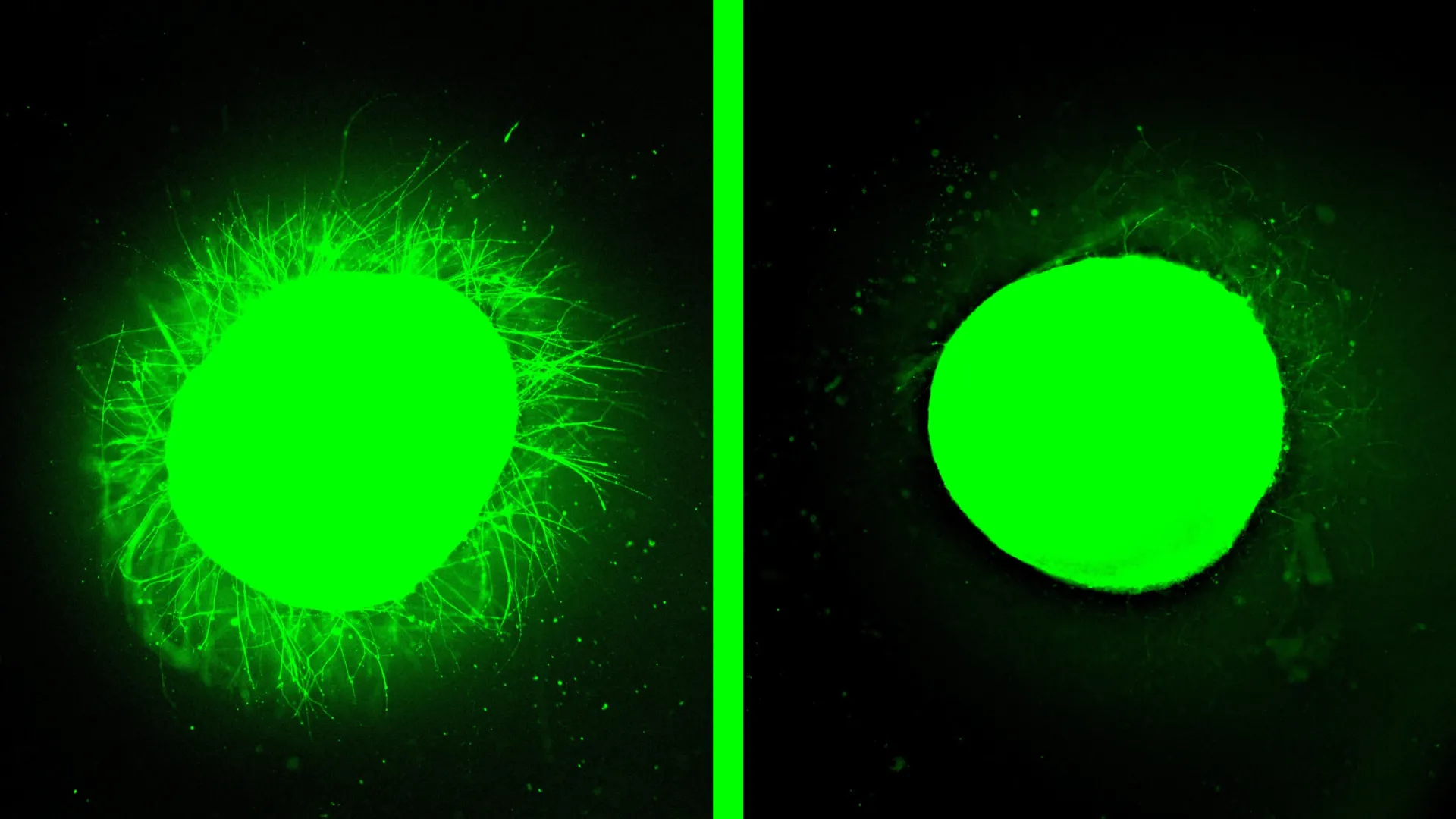
Northwestern University scientists have achieved a significant milestone in spinal cord injury research, developing the most sophisticated laboratory-grown model to date capable of simulating human spinal cord trauma and evaluating novel regenerative treatments. This breakthrough, detailed in the February 11th publication of Nature Biomedical Engineering, offers unprecedented opportunities for understanding the complex biological cascades triggered by injury and for accelerating the development of effective therapies.
The pioneering research centers on human spinal cord organoids – miniature, three-dimensional structures cultivated from stem cells that mimic the intricate architecture and cellular composition of the human spinal cord. For the first time, these organoids have demonstrated the capacity to faithfully reproduce the critical pathological hallmarks of spinal cord injury, including widespread cell death, acute inflammation, and the formation of glial scars. This scar tissue, a dense barrier of astrocytes and extracellular matrix components, is a primary impediment to nerve regeneration, physically and chemically blocking the regrowth of damaged neurons.
A New Era in Spinal Cord Injury Modeling
The creation of this advanced organoid model represents a substantial leap beyond previous efforts. While other research groups have successfully generated spinal cord organoids for fundamental biological studies, the Northwestern team has engineered theirs to be robust enough to withstand and accurately represent traumatic damage. These organoids, measuring several millimeters across, are mature enough to host complex cellular interactions and biological responses characteristic of injury.
A key innovation in this model is the incorporation of microglia, the resident immune cells of the central nervous system. By integrating these cells alongside neurons and astrocytes over several months of guided stem cell differentiation, the researchers have created a more physiologically relevant environment. This allows the organoid to accurately recapitulate the inflammatory cascade that is a hallmark of spinal cord injury, providing a more comprehensive platform for testing therapeutic interventions.
"The ability to introduce microglia into our human spinal cord organoid was a monumental achievement," stated Samuel I. Stupp, the study’s senior author and a leading figure in regenerative materials science at Northwestern. "This means our organoid now possesses all the biochemical signals that the native immune system produces in response to injury. It significantly enhances its realism and accuracy as a model for spinal cord injury."
Simulating Traumatic Injury with Unprecedented Fidelity
To validate the organoid’s capacity to mimic human injury, the researchers subjected the miniature spinal cords to two distinct types of trauma, mirroring common injury mechanisms in humans:
- Laceration Model: A scalpel was used to simulate a clean cut, akin to surgical trauma or penetrating injuries.
- Contusion Model: A controlled impact was applied to replicate compressive forces, comparable to those sustained in severe accidents such as car crashes or falls.
Both injury types elicited predictable and critical biological responses within the organoids. Researchers observed significant cell death, confirming the vulnerability of neural tissue to mechanical stress. Crucially, the formation of glial scars was documented, characterized by the hypertrophic growth and dense packing of astrocytes. This process also involved the production of chondroitin sulfate proteoglycans (CSPGs), extracellular molecules known to actively inhibit axonal regeneration in the injured central nervous system.
"We could clearly differentiate between the astrocytes forming the normal tissue and those constituting the glial scar, which are markedly enlarged and densely packed," explained Professor Stupp. "We also detected the production of chondroitin sulfate proteoglycans, indicating the organoid’s capacity to reproduce key molecular responses to injury."
Promising Results for "Dancing Molecules" Therapy
The true power of this advanced organoid model became evident when it was used to test a revolutionary regenerative treatment developed by Stupp’s laboratory: "dancing molecules." This therapy, a form of supramolecular therapeutic peptide (STP), utilizes the controlled motion of molecules to stimulate tissue repair and potentially reverse paralysis.
The dancing molecules therapy, first introduced in 2021, operates by delivering a liquid injection that rapidly self-assembles into a three-dimensional nanofiber scaffold. This scaffold mimics the extracellular matrix of the spinal cord and provides a dynamic environment for therapeutic molecules. The key to their efficacy lies in their "dancing" – their ability to move rapidly within this network, constantly interacting with cell receptors.
In the Northwestern study, the injured spinal cord organoids were treated with these dancing molecules. The results were profoundly encouraging:
- Reduced Glial Scarring: The dense glial scar tissue, a major obstacle to repair, significantly diminished, becoming barely detectable in treated organoids.
- Neurite Outgrowth: Substantial neurite extension was observed. Neurites are the long, slender projections of neurons, including axons, which are responsible for transmitting electrical and chemical signals. Their regrowth is essential for restoring neural communication.
- Organized Neuronal Growth: The therapy promoted neurons to grow in more organized patterns, suggesting a potential for functional reconnection.
"The glial scar faded significantly to become barely detectable, and we saw neurites growing, resembling the axon regeneration we observed in animals," Professor Stupp confirmed. "This is strong validation that our therapy has a high probability of efficacy in humans."
Background and Context: The Quest for Spinal Cord Repair
Spinal cord injury (SCI) remains one of the most devastating forms of trauma, often resulting in permanent paralysis, loss of sensation, and significant impairment of bodily functions. The current medical landscape offers limited options for true regeneration, with treatments primarily focused on managing symptoms and preventing further damage. The inherent challenges in repairing the central nervous system, particularly the formation of glial scars and the intrinsic inability of mature neurons to regenerate, have long frustrated researchers.
Previous research has relied heavily on animal models, which have provided valuable insights but often fail to fully replicate the complexities of human physiology. The development of human organoids offers a bridge between animal studies and human clinical trials, allowing for more accurate predictions of therapeutic responses in human tissue.
The "dancing molecules" therapy itself stems from years of research into supramolecular chemistry and its applications in medicine. STPs, the broader class to which dancing molecules belong, leverage the power of self-assembly and dynamic molecular motion. This principle is also finding application in other areas of medicine, such as current GLP-1 drugs for diabetes and weight loss, highlighting the broad potential of supramolecular approaches.
Timeline of the Research and Development
The journey leading to this breakthrough can be traced through several key phases:
- Initial Concept of Supramolecular Therapies (Early 2000s): Professor Stupp’s lab began exploring the potential of supramolecular assemblies for biological applications, including early investigations into concepts related to STPs.
- Development of "Dancing Molecules" (Circa 2021): The specific formulation of "dancing molecules" designed for tissue regeneration was first introduced, demonstrating promising results in preclinical animal models.
- Advancements in Spinal Cord Organoid Technology (Ongoing): Concurrent progress in stem cell biology and organoid engineering enabled the creation of increasingly complex and physiologically relevant human tissue models.
- Integration and Validation (Present Study): The Northwestern team successfully integrated the "dancing molecules" therapy with their advanced human spinal cord organoid model, leading to the findings published in Nature Biomedical Engineering.
- FDA Orphan Drug Designation (Recent): The "dancing molecules" therapy recently received Orphan Drug Designation from the U.S. Food and Drug Administration (FDA). This designation is granted to therapies that treat rare diseases or conditions, and it provides incentives for the development of such treatments. For spinal cord injury, this signifies a step towards potential clinical application.
Supporting Data and Scientific Rigor
The study’s findings are underpinned by rigorous experimental data:
- Reproducible Injury Markers: The organoid model consistently demonstrated key markers of SCI, including increased levels of reactive astrocytes (indicating scar formation), elevated inflammatory cytokines, and reduced neuronal viability.
- Quantifiable Neurite Growth: Advanced imaging techniques allowed researchers to quantify the extent of neurite outgrowth, showing a statistically significant increase in treated organoids compared to controls.
- Scar Reduction Metrics: Histological analysis and specific molecular assays confirmed a marked reduction in glial scar components, such as CSPGs, in the presence of the dancing molecules.
- Comparative Efficacy: The study also built upon previous animal experiments, where a single injection of dancing molecules 24 hours post-injury enabled mice to regain locomotion within four weeks. Formulations with faster molecular motion consistently outperformed slower variants, reinforcing the crucial role of dynamic molecular activity.
Expert Perspectives and Broader Implications
The development of this highly realistic human spinal cord injury model and the promising results of the dancing molecules therapy have significant implications for the future of SCI research and treatment.
"One of the most exciting aspects of organoids is that we can use them to test new therapies in human tissue," Professor Stupp emphasized. "Short of a clinical trial, it’s the only way you can achieve this objective. We decided to develop two different injury models in a human spinal cord organoid and test our therapy to see if the results resembled what we previously saw in the animal model."
The implications extend beyond immediate therapeutic development:
- Accelerated Drug Discovery: The organoid model can serve as a powerful platform for screening a wide range of potential SCI therapies, significantly reducing the time and cost associated with preclinical research.
- Personalized Medicine: In the future, it is envisioned that these organoids could be generated from a patient’s own stem cells. This would allow for personalized testing of therapies, tailoring treatments to individual patient biology and potentially reducing the risk of immune rejection if used for tissue transplantation.
- Understanding Disease Mechanisms: The model’s ability to accurately replicate SCI pathology can deepen our understanding of the complex cellular and molecular events that occur after injury, potentially revealing new therapeutic targets.
Future Directions and Challenges
While the current findings are exceptionally promising, the research team acknowledges the need for further development. Professor Stupp indicated plans to engineer even more advanced organoids that can better replicate chronic, long-standing injuries, which are characterized by more robust and persistent scar tissue. The development of such models is critical, as many human SCI cases involve chronic conditions.
The transition from organoid studies to human clinical trials will involve navigating regulatory pathways, scaling up therapy production, and conducting extensive safety and efficacy studies. The recent FDA Orphan Drug Designation for the dancing molecules therapy represents a crucial step in this direction.
The successful integration of cutting-edge organoid technology with a novel regenerative therapeutic marks a pivotal moment in the quest to restore function after spinal cord injury. This Northwestern University breakthrough offers a beacon of hope for millions worldwide affected by this debilitating condition, paving the way for more effective and personalized treatment strategies in the years to come.
