
 By Billy Candra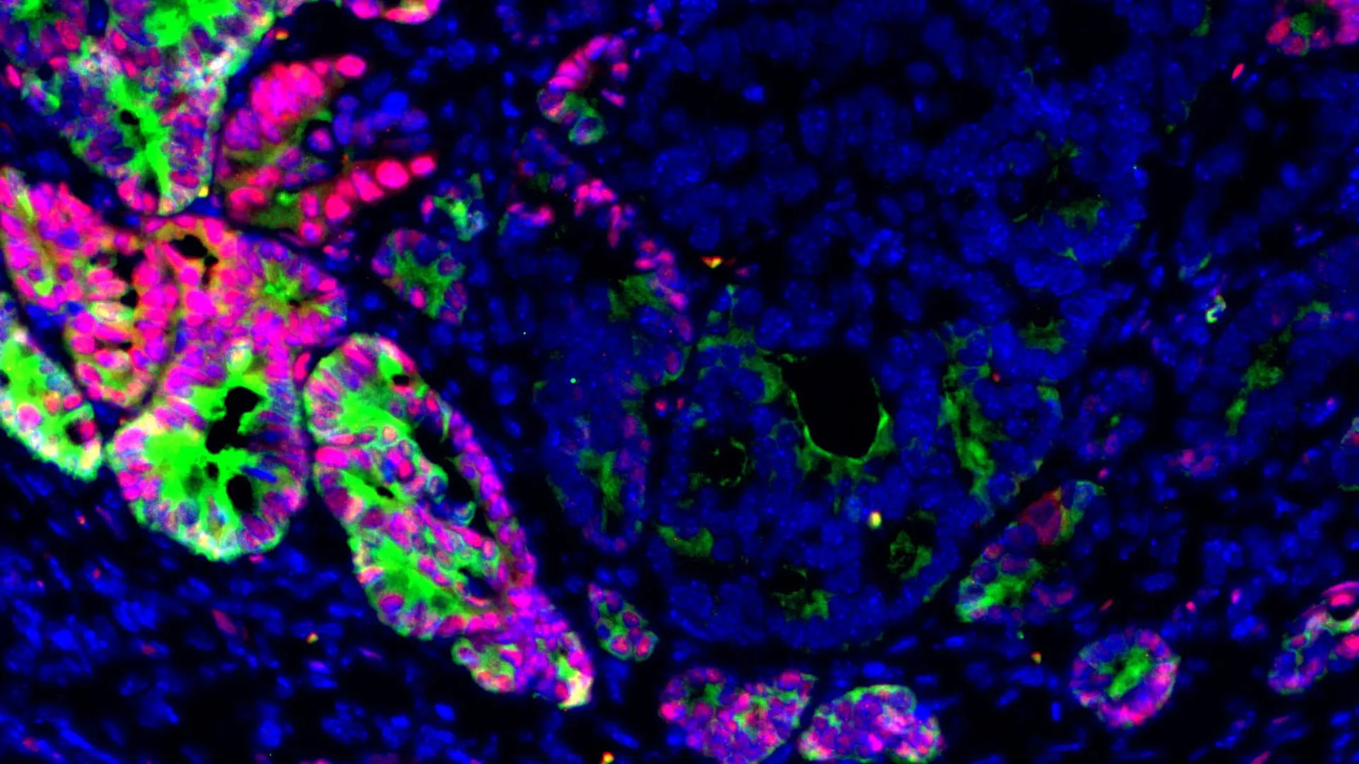
Colorectal cancer (CRC) remains a significant global health challenge, ranking as the third most common cancer and the second leading cause of cancer-related deaths worldwide. In the United States alone, the American Cancer Society estimates over 150,000 new cases and more than 53,000 deaths annually. A primary driver of this grim mortality statistic is metastasis—the process by which cancer cells break away from the primary tumor and establish new tumors in distant organs. For CRC, the liver is the most common site of metastasis, accounting for approximately 70% of all metastatic cases. Once CRC spreads beyond its original site, particularly to the liver, treatment becomes far more challenging, and the five-year survival rate drops precipitously from over 90% for localized disease to less than 15% for metastatic disease. This stark reality underscores the urgent need for a deeper understanding of the mechanisms driving metastasis and for novel strategies to prevent it.
A Novel Epigenetic Mechanism Unveiled
For decades, scientists have diligently searched for specific genetic mutations that might act as clear triggers for liver metastasis in colorectal cancer. However, this quest has largely yielded inconclusive results, with no singular "driver mutation" emerging as a consistent culprit. This lack of clear genetic drivers has prompted researchers to explore alternative mechanisms, leading to the groundbreaking discovery published June 22 in Cell Stem Cell. The study points to a fundamentally different mechanism: epigenetic changes, specifically the loss of the transcription factor GATA6.
GATA6 normally plays a crucial role as a molecular "identity keeper" within the cells lining the intestine. As a transcription factor, GATA6 binds to specific DNA sequences, thereby controlling the transcription of genetic information from DNA to messenger RNA. This intricate regulation dictates which genes are expressed or silenced, ensuring that intestinal cells maintain their specialized functions and differentiated state. It helps these cells perform their specific duties, from nutrient absorption to barrier protection.
The research revealed that GATA6 levels are significantly lower in liver metastases observed in both murine models and human patients with colorectal cancer. Furthermore, reduced GATA6 expression was directly correlated with poorer patient outcomes, reinforcing its critical role in disease progression. "We discovered that GATA6 loss acts as a critical switch that can change cancer cells in the primary tumor from non-metastatic to pro-metastatic," explained Dr. Norihiro Goto, assistant professor of medicine in the Division of Gastroenterology & Hepatology at Weill Cornell and co-leader of the research. "Our findings suggest that epigenetic changes may be more important for promoting liver metastasis than previously understood."
Unlike genetic mutations, which involve permanent alterations to the DNA sequence itself, epigenetic changes modify gene activity without changing the underlying DNA code. These modifications, such as DNA methylation or histone acetylation, influence how tightly DNA is packaged and thus how accessible genes are for transcription. In essence, epigenetics acts like a dimmer switch, turning genes on or off, or modulating their expression levels. This flexibility allows cells to adapt to various environmental cues, but in the context of cancer, it can be hijacked to promote aggressive behaviors like metastasis. Dr. Saori Goto, an instructor in medicine at Weill Cornell, served as the first author of the study, with Dr. Omer H. Yilmaz, associate professor of biology at the Massachusetts Institute of Technology, also co-leading the impactful work.
Innovative Organoid Models Pave the Way
A significant challenge in studying metastasis has been the difficulty in observing the early, critical events that enable cancer cells to spread. Traditional analyses of established liver metastases in patient samples provide only a snapshot of the end-stage process, obscuring the initial transformations. "When researchers analyze patient samples from liver metastases, we fail to capture the important signals occurring in the early stages of the metastatic process," Dr. Norihiro Goto noted.
To overcome this limitation and gain a dynamic understanding of metastatic progression, the research team developed a sophisticated laboratory model utilizing organoids. These miniature, three-dimensional cellular clusters are derived from patient or mouse tumors and meticulously cultured to reproduce many key characteristics of real tumors, including their complex architecture and cellular heterogeneity. The organoid model allowed the researchers to recreate and closely monitor the entire metastatic cascade.
The scientists implanted these organoids into the colons of mice. Over time, these organoids developed into increasingly aggressive primary tumors, which subsequently demonstrated the ability to spread to the liver. Crucially, the team repeated this entire process several times, serially transplanting metastatic cells from one mouse to another. This iterative approach allowed them to observe, in a controlled environment, how cancer cells progressively acquired and refined their metastatic capabilities. This novel methodology provided an unprecedented window into the adaptive changes cancer cells undergo during the early, formative stages of metastasis, offering insights unattainable through conventional tissue analysis.
GATA6: A Critical Switch for Cellular Plasticity
Through their meticulous organoid-based experiments, the researchers uncovered a profound consequence of GATA6 loss: it promotes "lineage plasticity." This term refers to the remarkable ability of cells to alter their identity, function, and behavior, essentially switching from one cell type to another or adopting a more undifferentiated state. In the context of cancer, this plasticity is a formidable adversary.
When GATA6 was absent, colorectal cancer cells activated alternative genetic programs that are typically dormant in differentiated intestinal cells. This activation led them to adopt a highly flexible, "fetal-like" state. Cells in this fetal-like state exhibit characteristics reminiscent of embryonic cells, possessing increased migratory capacity, enhanced survival under stress, and a reduced need for specific microenvironmental cues—all traits highly advantageous for navigating the bloodstream and establishing new colonies in distant organs. These transformed cells were demonstrably better equipped to travel through the bloodstream, survive the harsh conditions of circulation, and successfully colonize the liver, forming secondary tumors.
This type of cellular reshaping is not inherently pathological; it is a vital biological process normally utilized by the body during critical phases such such as embryonic development, wound healing, and tissue regeneration, or in response to severe stress. For instance, during injury, local cells might dedifferentiate or transdifferentiate to help repair damaged tissue. However, in the context of cancer, this same inherent cellular flexibility is co-opted and exploited, becoming a powerful driver of metastasis and disease progression.
One compelling indicator of this induced plasticity was the appearance of cells lacking LGR5 (Leucine-rich repeat-containing G-protein coupled receptor 5). LGR5 is a well-established marker for intestinal stem cells, which are responsible for continuously replenishing the intestinal lining. Earlier research had already indicated that LGR5-negative cells possess a unique ability to initiate liver metastases. The new study conclusively demonstrated that shutting down GATA6 causes cancer cells to undergo a phenotypic shift, transitioning from an LGR5-positive state to an LGR5-negative state. These LGR5-negative cells were found to display the characteristic fetal-like traits and the aggressive ability to disseminate and form tumors in other organs. Conversely, the researchers observed that when GATA6 activity was restored, or when related pathways that mimic GATA6 function were activated, the metastatic potential of colorectal cancer cells was significantly reduced.
Dr. Norihiro Goto further elaborated on these findings: "When we genetically delete GATA6, the frequency and burden of liver metastases in mouse models significantly increase, while having little effect on primary tumor growth." This observation is particularly profound because it suggests that metastasis may not simply be a consequence of a rapidly growing or large primary tumor. Instead, the study posits that metastasis may depend more on specific transitions between cellular states—a qualitative change in cell identity—rather than purely quantitative measures of tumor growth. This distinction represents a significant conceptual shift in understanding metastatic disease.
Implications for Diagnosis and Treatment
The groundbreaking findings from Weill Cornell Medicine and MIT carry profound implications for both the diagnosis and treatment of colorectal cancer, particularly in the context of preventing metastatic spread.
Firstly, the identification of GATA6 loss as a critical epigenetic switch raises the exciting possibility that GATA6 could serve as a novel biomarker for assessing metastatic risk. Clinically, this could translate into a powerful prognostic tool. For instance, biopsies from primary colorectal tumors could be analyzed for GATA6 expression levels. Tumors exhibiting low GATA6 levels might then be flagged as having a higher likelihood of containing cells capable of switching into a metastasis-promoting state. Such information would be invaluable for oncologists, enabling them to stratify patients more effectively. Patients identified as high-risk could then benefit from closer surveillance, more intensive imaging schedules, or even more aggressive adjuvant therapies designed to preemptively target potential micrometastases before they become clinically apparent. This personalized approach could significantly improve patient outcomes by intervening earlier in the disease course.
Secondly, the study points towards a compelling new therapeutic strategy. Instead of solely focusing on eliminating the primary tumor or established metastases, a novel approach could involve maintaining cellular identity or actively preventing cancer cells from entering these highly flexible, pro-metastatic states. This could involve developing drugs that upregulate GATA6 expression, modulate epigenetic pathways to restore a differentiated phenotype, or interfere with the signaling cascades that promote lineage plasticity. Such therapies would aim to "lock" cancer cells into a non-metastatic state, effectively disarming their ability to spread.
However, Dr. Norihiro Goto wisely cautioned that developing such therapies will present a significant challenge. The pathways and mechanisms involved in lineage plasticity and cellular adaptation are often shared with essential biological processes, such as normal tissue repair and regeneration. Therefore, future therapeutic interventions must be highly specific, targeting the cancer-specific exploitation of these programs without disrupting the body’s vital physiological functions. This necessitates a deep understanding of the unique vulnerabilities that arise in GATA6-deficient cancer cells, distinct from their counterparts in healthy tissues.
Future Directions and Collaborative Efforts
The current research serves as a critical stepping stone, opening numerous avenues for future investigation. The immediate focus will be on identifying these unique vulnerabilities within GATA6-deficient cancer cells that could be selectively exploited by novel therapeutic agents. This could involve high-throughput screening of drug libraries or targeted development of small molecules designed to interfere with the specific genetic programs activated in the fetal-like state.
Beyond the intrinsic cellular mechanisms, the research team also plans to delve into the intricate interplay between the tumor and its microenvironment. This includes investigating how surrounding immune cells, stromal components, and specific signals originating from the liver itself influence these critical cellular transitions. The liver, with its unique immunological and metabolic milieu, plays an active role in facilitating metastasis. Understanding these reciprocal interactions in preclinical models will be crucial for developing comprehensive anti-metastatic strategies.
"In addition to treating primary tumors, we need to find strategies to target the mechanism of liver metastasis," Dr. Norihiro Goto emphasized. "Our study is a step toward developing therapies that block the spread of cancer at the earliest stages." This collaborative research, supported by a diverse array of funding bodies including the Astellas Foundation, Japan Society for the Promotion of Science, the National Institutes of Health, Pew-Stewart Trust, AFAR and Glenn Foundation, Kenneth Rainin Foundation, Crohn’s & Colitis Foundation, and Mark Foundation for Cancer Research, underscores the global scientific community’s commitment to unraveling the complexities of cancer metastasis. The ultimate goal is to translate these fundamental discoveries into tangible clinical benefits, offering renewed hope to patients battling colorectal cancer and striving to transform a deadly diagnosis into a manageable condition.
