
 By George Ongkojoyo
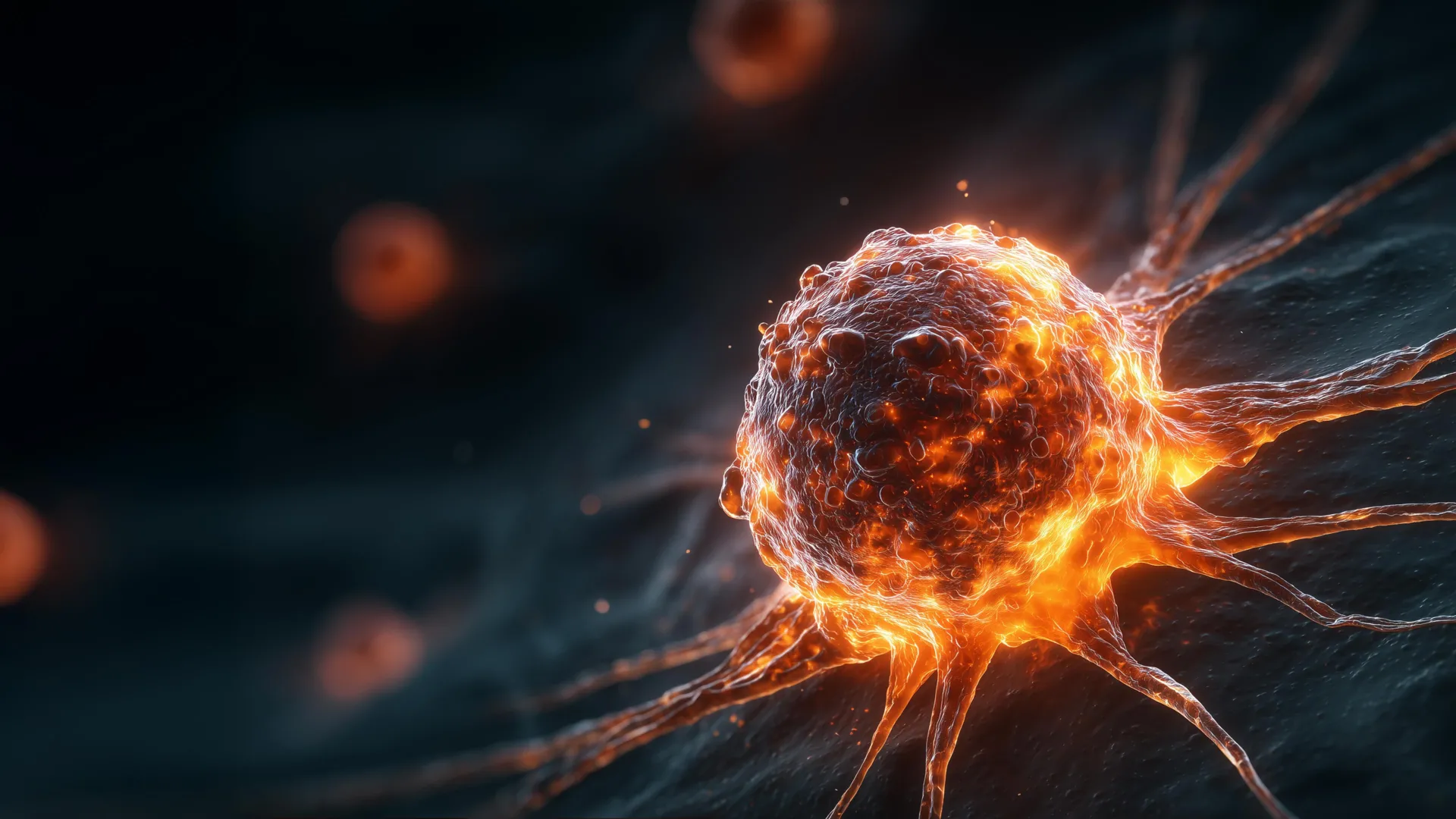
In a series of groundbreaking studies that challenge long-held assumptions in oncology, researchers at Virginia Tech have identified critical factors that drive the aggressiveness of certain tumors, focusing on the role of abnormal chromosome counts and the physical size of cancer cells. The research, spearheaded by cell biologist Daniela Cimini and her team at the Virginia Tech Department of Biological Sciences, reveals that cancer cells with four sets of chromosomes—known as tetraploid cells—act as master manipulators of their environment. These cells not only recruit healthy tissue to facilitate tumor growth but also exhibit a paradoxical relationship between their physical size and their level of malignancy. The findings, published in the Proceedings of the National Academy of Sciences (PNAS) and Cancer Research, provide a new framework for predicting patient outcomes and developing more targeted therapeutic interventions.
The Biological Foundation: Understanding Tetraploidy and Chromosomal Instability
To appreciate the significance of the Virginia Tech findings, one must first understand the standard blueprint of human biology. Most healthy human cells are diploid, containing two complete sets of chromosomes—one set of 23 from each parent, totaling 46. During the process of cell division, or mitosis, a cell duplicates its DNA and splits into two identical daughter cells. However, this process is prone to occasional, catastrophic errors. When a cell duplicates its genetic material but fails to complete the physical act of dividing, the result is a tetraploid cell, which possesses four sets of chromosomes.
While tetraploidy can occur naturally in specific tissues, such as the liver or heart muscle, its presence in other areas of the body is often a precursor to cancer. Chromosomal instability, characterized by the frequent gain or loss of chromosomes, is a hallmark of aggressive tumors. For decades, scientists have known that tetraploid cells are often found at the early stages of cancer development and are associated with a higher likelihood of the disease spreading. What remained a mystery, however, was the precise mechanism by which these abnormal cells influenced the broader tumor ecosystem, especially when they often represent only a minority of the total cell population within a mass.
A Five-Year Chronology of Discovery
The insights gained by the Virginia Tech team were not the result of a single "eureka" moment but rather the culmination of five years of meticulous laboratory investigation. The project involved a collaborative effort between senior faculty and graduate researchers, including Megan Sweet and Mat Bloomfield.
The research timeline began with the development of specialized cell lines. To isolate the effects of tetraploidy, the team took healthy diploid cancer cells and chemically induced them to skip the final stage of division, creating a controlled population of tetraploid cells. This allowed the researchers to compare "normal" cancer cells with their chromosomally doubled counterparts in a highly regulated environment.
The middle phase of the research involved transitioning from petri dishes to in vivo models. By implanting these cells into mice, the team could observe how different cell types interacted with a living host. It was during this stage that Megan Sweet performed the intensive work of histological analysis. Working within a cryostat—a refrigerated chamber designed for precision cutting—Sweet prepared tumor slices thinner than a human hair. These slices were then stained with specific dyes to highlight cellular structures, allowing the team to map the architecture of the growing tumors.
The Recruitment Mechanism: How Cancer Co-opts Healthy Tissue
One of the most significant findings of the Virginia Tech study involves the interaction between tetraploid cancer cells and the surrounding "stroma"—the non-cancerous connective tissue that provides structural support to organs.
In their experiments, Sweet and Bloomfield observed a puzzling phenomenon: as tumors grew, the percentage of tetraploid cells within the mass actually decreased over time. Conventionally, one might assume that a dwindling population of abnormal cells would lead to a less aggressive tumor. The opposite was true. These tumors continued to grow at an accelerated rate and reached much larger sizes than those composed entirely of diploid cells.
The data revealed that the tetraploid cells were functioning as "recruiters." Even in small numbers, these cells emitted biological signals that drew in an abundance of stromal cells. These healthy cells were essentially hijacked, forced to create a supportive scaffold and a nutrient-rich environment that fueled the rapid expansion of the remaining cancer cells. This discovery explains why even a small "sub-clone" of abnormal cells can dictate the clinical trajectory of an entire tumor, acting as a catalyst for malignancy that far outweighs its numerical presence.
The Size Paradox: Why Smaller Cells Pose a Greater Threat
While Sweet focused on the environment of the tumor, Mat Bloomfield investigated the physical characteristics of the tetraploid cells themselves. Because tetraploid cells contain double the genetic material of diploid cells, biological convention dictates that they should be roughly twice as large. Indeed, many of the laboratory-grown tetraploid cells met this expectation.
However, Bloomfield noticed a distinct subset of tetraploid colonies that were 25 to 30 percent smaller than their peers. Upon further testing, these smaller cells proved to be significantly more dangerous. The data indicated that these "small-clone" tetraploid cells possessed several lethal advantages:
- Accelerated Proliferation: They divided faster than their larger counterparts.
- Increased Invasiveness: They demonstrated a higher capacity to break through cellular barriers, a key step in metastasis.
- Drug Resistance: They showed a marked tolerance for common chemotherapy agents and stress-inducing drugs that typically kill cancer cells.
This size-based disparity was observed across multiple cancer types, including colorectal and breast cancer cells. The consistency of this pattern suggests that cellular morphometrics—the measurement of cell shape and size—could serve as a universal biomarker for cancer aggression.
Validating Findings with The Cancer Genome Atlas
To ensure that their observations in mice and lab cultures translated to human health, the Virginia Tech researchers turned to The Cancer Genome Atlas (TCGA). This landmark National Cancer Institute program contains genomic and clinical data from over 20,000 primary cancer and matched normal samples spanning 33 cancer types.
By cross-referencing their laboratory findings with the vast repository of human data in the TCGA, the team confirmed a clear correlation. Patients whose tumors contained smaller tetraploid cells consistently had worse prognoses and lower overall survival rates. This clinical validation elevates the research from a laboratory observation to a potentially life-saving diagnostic tool. If pathologists can incorporate cell size measurements into their standard evaluations, they may be able to identify high-risk patients who require more aggressive or alternative treatment strategies earlier in the process.
Expert Analysis and Future Implications
The implications of this research for the field of precision medicine are profound. For years, the focus of cancer diagnostics has been primarily genomic—identifying specific mutations in the DNA. While genomic sequencing remains vital, the Virginia Tech study suggests that the physical and structural properties of cells (phenotypes) provide a layer of information that genetic testing might miss.
"We already knew that tetraploidy can make cells more tumorigenic," Daniela Cimini noted in a statement regarding the findings. "But now we know that if you incorporate the size of the cells, it can be more predictive of tumorigenic potential."
This research opens several new avenues for investigation:
- Targeting the Recruitment Signal: If scientists can identify the specific chemical signals tetraploid cells use to recruit stromal cells, they may be able to develop drugs that "deafen" the healthy tissue, preventing the tumor from building its support network.
- Size-Specific Therapies: Understanding why smaller tetraploid cells are more resistant to drugs could lead to the development of new compounds that specifically target the metabolic or structural vulnerabilities of these smaller, more aggressive clones.
- Diagnostic Integration: Integrating automated image analysis into pathology workflows could allow for the rapid screening of tumor biopsies for cell size and chromosome count, providing a "danger score" for each patient.
Conclusion: A New Frontier in Oncology
The work being conducted in the laboratories of Virginia Tech represents a shift toward a more holistic understanding of cancer. By looking at the intersection of genetics, cell biology, and tissue ecology, researchers are beginning to understand not just what cancer is, but how it behaves as a complex, predatory system.
For researchers like Megan Sweet and Mat Bloomfield, the journey continues. The team plans to delve deeper into the molecular pathways that allow a cell with four sets of chromosomes to shrink in size while gaining in strength. As they continue to slice, stain, and analyze, their work provides a beacon of hope for more accurate diagnoses and more effective treatments in the ongoing battle against cancer. The discovery that the most dangerous threats can be found in the smallest packages—and that they rely on the recruitment of our own healthy cells—is a sobering but essential step forward in modern medical science.
