
 By Nana O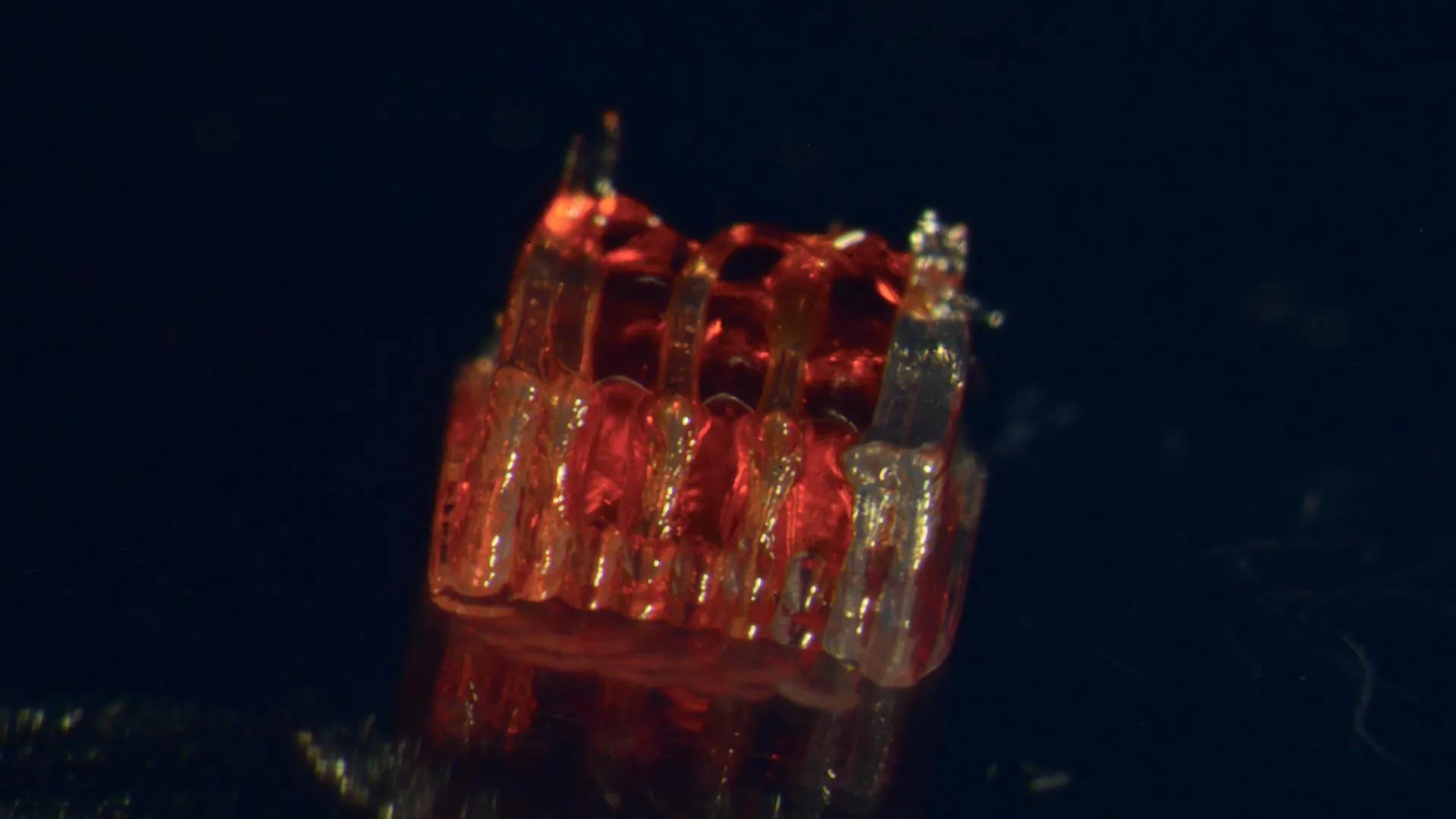
A pioneering research effort at the University of Minnesota Twin Cities has unveiled a transformative approach to spinal cord injury recovery, integrating for the first time 3D printing technology with advanced stem cell biology and the creation of lab-grown tissues. This groundbreaking work, detailed in the prestigious peer-reviewed journal Advanced Healthcare Materials, offers a beacon of hope for millions worldwide affected by debilitating spinal cord damage.
The Unmet Challenge of Spinal Cord Injury
Spinal cord injuries represent one of the most devastating forms of trauma, frequently resulting in permanent paralysis and a profound loss of function. According to the National Spinal Cord Injury Statistical Center, over 300,000 individuals in the United States live with the consequences of these injuries. Despite decades of research, the medical community has yet to achieve a complete reversal of the damage, a significant limitation attributed to the fundamental biological hurdles of nerve cell death and the inherent inability of severed nerve fibers to regenerate across the injury site. This new research directly confronts these long-standing obstacles.
A Novel Biomimetic Approach: The Organoid Scaffold
The core of this innovative method lies in the creation of a sophisticated, three-dimensional printed framework, termed an "organoid scaffold." This scaffold is meticulously designed with microscopic channels, mimicking the intricate architecture of the natural spinal cord. These channels serve as a precisely controlled environment to guide the growth of specialized cells.
The research team then populates these channels with regionally specific spinal neural progenitor cells (sNPCs). These are not ordinary cells; they are derived from human adult stem cells, endowing them with the remarkable plasticity to divide and differentiate into the specific types of mature cells required for neural function, including neurons and supporting glial cells.
"We leverage the 3D printed channels of the scaffold to meticulously direct the growth of the stem cells," explained Guebum Han, a former postdoctoral researcher in mechanical engineering at the University of Minnesota and the study’s first author, who has since moved to Intel Corporation. "This controlled environment ensures that the new nerve fibers extend in the desired, organized fashion. The system we’ve engineered effectively creates a biological relay, designed to bypass the damaged segment of the spinal cord when implanted."
Translating Lab Innovation to Biological Reality: The Rat Model
To validate their approach, the researchers conducted a series of critical experiments. They transplanted these custom-designed organoid scaffolds into rats that had sustained complete severing of their spinal cords, a severe injury model designed to mimic catastrophic human trauma. The results were profoundly encouraging.
The implanted sNPCs within the scaffold not only survived but also successfully differentiated into functional neurons. Crucially, these newly formed nerve fibers demonstrated the capacity to extend in both directions – rostral (towards the head) and caudal (towards the tail) – effectively bridging the severed gap. This bidirectional growth allowed them to establish new synaptic connections with the host’s existing, intact nerve circuitry.
Over time, the integration of these new nerve cells into the host spinal cord tissue was observed to be seamless. This integration was directly correlated with significant functional recovery in the experimental animals, indicating that the transplanted neural tissue was not merely surviving but actively participating in restoring lost motor and sensory pathways.
The Promise of Regenerative Medicine and Clinical Translation
The potential implications of this research are far-reaching, ushering in a new era of possibility within regenerative medicine. Ann Parr, a professor of neurosurgery at the University of Minnesota and a senior figure in the study, expressed optimism about the future. "Regenerative medicine has indeed brought about a new era in spinal cord injury research," Professor Parr stated. "Our laboratory is incredibly excited to explore the future potential of these ‘mini spinal cords’ and their capacity for clinical translation."
While acknowledging that this work is still in its nascent stages, the development represents a significant leap forward. The researchers are actively pursuing strategies to scale up the production of these sophisticated organoid scaffolds and are committed to refining this synergistic combination of technologies for eventual human clinical applications. The ultimate goal is to provide a viable therapeutic option for individuals currently facing the profound challenges of spinal cord injury.
Building on a Foundation of Expertise: The Research Team
This groundbreaking achievement is the culmination of a multidisciplinary effort, bringing together leading researchers from various departments at the University of Minnesota and beyond. In addition to Han and Parr, key contributors to this study include:
- From the University of Minnesota Department of Mechanical Engineering: Hyunjun Kim and Michael McAlpine. Their expertise in advanced manufacturing and 3D printing was instrumental in designing the intricate scaffold architecture.
- From the University of Minnesota Department of Neurosurgery: Nicolas S. Lavoie, Nandadevi Patil, and Olivia G. Korenfeld. Their profound understanding of spinal cord physiology and surgical techniques was critical for the implantation and functional assessment.
- From the University of Minnesota Department of Neuroscience: Manuel Esguerra. His insights into neural development and regeneration provided crucial biological guidance.
- From the Department of Physics at Virginia Commonwealth University: Daeha Joung. His contributions likely focused on the physical properties and interactions within the engineered tissue.
This collaborative spirit, bridging engineering, biology, and medicine, is a hallmark of modern scientific advancement and was essential to overcoming the complex challenges inherent in this research.
A Timeline of Innovation and Future Directions
The journey leading to this publication likely involved several years of dedicated research, building upon prior advancements in 3D printing and stem cell therapies. While a precise chronological breakdown of the study’s development is not provided, a typical research progression for such a project would involve:
- Initial Conceptualization and Design (Years 1-2): Researchers would have begun by conceptualizing the idea of combining 3D printing with stem cells for spinal cord repair. This phase would involve designing the specific geometry of the scaffold, selecting appropriate biomaterials, and identifying the optimal types of stem cells.
- Scaffold Fabrication and Optimization (Years 2-3): The 3D printing aspect would have been refined to achieve the necessary precision and create the microscopic channels required. Material biocompatibility testing would also be a key component.
- Cell Culture and Differentiation Protocols (Years 3-4): Establishing robust protocols for culturing and differentiating human adult stem cells into sNPCs would be a significant undertaking. Ensuring regional specificity of these cells would be crucial for success.
- In Vitro Testing and Proof of Concept (Years 4-5): Initial experiments would likely have been conducted in laboratory settings, testing the viability of cells within the scaffolds and observing their growth and differentiation patterns without implantation.
- In Vivo Animal Studies (Years 5-7): The successful transplantation into rat models, as described in the publication, would represent a major milestone. This phase would involve meticulous surgical procedures, long-term monitoring of the animals, and rigorous assessment of functional recovery.
- Data Analysis and Manuscript Preparation (Years 7-8): Once sufficient data was gathered, the researchers would have embarked on comprehensive data analysis, interpretation, and the writing of the scientific manuscript for peer review and publication.
The current publication marks a significant point in this timeline, serving as a validation of the core concept and a platform for future endeavors.
Funding and Support for Advancing Spinal Cord Injury Research
This vital research was made possible through substantial support from various funding bodies, underscoring the recognized importance of addressing spinal cord injuries. The study received financial backing from:
- The National Institutes of Health (NIH): A leading federal agency dedicated to advancing fundamental knowledge and improving health through biomedical research. NIH funding is critical for high-impact, long-term scientific investigations.
- The State of Minnesota Spinal Cord Injury and Traumatic Brain Injury Research Grant Program: This state-specific program highlights a commitment at the regional level to supporting research aimed at alleviating the suffering caused by these types of injuries.
- The Spinal Cord Society: A non-profit organization dedicated to funding research and supporting individuals affected by spinal cord injuries. Their contributions often play a crucial role in accelerating promising research projects.
The collective investment from these organizations reflects a broad recognition of the urgent need for innovative treatments for spinal cord injuries and the potential of novel approaches like the one developed at the University of Minnesota.
The Broader Impact and Future Outlook
The implications of this research extend far beyond the laboratory. For individuals living with spinal cord injuries, this work represents a tangible advancement towards regaining lost function and improving their quality of life. The development of "mini spinal cords" that can integrate with existing neural networks offers a paradigm shift from managing symptoms to actively repairing damage.
The success in animal models provides a strong foundation for future human clinical trials. However, the transition from animal studies to human application is a complex and lengthy process, involving stringent regulatory approvals, extensive safety testing, and further refinement of the technology.
Key areas for future development include:
- Scaling Up Production: Developing methods to produce these complex organoid scaffolds efficiently and cost-effectively for widespread use.
- Long-Term Efficacy and Safety: Conducting further studies to assess the long-term durability of the regenerated connections and ensure the absence of adverse effects.
- Personalized Medicine Approaches: Investigating whether the scaffolds and cell populations can be tailored to individual injury types and patient characteristics.
- Minimally Invasive Implantation Techniques: Exploring surgical methods that reduce the invasiveness of the transplantation procedure.
The scientific community will be closely watching the progress of this research, as it holds the potential to fundamentally alter the landscape of spinal cord injury treatment and offer a renewed sense of hope to millions. The University of Minnesota Twin Cities has positioned itself at the forefront of this exciting frontier in regenerative medicine.
