
 By Nana O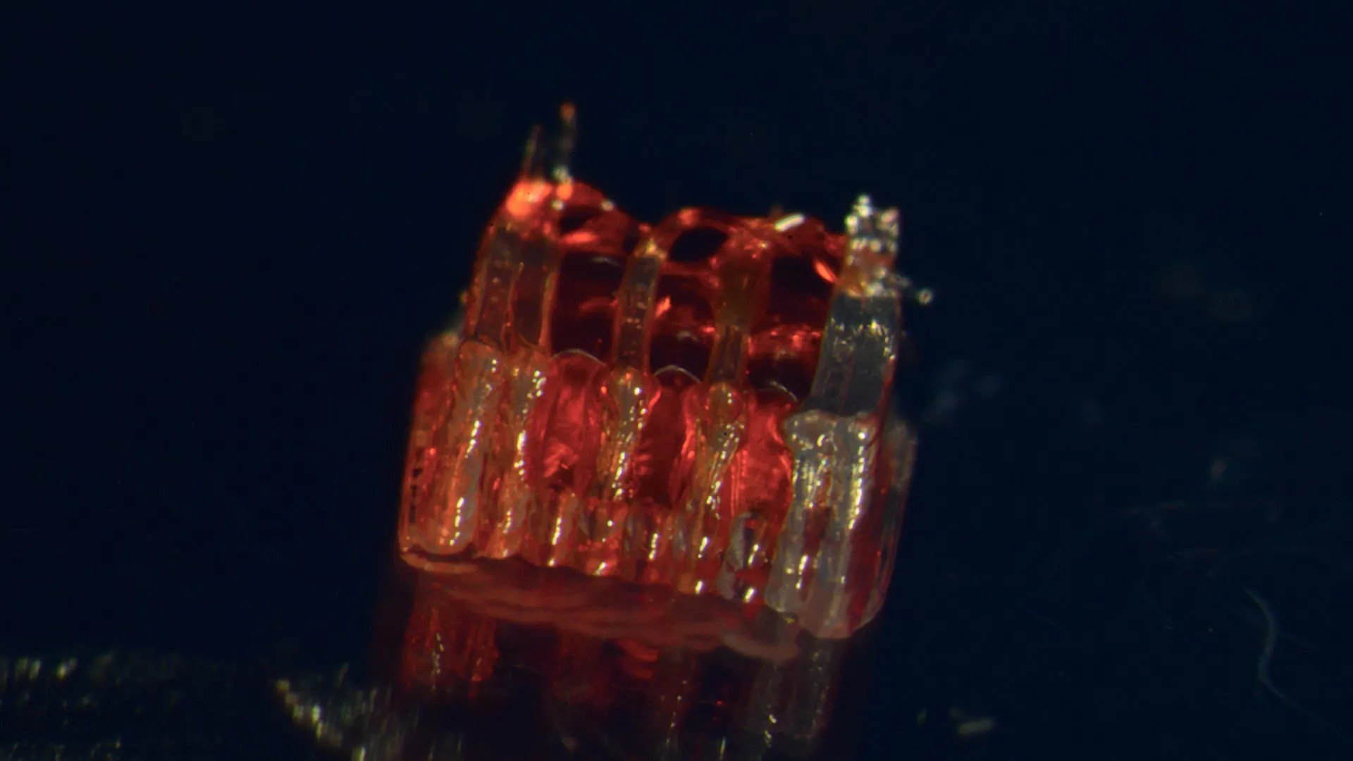
A pioneering research effort by a team at the University of Minnesota Twin Cities has unveiled a groundbreaking therapeutic strategy for spinal cord injuries, meticulously integrating advanced 3D printing technology with the intricate biology of stem cells and the creation of lab-grown tissues. This innovative approach, detailed in a recent publication in the esteemed peer-reviewed scientific journal Advanced Healthcare Materials, offers a novel pathway toward restoring function and potentially reversing the devastating effects of spinal cord damage.
The Unmet Challenge of Spinal Cord Injury
Spinal cord injuries represent a profound public health crisis, impacting hundreds of thousands of lives annually. In the United States alone, the National Spinal Cord Injury Statistical Center reports that over 300,000 individuals live with the consequences of such injuries, which often result in permanent paralysis and a drastic reduction in quality of life. The fundamental biological hurdles to recovery are formidable: the death of critical nerve cells and the intrinsic inability of damaged nerve fibers, known as axons, to regenerate across the injury site. This biological impasse has long frustrated medical science, leaving patients with limited options for regaining lost function. The University of Minnesota’s new research directly confronts these challenges, proposing a multifaceted solution designed to bridge the gap in the damaged nervous system.
A Novel Bio-Integrated Scaffold
At the core of this breakthrough is the creation of a sophisticated, three-dimensional printed framework, termed an organoid scaffold. This scaffold is not merely a passive structure; it is engineered with a network of microscopic channels, meticulously designed to guide cellular growth and organization. The ingenuity lies in populating these channels with regionally specific spinal neural progenitor cells (sNPCs). These sNPCs are derived from human adult stem cells, possessing the remarkable plasticity to divide and differentiate into the various specialized cell types that constitute the nervous system, including neurons.
Dr. Guebum Han, a former postdoctoral researcher in mechanical engineering at the University of Minnesota and the lead author of the study, explained the strategic importance of the 3D-printed channels. "We use the 3D printed channels of the scaffold to direct the growth of the stem cells, which ensures the new nerve fibers grow in the desired way," Dr. Han stated. He further elaborated on the functional outcome: "This method creates a relay system that when placed in the spinal cord bypasses the damaged area." This "relay system" is conceptually akin to creating a biological bridge, allowing neural signals to circumvent the site of injury and reconnect with the intact portions of the spinal cord.
Pre-Clinical Success in Rodent Models
To rigorously test the efficacy of their novel approach, the research team conducted pre-clinical trials. They transplanted these bio-engineered scaffolds into rats that had sustained complete transections of their spinal cords – a severe injury model that closely mimics the devastating functional loss seen in human patients. The results were profoundly encouraging. The sNPCs within the scaffolds not only survived but also successfully differentiated into mature neurons. Crucially, these newly formed neurons extended their axons in both directions – rostral (towards the head) and caudal (towards the tail) – effectively bridging the severed spinal cord.
The study’s findings indicate that these new nerve fibers formed functional connections with the host animal’s existing neural circuitry. Over time, the newly generated nerve cells integrated seamlessly into the host spinal cord tissue. This integration was accompanied by a significant recovery of motor function in the rats, a testament to the therapeutic potential of the engineered organoid scaffolds. The ability of these transplanted cells to not only survive and grow but also to functionally integrate and restore lost neurological pathways represents a significant leap forward in regenerative medicine for spinal cord injuries.
The Promise of Regenerative Medicine
Professor Ann Parr, a leading figure in neurosurgery at the University of Minnesota and a key contributor to the research, expressed optimism about the broader implications of their work. "Regenerative medicine has brought about a new era in spinal cord injury research," Professor Parr remarked. She further highlighted the team’s future aspirations: "Our laboratory is excited to explore the future potential of our ‘mini spinal cords’ for clinical translation." The term "mini spinal cords" aptly describes the complex, functional neural tissue engineered within the scaffolds, suggesting a future where such constructs could become standard therapeutic tools.
Chronology of a Breakthrough
The journey leading to this groundbreaking discovery is a testament to sustained scientific inquiry and interdisciplinary collaboration. While the exact timeline of the research project is not explicitly detailed in the initial report, the publication in Advanced Healthcare Materials signifies the culmination of years of foundational research in 3D bioprinting, stem cell biology, and neuroregeneration.
- Foundational Research: Years of independent research in 3D printing capabilities for biomedical applications, understanding the differentiation pathways of neural progenitor cells, and exploring biomaterials for tissue engineering likely preceded this integrated project.
- Conceptualization and Design: The development of the organoid scaffold concept, including the design of the micro-channel architecture, would have been a critical early phase. This likely involved extensive computational modeling and material science expertise.
- Cell Sourcing and Optimization: The isolation and characterization of human adult stem cells and their differentiation into sNPCs would have been a parallel and ongoing effort, ensuring a reliable and effective cell source.
- Scaffold-Cell Integration: The crucial step of populating the 3D-printed scaffolds with sNPCs and optimizing the culture conditions to promote directed growth and differentiation would have been a significant experimental phase.
- Pre-clinical Testing: The rigorous transplantation studies in rodent models, as described in the published paper, represent the validation phase of the technology. This would have involved meticulous surgical procedures, long-term observation of animal subjects, and extensive functional assessments.
- Data Analysis and Publication: The compilation and analysis of experimental data, followed by the rigorous peer-review process for publication in a high-impact journal like Advanced Healthcare Materials, marks the formal dissemination of the research findings.
This chronological progression underscores the complexity and iterative nature of scientific discovery, involving multiple stages of hypothesis, experimentation, and validation.
Supporting Data and Scientific Context
The statistical prevalence of spinal cord injuries underscores the urgent need for effective treatments. Each year, approximately 17,000 new cases of spinal cord injury occur in the United States, adding to the substantial population already living with this condition. These injuries can result from various causes, including motor vehicle accidents, falls, violence, and medical conditions such as tumors or vascular malformations. The economic burden is also immense, with lifetime costs for individuals with spinal cord injuries ranging from hundreds of thousands to millions of dollars, depending on the severity of the injury and the level of paralysis.
The biological mechanisms underlying spinal cord injury are complex and multi-faceted. Upon initial trauma, there is immediate cell death due to mechanical forces and disruption of blood supply. This is followed by a secondary injury cascade involving inflammation, excitotoxicity (overstimulation of neurons by neurotransmitters), and glial scar formation. Glial scars, primarily composed of astrocytes, form a physical and chemical barrier that actively inhibits axon regeneration. Traditional therapeutic approaches have focused on managing symptoms, rehabilitation, and in some cases, surgical decompression to prevent further damage. However, these methods do not address the fundamental problem of neural regeneration.
The University of Minnesota’s approach tackles these issues by:
- Providing a Guiding Structure: The 3D-printed scaffold offers a physical conduit that bypasses the scar tissue and guides regenerating axons along a defined path.
- Delivering Potent Neural Progenitors: The sNPCs are specifically chosen for their ability to differentiate into neurons and other supporting glial cells, providing the building blocks for new neural tissue.
- Directing Growth: The micro-channels within the scaffold are engineered to promote directional growth, preventing aberrant connections and ensuring that axons reach their intended targets.
- Promoting Integration: The study demonstrated that these new neurons can functionally integrate with the host nervous system, a critical step for restoring lost function.
Broader Impact and Future Directions
While this research is still in its nascent stages, it represents a significant beacon of hope for individuals affected by spinal cord injuries and for the broader field of regenerative medicine. The potential implications are far-reaching:
- Restoration of Motor Function: The primary goal is to enable individuals to regain control over their limbs, improving mobility and independence.
- Recovery of Sensory Function: Beyond motor control, the technology could potentially restore lost sensation, impacting areas such as touch, temperature, and pain perception.
- Management of Autonomic Dysfunction: Spinal cord injuries often disrupt autonomic functions like bladder and bowel control, as well as blood pressure regulation. Successful neural regeneration could help restore these vital functions.
- Advancement of Tissue Engineering: This work pushes the boundaries of what is possible in 3D bioprinting and stem cell applications, paving the way for similar approaches to treat other neurological conditions or organ damage.
The research team acknowledges the need for further development and scaling. Future research will likely focus on optimizing the scaffold materials, refining the cell differentiation protocols, and conducting more extensive long-term studies in larger animal models. The ultimate goal is to translate these promising pre-clinical findings into safe and effective clinical trials for human patients. The collaborative nature of this research, involving expertise from mechanical engineering, neurosurgery, neuroscience, and physics, is a model for addressing complex biomedical challenges.
The team’s collaborative spirit is evident in the diverse composition of the research group, which includes:
- University of Minnesota Department of Mechanical Engineering: Hyunjun Kim and Michael McAlpine.
- University of Minnesota Department of Neurosurgery: Nicolas S. Lavoie, Nandadevi Patil, and Olivia G. Korenfeld.
- University of Minnesota Department of Neuroscience: Manuel Esguerra.
- Virginia Commonwealth University, Department of Physics: Daeha Joung.
This interdisciplinary approach is crucial for integrating cutting-edge technologies and biological insights to tackle a problem as complex as spinal cord injury.
Funding and Acknowledgement
The progress made in this research was made possible through significant financial support from several key organizations:
- The National Institutes of Health (NIH), a primary funder of biomedical research in the United States, providing vital resources for innovative scientific endeavors.
- The State of Minnesota Spinal Cord Injury and Traumatic Brain Injury Research Grant Program, demonstrating a commitment at the state level to addressing these debilitating conditions.
- The Spinal Cord Society, a non-profit organization dedicated to advancing research and improving the lives of individuals with spinal cord injuries.
The full findings of this transformative research can be accessed in the paper titled "3D-Printed Scaffolds Promote Enhanced Spinal Organoid Formation for Use in Spinal Cord Injury" published on the Advanced Healthcare Materials website. This publication marks a significant milestone in the ongoing quest for effective treatments for spinal cord injuries, offering a tangible path toward restoring hope and function for millions worldwide.
