
 By Suherman Candra Maholtra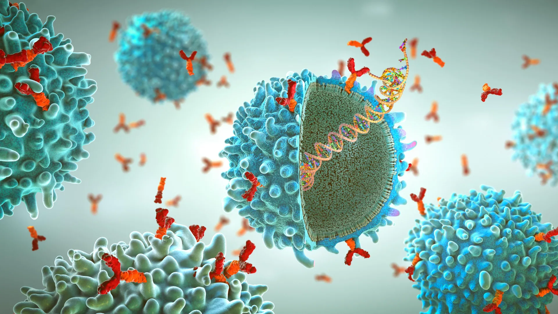
The field of vaccinology has long contended with a fundamental biological hurdle: the difficulty of studying viral surface proteins in their native, functional states. In a landmark study published in Nature Communications, a multidisciplinary team led by Scripps Research and IAVI (formerly the International AIDS Vaccine Initiative) has unveiled a sophisticated nanodisc platform designed to bridge the gap between laboratory models and real-world viral structures. By utilizing lipid-based nanodiscs to anchor these proteins, researchers can now observe how the human immune system interacts with pathogens like HIV and Ebola in a context that mirrors an actual infection. This advancement promises to streamline the vaccine development pipeline, reducing the time required for critical analytical processes from months to a single week, while providing unprecedented structural clarity for drug designers.
The Challenge of Membrane-Bound Proteins
To understand the significance of this new platform, one must first recognize the architectural complexity of viruses. Most viruses are enveloped in a lipid membrane—a fatty outer layer—studded with specialized proteins called glycoproteins. These proteins act as the "keys" that allow the virus to unlock and enter human cells. Consequently, they are the primary targets for vaccines, which aim to train the immune system to recognize and neutralize these keys before an infection can take hold.
However, studying these proteins in a laboratory setting is notoriously difficult. In their natural state, glycoproteins are deeply embedded within the viral membrane. The portion of the protein that sits inside the membrane is hydrophobic, meaning it repels water. For decades, scientists have struggled to isolate these proteins for study because, once removed from their lipid environment, they often lose their shape, become unstable, or fail to function correctly. To circumvent this, researchers frequently use "truncated" versions of the proteins—simplified models that have the membrane-anchoring section removed. While these versions are easier to handle and soluble in water, they lack the structural integrity of the real virus. Crucially, they often hide the "base" of the protein, a region near the membrane that is a prime target for some of the most powerful antibodies known to science.
The Nanodisc Solution: Mimicking the Viral Envelope
The breakthrough platform developed by Scripps Research utilizes nanodisc technology to solve this structural dilemma. Nanodiscs are synthetic, disc-shaped assemblies of lipids stabilized by "membrane scaffold proteins" (MSPs). These discs act as microscopic patches of a cell membrane, providing a stable, water-soluble environment for membrane-bound proteins.
By incorporating full-length viral glycoproteins into these nanodiscs, the research team, led by first author Kimmo Rantalainen and co-senior author William Schief, created a "mimic" of the virus’s outer surface. This setup allows the proteins to maintain their natural trimeric shape—the three-part structure typical of HIV and Ebola surface proteins. Unlike previous methods that relied on harsh detergents to stabilize proteins—which often interfered with delicate biochemical assays—the nanodisc platform is highly compatible with standard laboratory tools. This includes high-resolution imaging techniques like cryo-electron microscopy (cryo-EM), as well as immune cell sorting and antibody binding tests.
Chronology of Development and Testing
The development of this platform is the culmination of years of collaborative effort between Scripps Research, IAVI’s Neutralizing Antibody Center, and industrial partners like Moderna Inc. The research began with the recognition that traditional vaccine "baits"—the molecules used to find and study rare immune cells—were insufficient for targeting the most elusive parts of the HIV virus.
The team first applied the nanodisc platform to HIV-1, a virus that has evaded a successful vaccine for over forty years due to its rapid mutation and the complex "glycan shield" that hides its surface proteins. The researchers focused on the Membrane-Proximal External Region (MPER), a highly conserved area at the base of the HIV envelope protein. Because the MPER is located so close to the viral membrane, it is nearly impossible to study using truncated proteins. Using the nanodisc platform, the team was able to capture high-resolution structural views of how broadly neutralizing antibodies (bNAbs) bind to the MPER.
Following the success with HIV, the researchers expanded their testing to the Ebola virus. Ebola presents a different set of challenges, as its glycoprotein is structurally distinct from HIV’s. The platform successfully stabilized the Ebola proteins, allowing the team to confirm that antibodies could recognize the "natural" version of the protein within the lipid environment. This cross-virus success suggests that the platform is a "plug-and-play" system that can be adapted for a wide variety of pathogens, including influenza, SARS-CoV-2, and even emerging "Disease X" threats.
Supporting Data: Efficiency and Precision
One of the most striking outcomes of the study is the gain in operational efficiency. In traditional vaccine research, the process of characterizing how a candidate vaccine interacts with the immune system—a phase known as vaccine analytics—is a slow and iterative process. Isolating specific B-cells (the cells that produce antibodies) and testing their binding affinity to a viral protein often took upwards of four to six weeks.
The nanodisc platform has truncated this timeline significantly. By providing a stable and reliable "bait" for immune cells, the system allows researchers to complete these same analyses in approximately seven days. This 75% reduction in time is critical during a pandemic or when comparing dozens of different vaccine formulations simultaneously.
Furthermore, the data generated by the platform provides a higher degree of precision. In their HIV experiments, the researchers observed that the lipid environment of the nanodisc actually influenced how certain antibodies approached the protein. Some antibodies require a specific angle of attack that is only possible when the protein is anchored in a membrane. Truncated models, which lack this anchor, can lead to "false positives" or "false negatives" in the lab, where an antibody appears to work in a test tube but fails in a living organism. The nanodisc platform minimizes these discrepancies, providing a more accurate predictive model for vaccine efficacy.
Official Responses and Expert Insights
The implications of this research have been met with enthusiasm from the global scientific community. William Schief, a professor at Scripps Research and executive director of vaccine design at IAVI, emphasized the importance of context in structural biology. "For many years, we’ve had to rely on versions of viral proteins that are missing important pieces," Schief stated. "Our platform lets us study these proteins in a setting that better reflects their natural environment, which is critical if we want to understand how protective antibodies recognize a virus."
Kimmo Rantalainen, a senior scientist in Schief’s lab, highlighted the scalability of the discovery. "Putting all of these components together into a single, reliable system was the key," Rantalainen noted. "The individual pieces already existed, but making them work together in a way that’s reproducible and scalable opens up new possibilities for how vaccines are analyzed and designed."
The study also involved contributions from Andrew B. Ward’s lab at Scripps Research, renowned for its work in cryo-EM. The structural insights provided by the Ward lab were essential in proving that the nanodiscs were not just holding the proteins, but were holding them in the exact configuration they assume on the surface of a live virus.
Broader Impact and Future Implications
The development of the nanodisc platform arrives at a pivotal moment in global health. As the world moves toward "rational vaccine design"—a process where vaccines are engineered based on a precise understanding of molecular structures rather than through trial and error—tools that provide high-fidelity structural data are in high demand.
Beyond HIV and Ebola, the platform has immediate applications for the "universal flu vaccine," a long-held goal of the National Institutes of Health (NIH). Like HIV, the influenza virus has a conserved "stem" region near its membrane that could be the key to a vaccine that protects against all strains. The nanodisc platform is uniquely suited to help researchers target this stem.
Additionally, the platform’s ability to be used as a diagnostic tool should not be overlooked. It could be used to screen the blood of patients who have recovered from a virus to identify the most potent antibodies they produced, which can then be developed into monoclonal antibody therapies.
The research was supported by a diverse array of funding bodies, including the National Institute of Allergy and Infectious Diseases (NIAID), the Bill & Melinda Gates Foundation, and the Alexander von Humboldt Foundation. This broad support underscores the consensus that improving the foundational tools of vaccine science is a global priority.
In conclusion, while the nanodisc platform is not a vaccine itself, it represents a significant upgrade to the scientific "toolkit." By allowing researchers to see viruses as they truly are—embedded in their protective membranes—this technology removes a major blind spot in drug discovery. As this platform becomes more widely adopted, it is expected to accelerate the delivery of next-generation vaccines for some of the most persistent and dangerous viral threats facing humanity today.
